Downloaded 1,158 times


![29
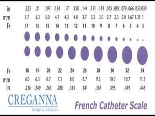
A catheter of 1 French has an
external diameter of 1⁄3 mm,[1] and therefore
the diameter of a round catheter in
millimeters can be determined by dividing the
French size by 3:
D (mm) = Fr / 3
or
Fr = D (mm) * 3
For example, if the French size is 9, the
diameter is 3 mm.](https://image.slidesharecdn.com/cathetersguidewires-150731190948-lva1-app6892/85/Catheters-guidewires-29-320.jpg)


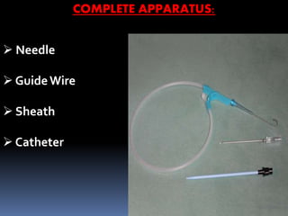


Guide wires and catheters are medical devices used in angiographic procedures. Guide wires are stainless steel wires that guide catheters through blood vessels. They have flexible tips and come in various lengths and diameters. Catheters are hollow tubes inserted into the body and come in different materials, sizes, and tip configurations. Common uses are angiography, drainage of fluids, and placement of stents and balloons. Both must be carefully sterilized between uses to prevent infection.


