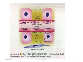
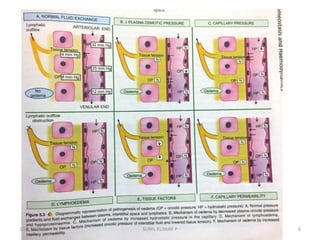
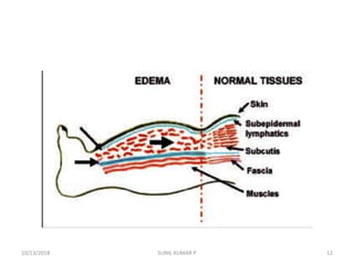
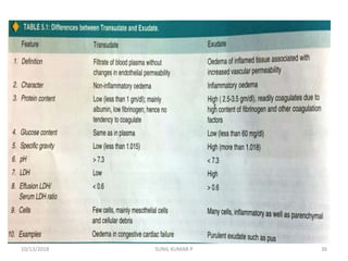
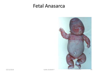
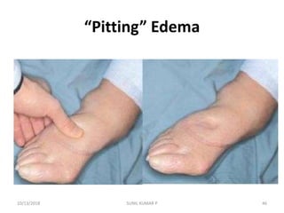
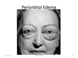
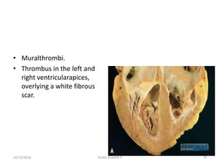
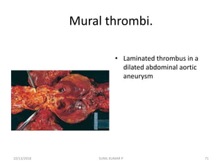
Hemodynamic Disorders
P SUNIL KUMAR
Haematology & Transfusion Medicine
St.John’s Medical College
Bangalore
10/13/2018 1SUNIL KUMAR P
Hemodynamic Disorders
 EDEMA
 THROMBOSIS
 EMBOLISM
 INFARCTION
10/13/2018 2SUNIL KUMAR P
• Normal fluid homeostasis is maintained by vessel
wall integrity, intravascular pressure and
osmolarity with in certain physiologic ranges
• Changes in intravascular volume,
• Pressure, or
• Protein content, or
• Alterations in endothelial function will affect the
movement of water across the vascular wall.
10/13/2018 3SUNIL KUMAR P
10/13/2018 4SUNIL KUMAR P
10/13/2018 5SUNIL KUMAR P
10/13/2018 6SUNIL KUMAR P
• Edema (increased fluid in the ECF)
• Hyperemia (INCREASED flow)
• Hemorrhage (extravasation)
• Hemostasis (opposite of thrombosis)
10/13/2018 7SUNIL KUMAR P
• Hemostasis (opposite of thrombosis)
• Thrombosis (inappropriate clotting blood)
• Embolism (downstream travel of a clot)
• Infarction (death of tissues w/o blood)
• Shock (circulatory failure/collapse)
10/13/2018 8SUNIL KUMAR P
Oedema
• Definition of Oedema
• Types of oedema
• Transudate & Exudate
• Differences B/w Trans .. & Exudate
• Pathogenesis of Edema
• Other types of oedema
10/13/2018 9SUNIL KUMAR P
DEFINITION………OF
Edema/Odema
• The Greek word “oidema” means swelling.
• Odema/ Edema may be defined as abnormal
and excessive accumulation of ‘free fluid’ in
the interstitial tissue spaces and serous
cavities.
10/13/2018 10SUNIL KUMAR P
Edema/oedema
• The presence of abnormal collection of fluid
within the cells…… is sometimes called
intracellular oedema or hydropic
degeneration.
10/13/2018 11SUNIL KUMAR P
10/13/2018 SUNIL KUMAR P 12
10/13/2018 SUNIL KUMAR P 13
• Oedema can be….
• 1. Free fluid in body fluid cavities
• 2. Free fluid in interstitial space
10/13/2018 SUNIL KUMAR P 14
Free fluid in body fluid cavities …..
• Depending upon the body cavity in which the
fluid accumulates , it correspondingly known
as …..
• Peritoneal cavity…. ( Ascites)
• Pleural cavity ….(Pleural effusion/Hydrothorax)
• Pericardial Cavity ……(Pericardial effusion
/Hydro pericardium)
10/13/2018 15SUNIL KUMAR P
Peritoneal cavity….
( Ascites)
10/13/2018 SUNIL KUMAR P 16
Peritoneal cavity…. ( Ascites)
10/13/2018 SUNIL KUMAR P 17
Radiological Features in Ascites
Normal US ABD Ascites…. Ultra Sound ABD
10/13/2018 SUNIL KUMAR P 18
Radiological Features in Ascites
10/13/2018 SUNIL KUMAR P 19
NORMAL CHEST X - RAY
NORMAL CHEST X- RAY
NORMAL CHEST X- RAY
10/13/2018 SUNIL KUMAR P 20
PERICARDIAL EFFUSION
OR
HYDRO PERICARDIUM
10/13/2018 SUNIL KUMAR P 21
Normal Pericardium VS Pericardial
Effusion
10/13/2018 SUNIL KUMAR P 22
PERICARDIAL EFFUSION
RADIOLOGICAL FEATURES
NORMAL CHEST X- RAY PERICARDIAL EFFUSION
10/13/2018 SUNIL KUMAR P 23
Pleural cavity
Pleural
effusion/Hydrothorax
10/13/2018 SUNIL KUMAR P 24
Pleural Effusion
10/13/2018 SUNIL KUMAR P 25
Pleural Effusion Radiological Features
10/13/2018 SUNIL KUMAR P 26
Free fluid in interstitial space…….
• The oedema fluid lies free in the interstitial
space between the cells and can be displaced
from one place to another.
10/13/2018 27SUNIL KUMAR P
10/13/2018 SUNIL KUMAR P 28
Types of oedema
• Mainly two types
• 1. Localised Oedema
• 2.Generalised Oedema
10/13/2018 29SUNIL KUMAR P
1. Localised Oedema
• Localised when limited to an organ or limb.
• Examples :
• 1. Lymphatic oedema.
• 2.Inflammatory oedema
• 3.Allergic oedema
10/13/2018 30SUNIL KUMAR P
2.Generalised Oedema
• Generalised Oedema ( Anasarca or dropsy)
• When it is systemic in distribution, particularly
noticeable in the subcutaneous tissues .
• Examples:
• 1.Renal oedema
• 2.Cardiac oedema
• 3.Nutritional oedema
10/13/2018 31SUNIL KUMAR P
Other forms of Oedema
• Pulmonary oedema
• Cerebral oedema
10/13/2018 32SUNIL KUMAR P
Depending upon fluid composition,
oedema fluid may be…..
• 1. Transudate
• 2. Exudate
10/13/2018 33SUNIL KUMAR P
1.Transudte
• A Transudate is a filtrate of blood.
• Transudate is extra vascular fluid with low protein
content and a low specific gravity (< 1.012).
• It has low nucleated cell counts
• It is due to increased pressure in the veins and
capillaries.
• That forces fluid through the vessel walls or to a low
level of protein in blood serum.
• Transudate accumulates in tissues outside the blood
vessels and causes edema (swelling).
10/13/2018 34SUNIL KUMAR P
2.Exudate
• An exudate is any fluid that filters from the
circulatory system into lesions or areas of
inflammation.
• a fluid with a high content of protein and
cellular debris
• That has escaped from blood vessels and has
been deposited in tissues or on tissue
surfaces, usually as a result of inflammation
10/13/2018 35SUNIL KUMAR P
10/13/2018 36SUNIL KUMAR P
Pathogenesis of Oedema
• Oedema is caused by mechanisms that
interfere with normal fluid balance…..
• of Plasma
• Interstitial fluid and
• Lymph flow
10/13/2018 37SUNIL KUMAR P
Pathogenesis of Oedema
• 1.Decreased plasma oncotic pressure.
• 2.Increased capillary Hydrostatic pressure
• 3.Lymphatic obstruction
• 4. Sodium and Water retention
• 5.Inflammation
10/13/2018 38SUNIL KUMAR P
Patho-physiologic Causes of Edema
• 1.Increased Capillary Hydrostatic Pressure :
Impaired venous return.
• Examples of oedema by this mechanisms …..
• 1.Oedema of cardiac diseases : congestive
cardiac failure
• 2.Ascites of liver disease : Cirrhosis of the liver
• 3.Passive congestion : Varicosities
• 4.Postural oedema
10/13/2018 39SUNIL KUMAR P
2.Reduced plasma osmotic pressure
( Hypo-proteinemia) :
• Examples of oedema by this mechanisms
• 1.Oedema of renal disease : Nephrotic
syndrome, acute glomerulonephritis
• 2. Ascites of liver disease: Liver cirrhosis,
• 3.Protein – losing enteropathy
10/13/2018 40SUNIL KUMAR P
3.Lymphatic Obstruction :
• Neoplastic,
• post surgical.
• Inflammation of the lymphatic's
10/13/2018 41SUNIL KUMAR P
• 4.Sodium and water Retention :
Excessive salt intake with renal insufficiency
• 5.Inflammation :
– Acute inflammation, chronic inflammation
10/13/2018 42SUNIL KUMAR P
Types of edema
• Peri orbital edema: is a characteristic finding
in severe renal disease.
• Pitting edema: finger pressure over
substantially edematous subcutaneous tissue
displaces the interstitial fluid and leaves a
finger shaped depression
• Pulmonary edema: most typically seen in the
setting of left ventricular failure
10/13/2018 43SUNIL KUMAR P
Fetal Anasarca
10/13/2018 44SUNIL KUMAR P
Subcutaneous Edema
• can be diffused but usually accumulates preferentially
in parts of the body positioned the greatest distance
below the heart where hydrostatic pressures are
highest
• Thus, edema typically is most pronounced in the legs
with standing and the sacrum with recumbency, a
relationship termed dependent edema.
• Pitting edema - a finger-shaped depression when the
finger leaves pressure over edematous subcutaneous
tissue that displaces the interstitial fluid
10/13/2018 45SUNIL KUMAR P
“Pitting” Edema
10/13/2018 46SUNIL KUMAR P
Periorbital Edema
10/13/2018 47SUNIL KUMAR P
Pulmonary Edema
• Acute pulmonary oedema is the most
important form of local oedema as it causes
serious functional impairment but has special
features….
• Pulmonary oedema due to
• 1.Elevation in pulmonary hydrostatic pressure
• 2.Increased Vascular permeability
10/13/2018 48SUNIL KUMAR P
Cerebral edema
• Cerebral oedema or swelling of brain is the
most threatening example of oedema.
• The mechanisms of fluid exchange in the brain
differs from elsewhere in the body …
• Since there are no draining lymphatic's in the
brain but instead the function of fluid
exchange is performed by the blood –brain
barrier located at endothelial cells of the
capillaries
10/13/2018 49SUNIL KUMAR P
Cerebral oedema can be of 3 types
• 1.Vasoenic oedema
• 2.Cytotoxic oedema
• 3.Interstital oedema
10/13/2018 SUNIL KUMAR P 50
Renal oedema
• Generalised oedema occurs in certain diseases
of renal origin such as….
• In nephrotic syndrome,
• Some types of glomerulonephritis and
• In renal failure
10/13/2018 SUNIL KUMAR P 51
HEMOSTASIS AND THROMBOSIS
• Normal hemostasis comprises a series of
regulated processes that maintain blood in a
fluid, clot-free state in normal vessels while
rapidly forming a localized haemostatic plug
at the site of vascular injury.
10/13/2018 52SUNIL KUMAR P
• The pathologic counter part of hemostasis is
thrombosis, the formation of blood clot
(thrombus) within intact vessels.
• Both hemostasis and thrombosis involve three
elements:
• the vascular wall,
• platelets, and
• the coagulation cascade.
10/13/2018 SUNIL KUMAR P 53
• Normal hemostasis result of a set of well-
regulated processes that accomplish two
important functions:
• (1)They maintain blood in a fluid, clot-free
state in normal vessels.
• (2)They are aimed to induce a rapid and
localized haemostatic plug at a site of vascular
injury
10/13/2018 54SUNIL KUMAR P
Thrombosis:
• Thrombosis is the process of formation of
solid mass in circulation from the constituents
of flowing blood; the mass itself is called a
thrombus.
10/13/2018 55SUNIL KUMAR P
PATHOPHYSIOLOGY
• Since the protective haemostatic plug formed
as a result of normal hemostasis is an example
of thrombosis.
• It is essential to describe the thrombogenesis
in relation to the normal haemostatic
mechanisms
10/13/2018 SUNIL KUMAR P 56
• Virchow described three primary events which
predispose to thrombus formation(virchow’s triad)
• Three primary causes for thrombus formation, the so-
called Virchow triad:
• 1.Endothelial injury
• 2.Altered blood flow
• 3.Hypercoagulability of blood.
• To this added the processes that follow these primary
events; activation of platelets & clotting sysytem
10/13/2018 SUNIL KUMAR P 57
Virchow triad in thrombosis.
• Endothelial integrity is the
single most important factor.
Note that injury to endothelial
cells can affect local blood
flow and/ or coagulability
;abnormal blood flow(stasis or
turbulence)can, inturn, cause
endothelial injury. The
elements of the triad may
actin dependently or may
combine to cause thrombus
formation.
10/13/2018 58SUNIL KUMAR P
• 1.Endothelial injury
• 2.Role of platelets
• 3.Role of coagulation system
• 4.Alteration of blood flow
• 5.Hypercoagulability of blood
10/13/2018 SUNIL KUMAR P 59
1.Endothelial injury
10/13/2018 60SUNIL KUMAR P
10/13/2018 61SUNIL KUMAR P
• Thrombi may develop any where in the
cardiovascular system, but stasis is a major
factor in the development of venous thrombi
• An area of attachment to the underlying
vessel or heart wall, frequently firmest at the
point of origin, is characteristic of all
thromboses.
10/13/2018 62SUNIL KUMAR P
• The propagating tail may not be well attached
and, particularly in veins, is prone to
fragmentation, creating an embolus.
• Mural thrombi-arterial thrombi that arise in
heart chambers or in the aortic lumen, that
usually adhere to the wall of the underlying
structure
10/13/2018 63SUNIL KUMAR P
2. Role of platelets
• Following endothelial injury, platelets come to
play a central role in normal hemostasis as
well as thrombosis .
• The sequence of events are
• 1.Platelet adhesion
• 2.Platelet release reaction
• 3.platelet aggregation
10/13/2018 SUNIL KUMAR P 64
3.Role of Coagulation System
• Coagulation mechanisms is the conversion of
the plasma fibrinogen into solid mass of fibrin.
• The coagulation system is involved in both
haemostatic process and thrombus formation.
• 1. In the intrinsic pathway
• 2.in the extrinsic pathway
• 3.in the common pathway
10/13/2018 SUNIL KUMAR P 65
4. Alteration of blood flow
• Turbulence : meaning unequal flow
• Stasis : meaning slowing
• Turbulence and slowing occur in thrombosis in
which the normal flow of blood is disturbed.
• When blood is slows thrombi is facilitated by
turbulence in the blood flow,
• While stasis initiates the venous thrombi even
without evidence of endothelial injury.
10/13/2018 SUNIL KUMAR P 66
5.Hypercoagulability of blood
• Hypercoagulablity of blood occur by the
following changes in the composition of
blood.
• 1.Increase in coagulation factors : eg;
fibrinogen, prothrombin, FVIIa, VIIIa nd Xa
• 2.increase in platelet count
• 3.Decreased levels of coagulation inhibitors :
eg; Antithrombin III , fibrin split products.
10/13/2018 SUNIL KUMAR P 67
Morphological features
• Thrombosis may occur in the heart, arteries,
veins and the capillaries.
• Arterial thrombi produce ischemia, and
infarction , whereas cardiac and venous
thrombi cause embolism.
10/13/2018 SUNIL KUMAR P 68
Thrombosis
10/13/2018 69SUNIL KUMAR P
• Muralthrombi.
• Thrombus in the left and
right ventricularapices,
overlying a white fibrous
scar.
10/13/2018 70SUNIL KUMAR P
Mural thrombi.
• Laminated thrombus in a
dilated abdominal aortic
aneurysm
10/13/2018 71SUNIL KUMAR P
Fate of the Thrombus.
• 1. Resolution
• 2.Organisation
• 3.Propagation
• 4.Thromboembolism
10/13/2018 72SUNIL KUMAR P
1. Resolution
• Thrombus activates the fibrinolytic system
with consequent release of plasmin which my
dissolve the thrombus completely resulting in
resolution.
• Eg; of activators of fibrinolytic activity are
• 1.urokinase
• 2.streptokinase
10/13/2018 SUNIL KUMAR P 73
2.Organisation
• 1. if the thrombus is not removed, it starts
getting organised.
• 2.Phagocytose cells appear and begin to
phagocytose fibrin and cell debris.
• 3.The proteolytic enzymes liberated by
leukocytes and endothelial cells start
digesting coagulum.
10/13/2018 SUNIL KUMAR P 74
3.propagation
• The thrombus may enlarge in size due to more
and more deposition from the constituents of
flowing blood.
• In this way , it may ultimately cause
obstruction of some important vessel.
10/13/2018 SUNIL KUMAR P 75
4.Thromboembolism
• 1. the thrombi in early stage and infected
thrombi are quite friable and may get
detached from the vessel wall.
• These are released in part or completely in
bloodstream as emboli which produce ill –
effects at the site of their lodgement.
10/13/2018 SUNIL KUMAR P 76
Clinical effects
• 1. cardiac thrombi
• 2.arterial thrombi
• 3.venous thrombi
• 4.capillary thrombi
10/13/2018 SUNIL KUMAR P 77
Embolism
• An embolus is a detached intravascular solid,
liquid, or gaseous mass that is carried by the
blood to as it distant from its point of origin.
• Emboli lodge in vessels too small to permit
further passage, resulting in partial or
complete vascular occlusion
10/13/2018 78SUNIL KUMAR P
Pulmonary Thrombo-embolism
• 95% of venous emboli originate from deep
leg vein thrombi
10/13/2018 79SUNIL KUMAR P
• Large embolus derived
from a lower extremity
deep venous thrombosis
and now impacted in a
pulmonary artery branch.
10/13/2018 80SUNIL KUMAR P
Systemic Thrombo embolism
• Emboli traveling within the arterial circulation.
• •Most (80%) arise from intra-cardiac mural
thrombi,
• •Two thirds of which are associated with left
ventricular wall infarcts
• •The major sites for arteriolar embolizationare:
• 1. Lower extremities (75%)
• 2. Brain (10%)
10/13/2018 81SUNIL KUMAR P
A-Fat Embolism
• Microscopic fat globules may be found in the
circulation after fractures of long bones
(which have fatty marrow) or, rarely, in the
setting of soft tissue trauma and burns.
10/13/2018 82SUNIL KUMAR P
Amniotic Fluid Embolism
• •Underlying cause is the infusion of amniotic
fluid or fetal tissue in to the maternal
circulation via a tear in the placental
membranes or rupture of uterine veins.
• Characterized by sudden severe dyspnea,
cyanosis, and hypotensive shock, followed by
seizures and coma.
10/13/2018 83SUNIL KUMAR P
Infarction
• An infract is an area of ischemic necrosis
caused by occlusion of either the arterial
supply or the venous drainage in a particular
tissue.
• Nearly 99% of all infarct result from
thrombotic or embolic events, and almost all
result from arterial occlusions
10/13/2018 84SUNIL KUMAR P
Infarction
• Infarcts are classified on the basis of their
color (reflecting the amount of hemorrhage )
and the presence or absence of microbial
infection.
10/13/2018 85SUNIL KUMAR P
Red(hemorrhagic)infarcts occur
• 1.withvenousocclusions (such as in ovarian
torsion)
• (2)in loose tissues(such as lung)
• (3)In tissues with dual circulations ( e.g./ lung
and small intestestine.
10/13/2018 86SUNIL KUMAR P
White(anemic)infarcts occur
• 1. With arterial occlusions in solid organs with
endo arterial circulation (such as heart,
spleen, and kidney.)
• 2.Solidtissues
10/13/2018 87SUNIL KUMAR P
Examples of infarcts.
• (A) Hemorrhagic, roughly
wedge-shaped pulmonary
infarct.
• (B) Sharply demarcated
white infarct in the spleen.
10/13/2018 88SUNIL KUMAR P
• The dominat histologic characteristics of
infarction is ischemic coagulative necrosis.
• Most infracts are ultimately replaced by scar
tissue.
• The brain is an exception in to theses
generalizations ischemic injury in the central
nervous system results in liquefactive necrosis
10/13/2018 89SUNIL KUMAR P
• Septic infarctions may develop when
embolization occurs by fragmentation of a
bacterial vegetation from a heart valve or
when microbes seed an area of necrotic
tissue.
10/13/2018 90SUNIL KUMAR P

Hemodynamic disorders

  • 1.
    Hemodynamic Disorders P SUNILKUMAR Haematology & Transfusion Medicine St.John’s Medical College Bangalore 10/13/2018 1SUNIL KUMAR P
  • 2.
    Hemodynamic Disorders  EDEMA THROMBOSIS  EMBOLISM  INFARCTION 10/13/2018 2SUNIL KUMAR P
  • 3.
    • Normal fluidhomeostasis is maintained by vessel wall integrity, intravascular pressure and osmolarity with in certain physiologic ranges • Changes in intravascular volume, • Pressure, or • Protein content, or • Alterations in endothelial function will affect the movement of water across the vascular wall. 10/13/2018 3SUNIL KUMAR P
  • 4.
  • 5.
  • 6.
  • 7.
    • Edema (increasedfluid in the ECF) • Hyperemia (INCREASED flow) • Hemorrhage (extravasation) • Hemostasis (opposite of thrombosis) 10/13/2018 7SUNIL KUMAR P
  • 8.
    • Hemostasis (oppositeof thrombosis) • Thrombosis (inappropriate clotting blood) • Embolism (downstream travel of a clot) • Infarction (death of tissues w/o blood) • Shock (circulatory failure/collapse) 10/13/2018 8SUNIL KUMAR P
  • 9.
    Oedema • Definition ofOedema • Types of oedema • Transudate & Exudate • Differences B/w Trans .. & Exudate • Pathogenesis of Edema • Other types of oedema 10/13/2018 9SUNIL KUMAR P
  • 10.
    DEFINITION………OF Edema/Odema • The Greekword “oidema” means swelling. • Odema/ Edema may be defined as abnormal and excessive accumulation of ‘free fluid’ in the interstitial tissue spaces and serous cavities. 10/13/2018 10SUNIL KUMAR P
  • 11.
    Edema/oedema • The presenceof abnormal collection of fluid within the cells…… is sometimes called intracellular oedema or hydropic degeneration. 10/13/2018 11SUNIL KUMAR P
  • 12.
  • 13.
  • 14.
    • Oedema canbe…. • 1. Free fluid in body fluid cavities • 2. Free fluid in interstitial space 10/13/2018 SUNIL KUMAR P 14
  • 15.
    Free fluid inbody fluid cavities ….. • Depending upon the body cavity in which the fluid accumulates , it correspondingly known as ….. • Peritoneal cavity…. ( Ascites) • Pleural cavity ….(Pleural effusion/Hydrothorax) • Pericardial Cavity ……(Pericardial effusion /Hydro pericardium) 10/13/2018 15SUNIL KUMAR P
  • 16.
  • 17.
    Peritoneal cavity…. (Ascites) 10/13/2018 SUNIL KUMAR P 17
  • 18.
    Radiological Features inAscites Normal US ABD Ascites…. Ultra Sound ABD 10/13/2018 SUNIL KUMAR P 18
  • 19.
    Radiological Features inAscites 10/13/2018 SUNIL KUMAR P 19
  • 20.
    NORMAL CHEST X- RAY NORMAL CHEST X- RAY NORMAL CHEST X- RAY 10/13/2018 SUNIL KUMAR P 20
  • 21.
  • 22.
    Normal Pericardium VSPericardial Effusion 10/13/2018 SUNIL KUMAR P 22
  • 23.
    PERICARDIAL EFFUSION RADIOLOGICAL FEATURES NORMALCHEST X- RAY PERICARDIAL EFFUSION 10/13/2018 SUNIL KUMAR P 23
  • 24.
  • 25.
  • 26.
    Pleural Effusion RadiologicalFeatures 10/13/2018 SUNIL KUMAR P 26
  • 27.
    Free fluid ininterstitial space……. • The oedema fluid lies free in the interstitial space between the cells and can be displaced from one place to another. 10/13/2018 27SUNIL KUMAR P
  • 28.
  • 29.
    Types of oedema •Mainly two types • 1. Localised Oedema • 2.Generalised Oedema 10/13/2018 29SUNIL KUMAR P
  • 30.
    1. Localised Oedema •Localised when limited to an organ or limb. • Examples : • 1. Lymphatic oedema. • 2.Inflammatory oedema • 3.Allergic oedema 10/13/2018 30SUNIL KUMAR P
  • 31.
    2.Generalised Oedema • GeneralisedOedema ( Anasarca or dropsy) • When it is systemic in distribution, particularly noticeable in the subcutaneous tissues . • Examples: • 1.Renal oedema • 2.Cardiac oedema • 3.Nutritional oedema 10/13/2018 31SUNIL KUMAR P
  • 32.
    Other forms ofOedema • Pulmonary oedema • Cerebral oedema 10/13/2018 32SUNIL KUMAR P
  • 33.
    Depending upon fluidcomposition, oedema fluid may be….. • 1. Transudate • 2. Exudate 10/13/2018 33SUNIL KUMAR P
  • 34.
    1.Transudte • A Transudateis a filtrate of blood. • Transudate is extra vascular fluid with low protein content and a low specific gravity (< 1.012). • It has low nucleated cell counts • It is due to increased pressure in the veins and capillaries. • That forces fluid through the vessel walls or to a low level of protein in blood serum. • Transudate accumulates in tissues outside the blood vessels and causes edema (swelling). 10/13/2018 34SUNIL KUMAR P
  • 35.
    2.Exudate • An exudateis any fluid that filters from the circulatory system into lesions or areas of inflammation. • a fluid with a high content of protein and cellular debris • That has escaped from blood vessels and has been deposited in tissues or on tissue surfaces, usually as a result of inflammation 10/13/2018 35SUNIL KUMAR P
  • 36.
  • 37.
    Pathogenesis of Oedema •Oedema is caused by mechanisms that interfere with normal fluid balance….. • of Plasma • Interstitial fluid and • Lymph flow 10/13/2018 37SUNIL KUMAR P
  • 38.
    Pathogenesis of Oedema •1.Decreased plasma oncotic pressure. • 2.Increased capillary Hydrostatic pressure • 3.Lymphatic obstruction • 4. Sodium and Water retention • 5.Inflammation 10/13/2018 38SUNIL KUMAR P
  • 39.
    Patho-physiologic Causes ofEdema • 1.Increased Capillary Hydrostatic Pressure : Impaired venous return. • Examples of oedema by this mechanisms ….. • 1.Oedema of cardiac diseases : congestive cardiac failure • 2.Ascites of liver disease : Cirrhosis of the liver • 3.Passive congestion : Varicosities • 4.Postural oedema 10/13/2018 39SUNIL KUMAR P
  • 40.
    2.Reduced plasma osmoticpressure ( Hypo-proteinemia) : • Examples of oedema by this mechanisms • 1.Oedema of renal disease : Nephrotic syndrome, acute glomerulonephritis • 2. Ascites of liver disease: Liver cirrhosis, • 3.Protein – losing enteropathy 10/13/2018 40SUNIL KUMAR P
  • 41.
    3.Lymphatic Obstruction : •Neoplastic, • post surgical. • Inflammation of the lymphatic's 10/13/2018 41SUNIL KUMAR P
  • 42.
    • 4.Sodium andwater Retention : Excessive salt intake with renal insufficiency • 5.Inflammation : – Acute inflammation, chronic inflammation 10/13/2018 42SUNIL KUMAR P
  • 43.
    Types of edema •Peri orbital edema: is a characteristic finding in severe renal disease. • Pitting edema: finger pressure over substantially edematous subcutaneous tissue displaces the interstitial fluid and leaves a finger shaped depression • Pulmonary edema: most typically seen in the setting of left ventricular failure 10/13/2018 43SUNIL KUMAR P
  • 44.
  • 45.
    Subcutaneous Edema • canbe diffused but usually accumulates preferentially in parts of the body positioned the greatest distance below the heart where hydrostatic pressures are highest • Thus, edema typically is most pronounced in the legs with standing and the sacrum with recumbency, a relationship termed dependent edema. • Pitting edema - a finger-shaped depression when the finger leaves pressure over edematous subcutaneous tissue that displaces the interstitial fluid 10/13/2018 45SUNIL KUMAR P
  • 46.
  • 47.
  • 48.
    Pulmonary Edema • Acutepulmonary oedema is the most important form of local oedema as it causes serious functional impairment but has special features…. • Pulmonary oedema due to • 1.Elevation in pulmonary hydrostatic pressure • 2.Increased Vascular permeability 10/13/2018 48SUNIL KUMAR P
  • 49.
    Cerebral edema • Cerebraloedema or swelling of brain is the most threatening example of oedema. • The mechanisms of fluid exchange in the brain differs from elsewhere in the body … • Since there are no draining lymphatic's in the brain but instead the function of fluid exchange is performed by the blood –brain barrier located at endothelial cells of the capillaries 10/13/2018 49SUNIL KUMAR P
  • 50.
    Cerebral oedema canbe of 3 types • 1.Vasoenic oedema • 2.Cytotoxic oedema • 3.Interstital oedema 10/13/2018 SUNIL KUMAR P 50
  • 51.
    Renal oedema • Generalisedoedema occurs in certain diseases of renal origin such as…. • In nephrotic syndrome, • Some types of glomerulonephritis and • In renal failure 10/13/2018 SUNIL KUMAR P 51
  • 52.
    HEMOSTASIS AND THROMBOSIS •Normal hemostasis comprises a series of regulated processes that maintain blood in a fluid, clot-free state in normal vessels while rapidly forming a localized haemostatic plug at the site of vascular injury. 10/13/2018 52SUNIL KUMAR P
  • 53.
    • The pathologiccounter part of hemostasis is thrombosis, the formation of blood clot (thrombus) within intact vessels. • Both hemostasis and thrombosis involve three elements: • the vascular wall, • platelets, and • the coagulation cascade. 10/13/2018 SUNIL KUMAR P 53
  • 54.
    • Normal hemostasisresult of a set of well- regulated processes that accomplish two important functions: • (1)They maintain blood in a fluid, clot-free state in normal vessels. • (2)They are aimed to induce a rapid and localized haemostatic plug at a site of vascular injury 10/13/2018 54SUNIL KUMAR P
  • 55.
    Thrombosis: • Thrombosis isthe process of formation of solid mass in circulation from the constituents of flowing blood; the mass itself is called a thrombus. 10/13/2018 55SUNIL KUMAR P
  • 56.
    PATHOPHYSIOLOGY • Since theprotective haemostatic plug formed as a result of normal hemostasis is an example of thrombosis. • It is essential to describe the thrombogenesis in relation to the normal haemostatic mechanisms 10/13/2018 SUNIL KUMAR P 56
  • 57.
    • Virchow describedthree primary events which predispose to thrombus formation(virchow’s triad) • Three primary causes for thrombus formation, the so- called Virchow triad: • 1.Endothelial injury • 2.Altered blood flow • 3.Hypercoagulability of blood. • To this added the processes that follow these primary events; activation of platelets & clotting sysytem 10/13/2018 SUNIL KUMAR P 57
  • 58.
    Virchow triad inthrombosis. • Endothelial integrity is the single most important factor. Note that injury to endothelial cells can affect local blood flow and/ or coagulability ;abnormal blood flow(stasis or turbulence)can, inturn, cause endothelial injury. The elements of the triad may actin dependently or may combine to cause thrombus formation. 10/13/2018 58SUNIL KUMAR P
  • 59.
    • 1.Endothelial injury •2.Role of platelets • 3.Role of coagulation system • 4.Alteration of blood flow • 5.Hypercoagulability of blood 10/13/2018 SUNIL KUMAR P 59
  • 60.
  • 61.
  • 62.
    • Thrombi maydevelop any where in the cardiovascular system, but stasis is a major factor in the development of venous thrombi • An area of attachment to the underlying vessel or heart wall, frequently firmest at the point of origin, is characteristic of all thromboses. 10/13/2018 62SUNIL KUMAR P
  • 63.
    • The propagatingtail may not be well attached and, particularly in veins, is prone to fragmentation, creating an embolus. • Mural thrombi-arterial thrombi that arise in heart chambers or in the aortic lumen, that usually adhere to the wall of the underlying structure 10/13/2018 63SUNIL KUMAR P
  • 64.
    2. Role ofplatelets • Following endothelial injury, platelets come to play a central role in normal hemostasis as well as thrombosis . • The sequence of events are • 1.Platelet adhesion • 2.Platelet release reaction • 3.platelet aggregation 10/13/2018 SUNIL KUMAR P 64
  • 65.
    3.Role of CoagulationSystem • Coagulation mechanisms is the conversion of the plasma fibrinogen into solid mass of fibrin. • The coagulation system is involved in both haemostatic process and thrombus formation. • 1. In the intrinsic pathway • 2.in the extrinsic pathway • 3.in the common pathway 10/13/2018 SUNIL KUMAR P 65
  • 66.
    4. Alteration ofblood flow • Turbulence : meaning unequal flow • Stasis : meaning slowing • Turbulence and slowing occur in thrombosis in which the normal flow of blood is disturbed. • When blood is slows thrombi is facilitated by turbulence in the blood flow, • While stasis initiates the venous thrombi even without evidence of endothelial injury. 10/13/2018 SUNIL KUMAR P 66
  • 67.
    5.Hypercoagulability of blood •Hypercoagulablity of blood occur by the following changes in the composition of blood. • 1.Increase in coagulation factors : eg; fibrinogen, prothrombin, FVIIa, VIIIa nd Xa • 2.increase in platelet count • 3.Decreased levels of coagulation inhibitors : eg; Antithrombin III , fibrin split products. 10/13/2018 SUNIL KUMAR P 67
  • 68.
    Morphological features • Thrombosismay occur in the heart, arteries, veins and the capillaries. • Arterial thrombi produce ischemia, and infarction , whereas cardiac and venous thrombi cause embolism. 10/13/2018 SUNIL KUMAR P 68
  • 69.
  • 70.
    • Muralthrombi. • Thrombusin the left and right ventricularapices, overlying a white fibrous scar. 10/13/2018 70SUNIL KUMAR P
  • 71.
    Mural thrombi. • Laminatedthrombus in a dilated abdominal aortic aneurysm 10/13/2018 71SUNIL KUMAR P
  • 72.
    Fate of theThrombus. • 1. Resolution • 2.Organisation • 3.Propagation • 4.Thromboembolism 10/13/2018 72SUNIL KUMAR P
  • 73.
    1. Resolution • Thrombusactivates the fibrinolytic system with consequent release of plasmin which my dissolve the thrombus completely resulting in resolution. • Eg; of activators of fibrinolytic activity are • 1.urokinase • 2.streptokinase 10/13/2018 SUNIL KUMAR P 73
  • 74.
    2.Organisation • 1. ifthe thrombus is not removed, it starts getting organised. • 2.Phagocytose cells appear and begin to phagocytose fibrin and cell debris. • 3.The proteolytic enzymes liberated by leukocytes and endothelial cells start digesting coagulum. 10/13/2018 SUNIL KUMAR P 74
  • 75.
    3.propagation • The thrombusmay enlarge in size due to more and more deposition from the constituents of flowing blood. • In this way , it may ultimately cause obstruction of some important vessel. 10/13/2018 SUNIL KUMAR P 75
  • 76.
    4.Thromboembolism • 1. thethrombi in early stage and infected thrombi are quite friable and may get detached from the vessel wall. • These are released in part or completely in bloodstream as emboli which produce ill – effects at the site of their lodgement. 10/13/2018 SUNIL KUMAR P 76
  • 77.
    Clinical effects • 1.cardiac thrombi • 2.arterial thrombi • 3.venous thrombi • 4.capillary thrombi 10/13/2018 SUNIL KUMAR P 77
  • 78.
    Embolism • An embolusis a detached intravascular solid, liquid, or gaseous mass that is carried by the blood to as it distant from its point of origin. • Emboli lodge in vessels too small to permit further passage, resulting in partial or complete vascular occlusion 10/13/2018 78SUNIL KUMAR P
  • 79.
    Pulmonary Thrombo-embolism • 95%of venous emboli originate from deep leg vein thrombi 10/13/2018 79SUNIL KUMAR P
  • 80.
    • Large embolusderived from a lower extremity deep venous thrombosis and now impacted in a pulmonary artery branch. 10/13/2018 80SUNIL KUMAR P
  • 81.
    Systemic Thrombo embolism •Emboli traveling within the arterial circulation. • •Most (80%) arise from intra-cardiac mural thrombi, • •Two thirds of which are associated with left ventricular wall infarcts • •The major sites for arteriolar embolizationare: • 1. Lower extremities (75%) • 2. Brain (10%) 10/13/2018 81SUNIL KUMAR P
  • 82.
    A-Fat Embolism • Microscopicfat globules may be found in the circulation after fractures of long bones (which have fatty marrow) or, rarely, in the setting of soft tissue trauma and burns. 10/13/2018 82SUNIL KUMAR P
  • 83.
    Amniotic Fluid Embolism ••Underlying cause is the infusion of amniotic fluid or fetal tissue in to the maternal circulation via a tear in the placental membranes or rupture of uterine veins. • Characterized by sudden severe dyspnea, cyanosis, and hypotensive shock, followed by seizures and coma. 10/13/2018 83SUNIL KUMAR P
  • 84.
    Infarction • An infractis an area of ischemic necrosis caused by occlusion of either the arterial supply or the venous drainage in a particular tissue. • Nearly 99% of all infarct result from thrombotic or embolic events, and almost all result from arterial occlusions 10/13/2018 84SUNIL KUMAR P
  • 85.
    Infarction • Infarcts areclassified on the basis of their color (reflecting the amount of hemorrhage ) and the presence or absence of microbial infection. 10/13/2018 85SUNIL KUMAR P
  • 86.
    Red(hemorrhagic)infarcts occur • 1.withvenousocclusions(such as in ovarian torsion) • (2)in loose tissues(such as lung) • (3)In tissues with dual circulations ( e.g./ lung and small intestestine. 10/13/2018 86SUNIL KUMAR P
  • 87.
    White(anemic)infarcts occur • 1.With arterial occlusions in solid organs with endo arterial circulation (such as heart, spleen, and kidney.) • 2.Solidtissues 10/13/2018 87SUNIL KUMAR P
  • 88.
    Examples of infarcts. •(A) Hemorrhagic, roughly wedge-shaped pulmonary infarct. • (B) Sharply demarcated white infarct in the spleen. 10/13/2018 88SUNIL KUMAR P
  • 89.
    • The dominathistologic characteristics of infarction is ischemic coagulative necrosis. • Most infracts are ultimately replaced by scar tissue. • The brain is an exception in to theses generalizations ischemic injury in the central nervous system results in liquefactive necrosis 10/13/2018 89SUNIL KUMAR P
  • 90.
    • Septic infarctionsmay develop when embolization occurs by fragmentation of a bacterial vegetation from a heart valve or when microbes seed an area of necrotic tissue. 10/13/2018 90SUNIL KUMAR P

Editor's Notes

  • #2 Relating to the flow of blood within the organs and tissues of the body
  • #4 The tendency of the body to seek and maintain a condition of balance or equilibrium within its internal environment, even when faced with external changes