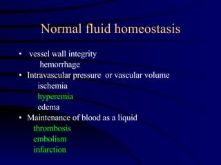
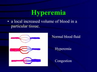
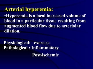
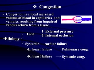
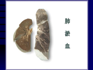
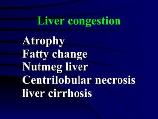
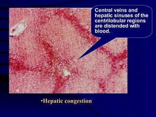
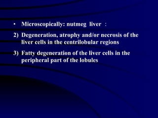
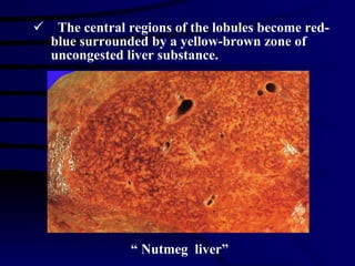
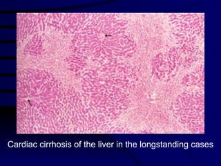
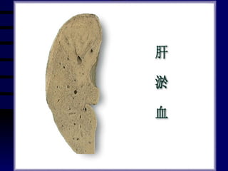
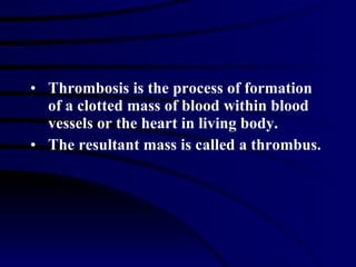
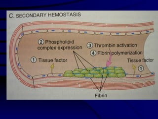
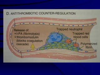
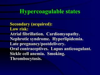
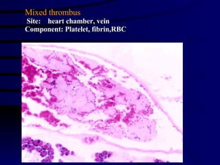
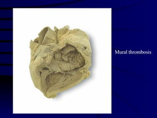
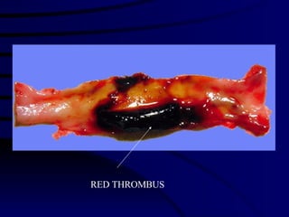
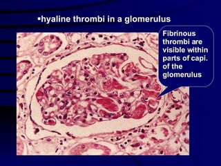
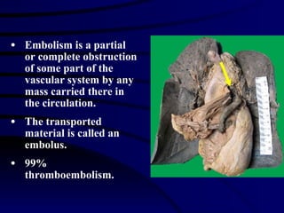
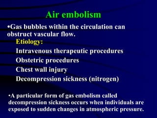
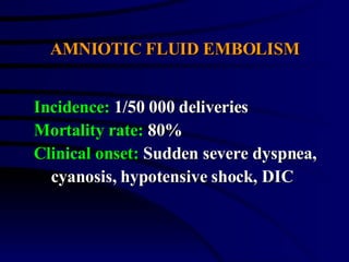
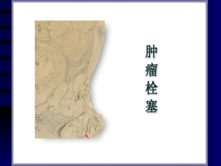
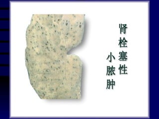
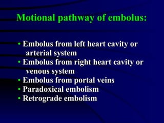
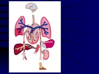
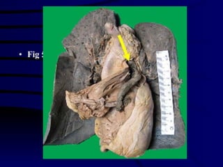
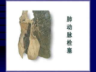
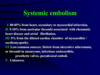
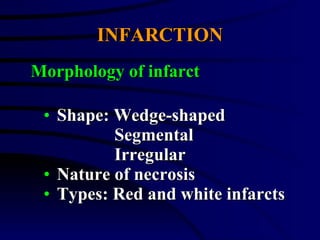
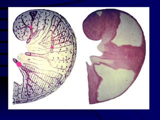
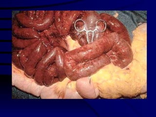
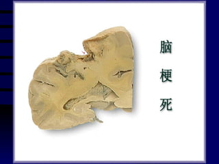
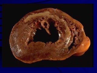
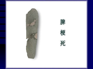
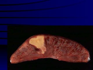
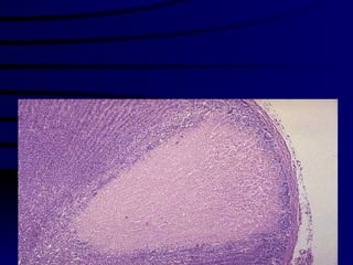
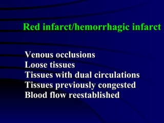
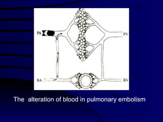
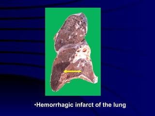
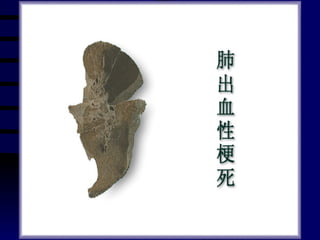
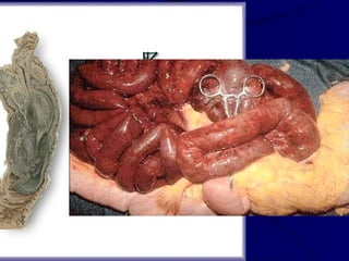
Chapter Three Hemodynamic Disorders
Normal fluid homeostasis vessel wall integrity  hemorrhage Intravascular pressure  or vascular volume ischemia hyperemia edema Maintenance of blood as a liquid thrombosis embolism infarction
Hyperemia: Arterial hyperemia Venous hyperemia Section A
a local increased volume of blood in a particular tissue.  Hyperemia Normal blood fluid Hyperemia Congestion
Physiological:  exercise Pathological : Inflammatory Post-ischemic   Arterial hyperemia: Hyperemia is a local increased volume of blood in a particular tissue resulting from augmented blood flow due to arteriolar dilation.
 
Gross: Larger; redder; increased temperature; cut surface is hemorrhage and wet. LM:  Dilatation of arteriole and capillary. Morphology of hyperemia
Congestion Congestion is a local increased volume of blood in capillaries  and veinules resulting from impaired venous return from a tissue. Systemic  —cardiac failure Etiology -L. heart failure -R. heart failure Pulmonary cong. Systemic cong. 1. External pressure 2. Internal occlusion  Local
Pulmonary congestion Edema Hemorrhage Heart failure cells Brown induration
Gross: Increased volume and weight of organs; blue-red color (cyanosis); reduced temp.; wetness and excessive blood on the cut surface. Morphology
Acute pulmonary congestion
Chronic pulmonary congestion
 
Liver congestion Atrophy Fatty change Nutmeg liver Centrilobular necrosis liver cirrhosis
Hepatic congestion Central veins and hepatic sinuses of the centrilobular regions are distended with blood.
 
Microscopically: nutmeg  liver : Degeneration, atrophy and/or necrosis of the liver cells in the centrilobular regions Fatty degeneration of the liver cells in the peripheral part of the lobules
The central regions of the lobules become red-blue surrounded by a yellow-brown zone of uncongested liver substance. “  Nutmeg  liver”
 
Cardiac cirrhosis of the liver in the longstanding cases
 
Thrombosis Thrombus  Section B
Thrombosis is the process of formation  of a clotted mass of blood within blood vessels or the heart in living body. The resultant mass is called a thrombus.
Normal hemostatic process


Endothelium Platelets Coagulation cascade
Fig 5-6 Pro- and anticoagulant activities of endothelial cells
Platelet adhesion and aggregation
Fig 5-8 The coagulation cascade
Fibrinolytic system
THREE INFLUENCES OF THROMBOSIS Endothelial injury (most important).  Alone can induce thrombosis. Alterations in normal flow. Hypercoagulability. When the last two are both present,  endothelial injury is not requisite.
Fig 5-6 Pro- and anticoagulant activities of endothelial cells
Endothelial injury Ulcerative atherosclerosis Transmural myocardial infarction Vasculitis Trauma Radiation Bacterial toxins
Alterations in normal blood flow Platelets activated by contact with  endothelium. Slowed flow retards dilution of activated  clotting factors and hepatic clearance. Stasis or turbulence retards the inflow of  inhibitors. Turbulence may induce endothelial injury
Hypercoagulable states Primary (genetic): Antithrombin III deficiency Protein C deficiency Protein S deficiency Other combined deficiency
Hypercoagulable states Secondary (acquired): High risk: Prolonged bed rest or immobilization. Myocardial infarction.  Tissue damage  (surgery, fractures, burns).  Cardiac failure.  Cancer.  Acute leukemia.  DIC.  Thrombotic thrombocytopenia.
Hypercoagulable states Secondary (acquired): Low risk: Atrial fibrillation.  Cardiomyopathy.  Nephrotic syndrome.  Hyperlipidemia.  Late pregnancy/postdelivery.  Oral contraceptives.  Lupus anticoagulant.  Sickle cell anemia.  Smoking.  Thrombocytosis.
 
 
White thrombus Red thrombus Mixed thrombus Fibrin thrombus Types of thrombus
White thrombus Site: heart valve ,  artery Component: Platelet, fibrin
Mixed thrombus Site:  heart chamber, vein Component: Platelet, fibrin,RBC
 
Mural thrombosis
Mural thrombosis
RED THROMBUS
Fibrinous thrombi are visible within parts of capi. of the glomerulus hyaline thrombi in a glomerulus
Absorption Organization Calcification Detachment Fate of thrombus
 
Organization and recanalization of thrombus During organization, the thrombus dissolved and blood could flow again.
 
Ischemia Congestion Heart valve disease DIC Embolism Effects of thrombosis
Embolism Embolus Section C
Embolism is a partial or complete obstruction of some part of the vascular system by any mass carried there in the circulation. The transported material is called an embolus. 99% thromboembolism.
Types of embolus Thromboembolism Fat embolism Air embolism Amniotic fluid embolism Other types
Etiology: Fractures of long bones Soft tissue trauma Burns Fat embolism
90% of individuals with severe skeletal injuries 10% with clinical findings(1-3 days) Pulmonary insufficiency Neurologic symptoms  Fat embolism
Fig 5-17
 
Etiology: Intravenous therapeutic procedures Obstetric procedures Chest wall injury Decompression sickness (nitrogen) Air embolism Gas bubbles within the circulation can obstruct vascular flow. A particular form of gas embolism called decompression sickness occurs when individuals are exposed to sudden changes in atmospheric pressure.
AMNIOTIC FLUID EMBOLISM Incidence:  1/50 000 deliveries Mortality rate:  80% Clinical onset:  Sudden severe dyspnea, cyanosis, hypotensive shock, DIC


Embolus from left heart cavity or  arterial system Embolus from right heart cavity or  venous system Embolus from portal veins Paradoxical embolism Retrograde embolism Motional pathway of embolus:
 
Fig 5-16
 
THROMBOEMBOLISM Instantaneous death (>60%). Cardiovascular collapse. Right heart failure Pulmonary 1.Large emboli (5%):
 
2.Small emboli (60-80%): Clinical silent in patients without  cardiovascular failure. blood flow from bronchial arteries (collateral vascular supply)
3. Between the extremes of large and  small emboli (10-15%): Pulmonary hemorrhage. 4. Multiple  small emboli: Pulmonary hypertension and vascular sclerosis.
Systemic embolism I .  80-85% from heart, secondary to myocardial infarction. II .  5-10% from auricular thrombi associated  with rheumatic  heart disease and atrial  fibrillation. III .  5% from the dilated cardiac chamber  of myocarditis /  cardiomyopathy. VI .  Less common sources: Debris from ulcerative atheromata,  or thrombi in aneurysms, infectious endocarditis,  prosthetic valves, paradoxical emboli. V .   Unknown.
INFARCTION (INFARCT) Section D
infarct/infarction An infarct is a localized area of ischemic necrosis in a tissue or organ produced by occlusion of either its arterial supply or its venous drainage. The process whereby the infarct is developed is known as infarction.
Intrinsic occlusion for example, thrombosis,  embolism expansion of atheroma Vasospasm Extrinsic compression for example, twisting of the vessels Etiology
INFARCTION Shape: Wedge-shaped Segmental Irregular Nature of necrosis Types: Red and white infarcts Morphology of infarct


LM: Ischemic coagulative necrosis Anemic infarct with few RBC Hemorrhagic infarct has engorgement and hemorrhage The pathology changes secondary to infarct such as hyperemia, hemorrhage, infla., organization and so on.
Myocardial infarct The myocardial cells shows coagulative necrosis with the outline of the myocardium. In the margin of the infarct there are numerous inflammatory exudation and connective tissue.
white infarct/anemic  infarct arterial occlusions firm tissues


Venous occlusions Loose tissues Tissues with dual circulations Tissues previously congested Blood flow reestablished Red infarct/hemorrhagic infarct
The  alteration of blood in pulmonary embolism
Hemorrhagic infarct of the lung


Hemorrhage Hemorrhage denotes an escape of blood from the cardiovascular system, usually is the result of the rupture of a blood vessel or the heart.

Hemodynamic Disorders

  • 1.
  • 2.
    Normal fluid homeostasisvessel wall integrity hemorrhage Intravascular pressure or vascular volume ischemia hyperemia edema Maintenance of blood as a liquid thrombosis embolism infarction
  • 3.
    Hyperemia: Arterial hyperemiaVenous hyperemia Section A
  • 4.
    a local increasedvolume of blood in a particular tissue. Hyperemia Normal blood fluid Hyperemia Congestion
  • 5.
    Physiological: exercisePathological : Inflammatory Post-ischemic Arterial hyperemia: Hyperemia is a local increased volume of blood in a particular tissue resulting from augmented blood flow due to arteriolar dilation.
  • 6.
  • 7.
    Gross: Larger; redder;increased temperature; cut surface is hemorrhage and wet. LM: Dilatation of arteriole and capillary. Morphology of hyperemia
  • 8.
    Congestion Congestion isa local increased volume of blood in capillaries and veinules resulting from impaired venous return from a tissue. Systemic —cardiac failure Etiology -L. heart failure -R. heart failure Pulmonary cong. Systemic cong. 1. External pressure 2. Internal occlusion Local
  • 9.
    Pulmonary congestion EdemaHemorrhage Heart failure cells Brown induration
  • 10.
    Gross: Increased volumeand weight of organs; blue-red color (cyanosis); reduced temp.; wetness and excessive blood on the cut surface. Morphology
  • 11.
  • 12.
  • 13.
  • 14.
    Liver congestion AtrophyFatty change Nutmeg liver Centrilobular necrosis liver cirrhosis
  • 15.
    Hepatic congestion Centralveins and hepatic sinuses of the centrilobular regions are distended with blood.
  • 16.
  • 17.
    Microscopically: nutmeg liver : Degeneration, atrophy and/or necrosis of the liver cells in the centrilobular regions Fatty degeneration of the liver cells in the peripheral part of the lobules
  • 18.
    The central regionsof the lobules become red-blue surrounded by a yellow-brown zone of uncongested liver substance. “ Nutmeg liver”
  • 19.
  • 20.
    Cardiac cirrhosis ofthe liver in the longstanding cases
  • 21.
  • 22.
  • 23.
    Thrombosis is theprocess of formation of a clotted mass of blood within blood vessels or the heart in living body. The resultant mass is called a thrombus.
  • 24.
  • 25.
  • 26.
  • 27.
  • 28.
  • 29.
    Fig 5-6 Pro-and anticoagulant activities of endothelial cells
  • 30.
  • 31.
    Fig 5-8 Thecoagulation cascade
  • 32.
  • 33.
    THREE INFLUENCES OFTHROMBOSIS Endothelial injury (most important). Alone can induce thrombosis. Alterations in normal flow. Hypercoagulability. When the last two are both present, endothelial injury is not requisite.
  • 34.
    Fig 5-6 Pro-and anticoagulant activities of endothelial cells
  • 35.
    Endothelial injury Ulcerativeatherosclerosis Transmural myocardial infarction Vasculitis Trauma Radiation Bacterial toxins
  • 36.
    Alterations in normalblood flow Platelets activated by contact with endothelium. Slowed flow retards dilution of activated clotting factors and hepatic clearance. Stasis or turbulence retards the inflow of inhibitors. Turbulence may induce endothelial injury
  • 37.
    Hypercoagulable states Primary(genetic): Antithrombin III deficiency Protein C deficiency Protein S deficiency Other combined deficiency
  • 38.
    Hypercoagulable states Secondary(acquired): High risk: Prolonged bed rest or immobilization. Myocardial infarction. Tissue damage (surgery, fractures, burns). Cardiac failure. Cancer. Acute leukemia. DIC. Thrombotic thrombocytopenia.
  • 39.
    Hypercoagulable states Secondary(acquired): Low risk: Atrial fibrillation. Cardiomyopathy. Nephrotic syndrome. Hyperlipidemia. Late pregnancy/postdelivery. Oral contraceptives. Lupus anticoagulant. Sickle cell anemia. Smoking. Thrombocytosis.
  • 40.
  • 41.
  • 42.
    White thrombus Redthrombus Mixed thrombus Fibrin thrombus Types of thrombus
  • 43.
    White thrombus Site:heart valve , artery Component: Platelet, fibrin
  • 44.
    Mixed thrombus Site: heart chamber, vein Component: Platelet, fibrin,RBC
  • 45.
  • 46.
  • 47.
  • 48.
  • 49.
    Fibrinous thrombi arevisible within parts of capi. of the glomerulus hyaline thrombi in a glomerulus
  • 50.
    Absorption Organization CalcificationDetachment Fate of thrombus
  • 51.
  • 52.
    Organization and recanalizationof thrombus During organization, the thrombus dissolved and blood could flow again.
  • 53.
  • 54.
    Ischemia Congestion Heartvalve disease DIC Embolism Effects of thrombosis
  • 55.
  • 56.
    Embolism is apartial or complete obstruction of some part of the vascular system by any mass carried there in the circulation. The transported material is called an embolus. 99% thromboembolism.
  • 57.
    Types of embolusThromboembolism Fat embolism Air embolism Amniotic fluid embolism Other types
  • 58.
    Etiology: Fractures oflong bones Soft tissue trauma Burns Fat embolism
  • 59.
    90% of individualswith severe skeletal injuries 10% with clinical findings(1-3 days) Pulmonary insufficiency Neurologic symptoms Fat embolism
  • 60.
  • 61.
  • 62.
    Etiology: Intravenous therapeuticprocedures Obstetric procedures Chest wall injury Decompression sickness (nitrogen) Air embolism Gas bubbles within the circulation can obstruct vascular flow. A particular form of gas embolism called decompression sickness occurs when individuals are exposed to sudden changes in atmospheric pressure.
  • 63.
    AMNIOTIC FLUID EMBOLISMIncidence: 1/50 000 deliveries Mortality rate: 80% Clinical onset: Sudden severe dyspnea, cyanosis, hypotensive shock, DIC
  • 64.
  • 65.
  • 66.
  • 67.
  • 68.
  • 69.
  • 70.
    Embolus from leftheart cavity or arterial system Embolus from right heart cavity or venous system Embolus from portal veins Paradoxical embolism Retrograde embolism Motional pathway of embolus:
  • 71.
  • 72.
  • 73.
  • 74.
    THROMBOEMBOLISM Instantaneous death(>60%). Cardiovascular collapse. Right heart failure Pulmonary 1.Large emboli (5%):
  • 75.
  • 76.
    2.Small emboli (60-80%):Clinical silent in patients without cardiovascular failure. blood flow from bronchial arteries (collateral vascular supply)
  • 77.
    3. Between theextremes of large and small emboli (10-15%): Pulmonary hemorrhage. 4. Multiple small emboli: Pulmonary hypertension and vascular sclerosis.
  • 78.
    Systemic embolism I. 80-85% from heart, secondary to myocardial infarction. II . 5-10% from auricular thrombi associated with rheumatic heart disease and atrial fibrillation. III . 5% from the dilated cardiac chamber of myocarditis / cardiomyopathy. VI . Less common sources: Debris from ulcerative atheromata, or thrombi in aneurysms, infectious endocarditis, prosthetic valves, paradoxical emboli. V . Unknown.
  • 79.
  • 80.
    infarct/infarction An infarctis a localized area of ischemic necrosis in a tissue or organ produced by occlusion of either its arterial supply or its venous drainage. The process whereby the infarct is developed is known as infarction.
  • 81.
    Intrinsic occlusion forexample, thrombosis, embolism expansion of atheroma Vasospasm Extrinsic compression for example, twisting of the vessels Etiology
  • 82.
    INFARCTION Shape: Wedge-shapedSegmental Irregular Nature of necrosis Types: Red and white infarcts Morphology of infarct
  • 83.
  • 84.
  • 85.
  • 86.
  • 87.
    LM: Ischemic coagulativenecrosis Anemic infarct with few RBC Hemorrhagic infarct has engorgement and hemorrhage The pathology changes secondary to infarct such as hyperemia, hemorrhage, infla., organization and so on.
  • 88.
    Myocardial infarct Themyocardial cells shows coagulative necrosis with the outline of the myocardium. In the margin of the infarct there are numerous inflammatory exudation and connective tissue.
  • 89.
    white infarct/anemic infarct arterial occlusions firm tissues
  • 90.
  • 91.
  • 92.
  • 93.
  • 94.
    Venous occlusions Loosetissues Tissues with dual circulations Tissues previously congested Blood flow reestablished Red infarct/hemorrhagic infarct
  • 95.
    The alterationof blood in pulmonary embolism
  • 96.
  • 97.
  • 98.
  • 99.
  • 100.
  • 101.
    Hemorrhage Hemorrhage denotesan escape of blood from the cardiovascular system, usually is the result of the rupture of a blood vessel or the heart.

Editor's Notes