SlideShare a Scribd company logo
BRAIN HERNIATION SYNDROME A Pictorial Review 
Thorsang Chayovan R1/Aj.Nuttha 
22.11.2014
BRAIN HERNIATION 
• most common types 
–Subfalcine herniation 
–descending transtentorial herniation 
•Others 
–Posterior fossa herniations 
•ascending transtentorial herniation 
•tonsillar herniation 
–Transalar herniation 
•Rare but important types 
–transdural/transcranial herniations 
–brain displacements across the sphenoid wing
SUBFALCINE HERNIATION
Subfalcine herniation 
•most common 
•supratentorial mass in one hemicranium 
•affected hemisphere pushes across the midline under the inferior "free" margin of the falx, extending into the contralateral hemicranium
Subfalcine herniation: imaging 
Axial and coronal images show that 
•cingulate gyrus 
•anterior cerebral artery (ACA) 
•internal cerebral vein (ICV) 
are pushed from one side to the other under the falx cerebri. 
The ipsilateral ventricle appears compressed and displaced across the midline
Complications 
•unilateral obstructive hydrocephalus 
–foramen of Monro occlusion 
•Periventricular hypodensity with "blurred" margins of the lateral ventricle 
–Fluid accumulates in the periventricular white matter
Complications 
•When severe, the herniating ACA can be pinned against the inferior "free" margin of the falx cerebri 
 secondary infarction of the cingulate gyrus
TRANSTENTORIAL HERNIATION
Transtentorial herniations 
descending herniations 
ascending herniations
Descending transtentorial herniations 
•the second most common 
•a hemispheric mass 
•initially produces subfalcine herniation 
•As the mass effect increases, the uncus of the temporal lobe is pushed medially 
begins to encroach on the suprasellar cistern 
hippocampus follows 
hippocampus effaces the ipsilateral quadrigeminal cistern 
both the uncus and hippocampus herniate inferiorly through the tentorial incisura
"Dysautonomia, Multisystem Atrophy and Parkinson's." Dysautonomia, Multisystem Atrophy and Parkinson's. N.p., n.d. Web. 18 Nov. 2014
Descending transtentorial herniation 
•Unilateral 
•Bilateral ("central“) 
–Severe
unilateral DTH: imaging 
early 
uncus is displaced medially 
Ipsilateral aspect of the suprasellar cistern effaced 
Ipsilateral prepontine + cerebellopontine angle cistern enlarged
Descending transtentorial herniation 
As DTH increases 
hippocampus also herniates medially 
quadrigeminal cistern compression 
midbrain pushed toward the opposite side of the incisura
Descending transtentorial herniation 
severe cases 
entire suprasellar and quadrigeminal cisterns are effaced. 
The temporal horn can even be displaced almost into the midline
bilateral DTH 
both hemispheres become swollen 
the whole central brain is flattened against the skull base 
All the basal cisterns are obliterated 
hypothalamus and optic chiasm are crushed against the sella turcica
Complete bilateral DTH 
both temporal lobes herniate medially into the tentorial hiatus 
midbrain and pons displaced inferiorly through the tentorial incisura 
The angle between the midbrain and pons 
is progressively reduced from 90° to almost 0°
Complications 
•CN III (oculomotor) nerve compression 
–CN III palsy 
•PCA occlusion as it passes back up over the medial edge of the tentorium 
–secondary PCA (occipital) infarct
Kernohan notch 
•As the herniating temporal lobe pushes the midbrain toward the opposite side of the incisura 
–contralateral cerebral peduncle is forced against the hard edge of the tentorium 
•Pressure ischemia  ipsilateral hemiplegia 
–the "false localizing" sign
Duret hemorrhage 
"Top-down" mass effect displaces the midbrain inferiorly 
closes the midbrain-pontine angle 
Perforating arteries from basilar artery 
are compressed and buckled 
secondary hemorrhagic midbrain infarct
Brainstem hemorrhage 
Dorsolateral 
Primary injury 
Severe DAI 
Ventral paramedian 
Duret
Hemorrhage in diffuse axonal injury 
•Gray-white junction 
•Corpus callosum 
•Brainstem
hypothalamic and basal ganglia infarcts 
complete bilateral DTH 
perforating arteries from the circle of Willis compression against the central skull base 
hypothalamic and basal ganglia infarcts
POSTERIOR FOSSA MASS: TONSILLAR HERNIATION ASCENDING TRANSTENTORIAL HERNIATION
Tonsillar herniation 
•The cerebellar tonsils are displaced inferiorly and become impacted into the foramen magnum. 
•congenital (e.g., Chiari 1 malformation) 
– mismatch between size and content of the posterior fossa 
•Acquired 
–an expanding posterior fossa mass pushing the tonsils downward—more common 
–intracranial hypotension: abnormally low intraspinal CSF pressure 
•tonsils are pulled downward
Tonsillar herniation: imaging 
•Diagnosing tonsillar herniation on NECT scans may be problematic. 
Cisterna magna obliteration
Tonsillar herniation: imaging 
•MR: much more easily diagnosed 
•In the sagittal plane 
–the tonsillar folia become vertically oriented 
–the inferior aspect of the tonsils becomes pointed 
–Tonsils > 5 mm (or 7 mm in children) below the foramen magnum are generally abnormal 
•especially if they are peg-like or pointed (rather than rounded)
Tonsillar herniation: imaging 
•In the axial plane, T2 scans show that the tonsils are impacted into the foramen magnum 
–obliterating CSF in the cisterna magna 
–displacing the medulla anteriorly
Complications 
•obstructive hydrocephalus 
•tonsillar necrosis
ASCENDING TRANSTENTORIAL HERNIATION
Ascending transtentorial herniation 
•caused by any expanding posterior fossa mass 
–Neoplasms > trauma
Complications 
•Acute intraventricular obstructive hydrocephalus 
–caused by compression of the cerebral aqueduct
OTHER LESS COMMON HERNIATION: TRANSALAR TRANSDURAL/TRANSCRANIAL HERNIATIONS
Transalar Herniation 
•brain herniates across the greater sphenoid wing (GSW) or "ala" 
•ascending > descending
Ascending transalar herniation 
•caused by a large middle cranial fossa mass 
•An intratemporal or large extraaxial mass 
Temporal lobe + sylvian fissure + MCA 
up and over the greater sphenoid wing
Descending transalar herniation 
•caused by a large anterior cranial fossa mass 
Gyrus rectus is forced posteroinferiorly over the GSW 
displacing the sylvian fissure and shifting the MCA backward
Transdural/Transcranial Herniation 
•Rare 
•Sometimes called a "brain fungus" 
•can be life-threatening 
Lacerated dura + a skull defect + increased ICP
Transdural/Transcranial Herniation 
•Traumatic 
–infants or young children with a comminuted inward skull fracture 
•Iatrogenic 
–a burr hole, craniotomy, or craniectomy
Transdural/Transcranial Herniation 
•MR best depicts these unusual herniations. 
•The disrupted dura 
–discontinuous black line on T2WI 
–Brain tissue, blood vessels, and CSF, are extruded through the defects into the subgaleal space
Kaewlai, R. Imaging of Traumatic Brain Injury. 2013.
Wikipedia
References 
•Osborn, Anne G. "Secondary Effects and Sequellae of CNS Trauma."Osborn's Brain: Imaging, Pathology, and Anatomy. Salt Lake City, UT: Amirsys Pub., 2013. N. pag. Print. 
•Osborn, Anne G. "Cerebral Vasculature: Normal Anatomy and Pathology."Diagnostic Neuroradiology. St. Louis: Mosby, 1994. N. pag. Print. 
•Kaewlai, R. Imaging of Traumatic Brain Injury. 2013. Web.

More Related Content

What's hot

Presentation2.pptx. posterior fossa tumour
Presentation2.pptx. posterior fossa tumourPresentation2.pptx. posterior fossa tumour
Presentation2.pptx. posterior fossa tumour
Abdellah Nazeer
 
CT Brain interpretation
CT Brain interpretationCT Brain interpretation
CT Brain interpretation
TaibaSuleman1
 
Presentation1.pptx, radiological imaging of cerebello pontine angle mass lesi...
Presentation1.pptx, radiological imaging of cerebello pontine angle mass lesi...Presentation1.pptx, radiological imaging of cerebello pontine angle mass lesi...
Presentation1.pptx, radiological imaging of cerebello pontine angle mass lesi...
Abdellah Nazeer
 
Imaging of Traumatic Brain Injury
Imaging of Traumatic Brain InjuryImaging of Traumatic Brain Injury
Imaging of Traumatic Brain Injury
Rathachai Kaewlai
 
Cv junction
Cv junctionCv junction
Anatomy of sellar suprasellar region
Anatomy of sellar suprasellar regionAnatomy of sellar suprasellar region
Anatomy of sellar suprasellar region
PGINeurosurgery
 
Imaging in stroke
Imaging in strokeImaging in stroke
Imaging in stroke
Navni Garg
 
Normal mri brain
Normal mri brainNormal mri brain
Normal mri brain
NeurologyKota
 
Meningioma of brain
Meningioma of brainMeningioma of brain
Meningioma of brain
suresh Bishokarma
 
Cisterns of brain and its contents along with its classification and approach...
Cisterns of brain and its contents along with its classification and approach...Cisterns of brain and its contents along with its classification and approach...
Cisterns of brain and its contents along with its classification and approach...
Rajeev Bhandari
 
Radiological anatomy & Techniques of the Ventricular system
Radiological anatomy & Techniques of the Ventricular systemRadiological anatomy & Techniques of the Ventricular system
Radiological anatomy & Techniques of the Ventricular system
Mohamed M.A. Zaitoun
 
How to read a Head CT, CT Brain
How to read a Head CT, CT BrainHow to read a Head CT, CT Brain
How to read a Head CT, CT Brain
Gauhar Azeem
 
Imaging of white matter diseases
Imaging of white matter diseasesImaging of white matter diseases
Imaging of white matter diseases
Navni Garg
 
Larynx anatomy ct and mri
Larynx anatomy ct and mriLarynx anatomy ct and mri
Larynx anatomy ct and mri
Anish Choudhary
 
Diagnostic Imaging of Subarachnoid Hemorrhage
Diagnostic Imaging of Subarachnoid HemorrhageDiagnostic Imaging of Subarachnoid Hemorrhage
Diagnostic Imaging of Subarachnoid Hemorrhage
Mohamed M.A. Zaitoun
 
SPINAL CORD ARTERIOVENOUS MALFORMATIONS
SPINAL CORD ARTERIOVENOUS MALFORMATIONSSPINAL CORD ARTERIOVENOUS MALFORMATIONS
SPINAL CORD ARTERIOVENOUS MALFORMATIONS
Dr Praveen kumar tripathi
 
Sulcal and gyral neuroanatomy
Sulcal and gyral neuroanatomySulcal and gyral neuroanatomy
Sulcal and gyral neuroanatomy
Navni Garg
 
Normal CT BRAIN
Normal CT BRAINNormal CT BRAIN
Normal CT BRAIN
NeurologyKota
 
Growing skull fracture
Growing skull fractureGrowing skull fracture
Growing skull fracture
Abhishek Rai
 
Basic approach to brain tumor
Basic approach to brain tumorBasic approach to brain tumor
Basic approach to brain tumor
Khon Kaen university
 

What's hot (20)

Presentation2.pptx. posterior fossa tumour
Presentation2.pptx. posterior fossa tumourPresentation2.pptx. posterior fossa tumour
Presentation2.pptx. posterior fossa tumour
 
CT Brain interpretation
CT Brain interpretationCT Brain interpretation
CT Brain interpretation
 
Presentation1.pptx, radiological imaging of cerebello pontine angle mass lesi...
Presentation1.pptx, radiological imaging of cerebello pontine angle mass lesi...Presentation1.pptx, radiological imaging of cerebello pontine angle mass lesi...
Presentation1.pptx, radiological imaging of cerebello pontine angle mass lesi...
 
Imaging of Traumatic Brain Injury
Imaging of Traumatic Brain InjuryImaging of Traumatic Brain Injury
Imaging of Traumatic Brain Injury
 
Cv junction
Cv junctionCv junction
Cv junction
 
Anatomy of sellar suprasellar region
Anatomy of sellar suprasellar regionAnatomy of sellar suprasellar region
Anatomy of sellar suprasellar region
 
Imaging in stroke
Imaging in strokeImaging in stroke
Imaging in stroke
 
Normal mri brain
Normal mri brainNormal mri brain
Normal mri brain
 
Meningioma of brain
Meningioma of brainMeningioma of brain
Meningioma of brain
 
Cisterns of brain and its contents along with its classification and approach...
Cisterns of brain and its contents along with its classification and approach...Cisterns of brain and its contents along with its classification and approach...
Cisterns of brain and its contents along with its classification and approach...
 
Radiological anatomy & Techniques of the Ventricular system
Radiological anatomy & Techniques of the Ventricular systemRadiological anatomy & Techniques of the Ventricular system
Radiological anatomy & Techniques of the Ventricular system
 
How to read a Head CT, CT Brain
How to read a Head CT, CT BrainHow to read a Head CT, CT Brain
How to read a Head CT, CT Brain
 
Imaging of white matter diseases
Imaging of white matter diseasesImaging of white matter diseases
Imaging of white matter diseases
 
Larynx anatomy ct and mri
Larynx anatomy ct and mriLarynx anatomy ct and mri
Larynx anatomy ct and mri
 
Diagnostic Imaging of Subarachnoid Hemorrhage
Diagnostic Imaging of Subarachnoid HemorrhageDiagnostic Imaging of Subarachnoid Hemorrhage
Diagnostic Imaging of Subarachnoid Hemorrhage
 
SPINAL CORD ARTERIOVENOUS MALFORMATIONS
SPINAL CORD ARTERIOVENOUS MALFORMATIONSSPINAL CORD ARTERIOVENOUS MALFORMATIONS
SPINAL CORD ARTERIOVENOUS MALFORMATIONS
 
Sulcal and gyral neuroanatomy
Sulcal and gyral neuroanatomySulcal and gyral neuroanatomy
Sulcal and gyral neuroanatomy
 
Normal CT BRAIN
Normal CT BRAINNormal CT BRAIN
Normal CT BRAIN
 
Growing skull fracture
Growing skull fractureGrowing skull fracture
Growing skull fracture
 
Basic approach to brain tumor
Basic approach to brain tumorBasic approach to brain tumor
Basic approach to brain tumor
 

Viewers also liked

Anatomy of brain sulcus and gyrus - Dr.Sajith MD RD
Anatomy of brain sulcus and gyrus - Dr.Sajith MD RDAnatomy of brain sulcus and gyrus - Dr.Sajith MD RD
Anatomy of brain sulcus and gyrus - Dr.Sajith MD RD
Sajith Selvaganesan
 
Radiology Spotters
Radiology Spotters Radiology Spotters
Radiology Spotters
Anish Choudhary
 
Ct scan in head and spine injuries
Ct scan in head and spine injuriesCt scan in head and spine injuries
Ct scan in head and spine injuries
Ahmed Debes
 
N806 and ct head
N806 and ct headN806 and ct head
N806 and ct head
jbmann
 
radiology Spotters mixed bag
radiology Spotters mixed bagradiology Spotters mixed bag
radiology Spotters mixed bag
Anish Choudhary
 
Sulcal anatomy supratentorial brain, excluding the temporal lobe.
Sulcal anatomy supratentorial brain, excluding the temporal lobe.Sulcal anatomy supratentorial brain, excluding the temporal lobe.
Sulcal anatomy supratentorial brain, excluding the temporal lobe.
Sarbesh Tiwari
 
Ct and mri interpretation
Ct and mri interpretationCt and mri interpretation
Ct and mri interpretation
Dr. Mohamed Maged Kharabish
 
MRI SECTIONAL ANATOMY OF BRAIN
MRI SECTIONAL ANATOMY OF BRAIN MRI SECTIONAL ANATOMY OF BRAIN
MRI SECTIONAL ANATOMY OF BRAIN
Vipin Kumar
 
Head injury types, clinical manifestations, diagnosis and management
Head injury  types, clinical manifestations, diagnosis and managementHead injury  types, clinical manifestations, diagnosis and management
Head injury types, clinical manifestations, diagnosis and management
Vibha Amblihalli
 
Pupillary dilatation in head injury
Pupillary dilatation in head injuryPupillary dilatation in head injury
Pupillary dilatation in head injury
SCGH ED CME
 
Visual and auditory cortex
Visual and auditory cortexVisual and auditory cortex
Visual and auditory cortex
Matt Roberts
 
Sulci,Gyri & Functional areas of cerebrum Dr.N.Mugunthan.M.S
Sulci,Gyri & Functional areas of cerebrum Dr.N.Mugunthan.M.SSulci,Gyri & Functional areas of cerebrum Dr.N.Mugunthan.M.S
Sulci,Gyri & Functional areas of cerebrum Dr.N.Mugunthan.M.S
mgmcri1234
 
Cns infections radiology.
Cns infections radiology.Cns infections radiology.
Cns infections radiology.
Raeez Basheer
 
Head injury ppt
Head injury pptHead injury ppt
Head injury ppt
Manali Solanki
 
MRI brain; Basics and Radiological Anatomy
MRI brain; Basics and Radiological AnatomyMRI brain; Basics and Radiological Anatomy
MRI brain; Basics and Radiological Anatomy
Imran Rizvi
 
Diagnostic Neuroradiology
Diagnostic NeuroradiologyDiagnostic Neuroradiology
Diagnostic Neuroradiology
Sumit Prajapati
 
Cerebrum
CerebrumCerebrum
Cerebrum
Anoop Nair
 
Spots with keys
Spots with keysSpots with keys
Spots with keys
Anish Choudhary
 
Spot Radiological Diagnosis
Spot Radiological DiagnosisSpot Radiological Diagnosis
Spot Radiological Diagnosis
AR Muhamad Na'im
 
Spot diagnosis by ^^
Spot diagnosis by ^^Spot diagnosis by ^^
Spot diagnosis by ^^
ethan
 

Viewers also liked (20)

Anatomy of brain sulcus and gyrus - Dr.Sajith MD RD
Anatomy of brain sulcus and gyrus - Dr.Sajith MD RDAnatomy of brain sulcus and gyrus - Dr.Sajith MD RD
Anatomy of brain sulcus and gyrus - Dr.Sajith MD RD
 
Radiology Spotters
Radiology Spotters Radiology Spotters
Radiology Spotters
 
Ct scan in head and spine injuries
Ct scan in head and spine injuriesCt scan in head and spine injuries
Ct scan in head and spine injuries
 
N806 and ct head
N806 and ct headN806 and ct head
N806 and ct head
 
radiology Spotters mixed bag
radiology Spotters mixed bagradiology Spotters mixed bag
radiology Spotters mixed bag
 
Sulcal anatomy supratentorial brain, excluding the temporal lobe.
Sulcal anatomy supratentorial brain, excluding the temporal lobe.Sulcal anatomy supratentorial brain, excluding the temporal lobe.
Sulcal anatomy supratentorial brain, excluding the temporal lobe.
 
Ct and mri interpretation
Ct and mri interpretationCt and mri interpretation
Ct and mri interpretation
 
MRI SECTIONAL ANATOMY OF BRAIN
MRI SECTIONAL ANATOMY OF BRAIN MRI SECTIONAL ANATOMY OF BRAIN
MRI SECTIONAL ANATOMY OF BRAIN
 
Head injury types, clinical manifestations, diagnosis and management
Head injury  types, clinical manifestations, diagnosis and managementHead injury  types, clinical manifestations, diagnosis and management
Head injury types, clinical manifestations, diagnosis and management
 
Pupillary dilatation in head injury
Pupillary dilatation in head injuryPupillary dilatation in head injury
Pupillary dilatation in head injury
 
Visual and auditory cortex
Visual and auditory cortexVisual and auditory cortex
Visual and auditory cortex
 
Sulci,Gyri & Functional areas of cerebrum Dr.N.Mugunthan.M.S
Sulci,Gyri & Functional areas of cerebrum Dr.N.Mugunthan.M.SSulci,Gyri & Functional areas of cerebrum Dr.N.Mugunthan.M.S
Sulci,Gyri & Functional areas of cerebrum Dr.N.Mugunthan.M.S
 
Cns infections radiology.
Cns infections radiology.Cns infections radiology.
Cns infections radiology.
 
Head injury ppt
Head injury pptHead injury ppt
Head injury ppt
 
MRI brain; Basics and Radiological Anatomy
MRI brain; Basics and Radiological AnatomyMRI brain; Basics and Radiological Anatomy
MRI brain; Basics and Radiological Anatomy
 
Diagnostic Neuroradiology
Diagnostic NeuroradiologyDiagnostic Neuroradiology
Diagnostic Neuroradiology
 
Cerebrum
CerebrumCerebrum
Cerebrum
 
Spots with keys
Spots with keysSpots with keys
Spots with keys
 
Spot Radiological Diagnosis
Spot Radiological DiagnosisSpot Radiological Diagnosis
Spot Radiological Diagnosis
 
Spot diagnosis by ^^
Spot diagnosis by ^^Spot diagnosis by ^^
Spot diagnosis by ^^
 

Similar to Brain herniation imaging

Intra cranial hemorrhages agp
Intra cranial hemorrhages agpIntra cranial hemorrhages agp
Intra cranial hemorrhages agp
Akshai George Paul
 
Cerebral herniation syndromes
Cerebral herniation syndromesCerebral herniation syndromes
Cerebral herniation syndromes
Dr Himanshu Soni
 
Head injuries
Head injuriesHead injuries
Head injuries
yasin jamal
 
headinjury-typesclinicalmanifestationsdiagnosisandmanagement-130918094211-php...
headinjury-typesclinicalmanifestationsdiagnosisandmanagement-130918094211-php...headinjury-typesclinicalmanifestationsdiagnosisandmanagement-130918094211-php...
headinjury-typesclinicalmanifestationsdiagnosisandmanagement-130918094211-php...
savitri49
 
Full story brain herniation imaging Dr Ahmed Esawy
Full story brain herniation imaging Dr Ahmed EsawyFull story brain herniation imaging Dr Ahmed Esawy
Full story brain herniation imaging Dr Ahmed Esawy
AHMED ESAWY
 
Basics of brain hemorrhage
Basics of brain hemorrhageBasics of brain hemorrhage
Basics of brain hemorrhage
Rengarajan Rajagopal
 
Basic approach to brain CT Dr. Muhammad Bin Zulfiqar
Basic approach to brain CT Dr. Muhammad Bin ZulfiqarBasic approach to brain CT Dr. Muhammad Bin Zulfiqar
Basic approach to brain CT Dr. Muhammad Bin Zulfiqar
Dr. Muhammad Bin Zulfiqar
 
radiology ppt on mri sequences how to read basic mri sequences and basic path...
radiology ppt on mri sequences how to read basic mri sequences and basic path...radiology ppt on mri sequences how to read basic mri sequences and basic path...
radiology ppt on mri sequences how to read basic mri sequences and basic path...
drashish05
 
posterior triangle of neck
posterior triangle of neck posterior triangle of neck
posterior triangle of neck
drhimanshuupadhyay
 
Imaging in head trauma
Imaging in head traumaImaging in head trauma
Imaging in head trauma
SCGH ED CME
 
Hydrocephalus by DR.ARSHAD
Hydrocephalus by DR.ARSHADHydrocephalus by DR.ARSHAD
Hydrocephalus by DR.ARSHAD
SMS_2015
 
Cerebral herniation
Cerebral herniationCerebral herniation
Cerebral herniation
Sumiya Arshad
 
Neurosurgery 1.pptx
Neurosurgery 1.pptxNeurosurgery 1.pptx
Neurosurgery 1.pptx
joendesh
 
Lesions of parapharyngeal region
Lesions of parapharyngeal regionLesions of parapharyngeal region
Lesions of parapharyngeal region
airwave12
 
Spinal tumors
Spinal tumorsSpinal tumors
Spinal tumors
DinaFerasHaddad
 
Hydrocephalus
HydrocephalusHydrocephalus
Hydrocephalus
abhilashasaha1
 
Birth Injuries.pptx
Birth Injuries.pptxBirth Injuries.pptx
Birth Injuries.pptx
AtanasOkumu
 
Radiology of Spine
Radiology of Spine Radiology of Spine
Radiology of Spine
Zeeshan Nasir
 
VESTIBULAR SCHWANNOMA.pptx
VESTIBULAR SCHWANNOMA.pptxVESTIBULAR SCHWANNOMA.pptx
VESTIBULAR SCHWANNOMA.pptx
KarishmaMishra13
 
Acute brain
Acute brainAcute brain
Acute brain
Musa Abusabha
 

Similar to Brain herniation imaging (20)

Intra cranial hemorrhages agp
Intra cranial hemorrhages agpIntra cranial hemorrhages agp
Intra cranial hemorrhages agp
 
Cerebral herniation syndromes
Cerebral herniation syndromesCerebral herniation syndromes
Cerebral herniation syndromes
 
Head injuries
Head injuriesHead injuries
Head injuries
 
headinjury-typesclinicalmanifestationsdiagnosisandmanagement-130918094211-php...
headinjury-typesclinicalmanifestationsdiagnosisandmanagement-130918094211-php...headinjury-typesclinicalmanifestationsdiagnosisandmanagement-130918094211-php...
headinjury-typesclinicalmanifestationsdiagnosisandmanagement-130918094211-php...
 
Full story brain herniation imaging Dr Ahmed Esawy
Full story brain herniation imaging Dr Ahmed EsawyFull story brain herniation imaging Dr Ahmed Esawy
Full story brain herniation imaging Dr Ahmed Esawy
 
Basics of brain hemorrhage
Basics of brain hemorrhageBasics of brain hemorrhage
Basics of brain hemorrhage
 
Basic approach to brain CT Dr. Muhammad Bin Zulfiqar
Basic approach to brain CT Dr. Muhammad Bin ZulfiqarBasic approach to brain CT Dr. Muhammad Bin Zulfiqar
Basic approach to brain CT Dr. Muhammad Bin Zulfiqar
 
radiology ppt on mri sequences how to read basic mri sequences and basic path...
radiology ppt on mri sequences how to read basic mri sequences and basic path...radiology ppt on mri sequences how to read basic mri sequences and basic path...
radiology ppt on mri sequences how to read basic mri sequences and basic path...
 
posterior triangle of neck
posterior triangle of neck posterior triangle of neck
posterior triangle of neck
 
Imaging in head trauma
Imaging in head traumaImaging in head trauma
Imaging in head trauma
 
Hydrocephalus by DR.ARSHAD
Hydrocephalus by DR.ARSHADHydrocephalus by DR.ARSHAD
Hydrocephalus by DR.ARSHAD
 
Cerebral herniation
Cerebral herniationCerebral herniation
Cerebral herniation
 
Neurosurgery 1.pptx
Neurosurgery 1.pptxNeurosurgery 1.pptx
Neurosurgery 1.pptx
 
Lesions of parapharyngeal region
Lesions of parapharyngeal regionLesions of parapharyngeal region
Lesions of parapharyngeal region
 
Spinal tumors
Spinal tumorsSpinal tumors
Spinal tumors
 
Hydrocephalus
HydrocephalusHydrocephalus
Hydrocephalus
 
Birth Injuries.pptx
Birth Injuries.pptxBirth Injuries.pptx
Birth Injuries.pptx
 
Radiology of Spine
Radiology of Spine Radiology of Spine
Radiology of Spine
 
VESTIBULAR SCHWANNOMA.pptx
VESTIBULAR SCHWANNOMA.pptxVESTIBULAR SCHWANNOMA.pptx
VESTIBULAR SCHWANNOMA.pptx
 
Acute brain
Acute brainAcute brain
Acute brain
 

More from Thorsang Chayovan

COVID-19 Findings on Chest CT
COVID-19 Findings on Chest CTCOVID-19 Findings on Chest CT
COVID-19 Findings on Chest CT
Thorsang Chayovan
 
Dyspepsia endoscopy guideline
Dyspepsia endoscopy guidelineDyspepsia endoscopy guideline
Dyspepsia endoscopy guideline
Thorsang Chayovan
 
Common respiratory problems
Common respiratory problemsCommon respiratory problems
Common respiratory problems
Thorsang Chayovan
 
Avascular necrosis
Avascular necrosisAvascular necrosis
Avascular necrosis
Thorsang Chayovan
 
Shoulder injury
Shoulder injuryShoulder injury
Shoulder injury
Thorsang Chayovan
 
Pediatric pneumonia Thai
Pediatric pneumonia ThaiPediatric pneumonia Thai
Pediatric pneumonia Thai
Thorsang Chayovan
 
Pediatric upper urinary tract infection in Thai
Pediatric upper urinary tract infection in ThaiPediatric upper urinary tract infection in Thai
Pediatric upper urinary tract infection in Thai
Thorsang Chayovan
 
Tokyo guidelines for cholangitis and cholecystitis
Tokyo guidelines for cholangitis and cholecystitis Tokyo guidelines for cholangitis and cholecystitis
Tokyo guidelines for cholangitis and cholecystitis
Thorsang Chayovan
 
The role of ercp in diseases of the biliary tract and pancreas
The role of ercp in diseases of the biliary tract and pancreasThe role of ercp in diseases of the biliary tract and pancreas
The role of ercp in diseases of the biliary tract and pancreas
Thorsang Chayovan
 
Role of endoscopy in dyspepsia
Role of endoscopy in dyspepsiaRole of endoscopy in dyspepsia
Role of endoscopy in dyspepsia
Thorsang Chayovan
 
Role of endoscopy in choledocholithiasis
Role of endoscopy in choledocholithiasis Role of endoscopy in choledocholithiasis
Role of endoscopy in choledocholithiasis
Thorsang Chayovan
 
Pneumonia in children
Pneumonia in childrenPneumonia in children
Pneumonia in children
Thorsang Chayovan
 
คู่มือเวชปฏิบัติหัตถการ
คู่มือเวชปฏิบัติหัตถการคู่มือเวชปฏิบัติหัตถการ
คู่มือเวชปฏิบัติหัตถการ
Thorsang Chayovan
 
Febrile neutropenia in children
Febrile neutropenia in childrenFebrile neutropenia in children
Febrile neutropenia in children
Thorsang Chayovan
 
Exanthematous fever in children
Exanthematous fever in childrenExanthematous fever in children
Exanthematous fever in children
Thorsang Chayovan
 
Diseases of the rectum and anal canal in Thai
Diseases of the rectum and anal canal in ThaiDiseases of the rectum and anal canal in Thai
Diseases of the rectum and anal canal in Thai
Thorsang Chayovan
 
Thai PALS manual 2009
Thai PALS manual 2009Thai PALS manual 2009
Thai PALS manual 2009
Thorsang Chayovan
 
NEJM Cholecystitis
NEJM CholecystitisNEJM Cholecystitis
NEJM Cholecystitis
Thorsang Chayovan
 
Thai hemorrhagic stroke guideline 2008
Thai hemorrhagic stroke guideline 2008Thai hemorrhagic stroke guideline 2008
Thai hemorrhagic stroke guideline 2008
Thorsang Chayovan
 

More from Thorsang Chayovan (20)

COVID-19 Findings on Chest CT
COVID-19 Findings on Chest CTCOVID-19 Findings on Chest CT
COVID-19 Findings on Chest CT
 
Dyspepsia endoscopy guideline
Dyspepsia endoscopy guidelineDyspepsia endoscopy guideline
Dyspepsia endoscopy guideline
 
Common respiratory problems
Common respiratory problemsCommon respiratory problems
Common respiratory problems
 
Avascular necrosis
Avascular necrosisAvascular necrosis
Avascular necrosis
 
Shoulder injury
Shoulder injuryShoulder injury
Shoulder injury
 
Pediatric pneumonia Thai
Pediatric pneumonia ThaiPediatric pneumonia Thai
Pediatric pneumonia Thai
 
Pediatric upper urinary tract infection in Thai
Pediatric upper urinary tract infection in ThaiPediatric upper urinary tract infection in Thai
Pediatric upper urinary tract infection in Thai
 
Tokyo guidelines for cholangitis and cholecystitis
Tokyo guidelines for cholangitis and cholecystitis Tokyo guidelines for cholangitis and cholecystitis
Tokyo guidelines for cholangitis and cholecystitis
 
The role of ercp in diseases of the biliary tract and pancreas
The role of ercp in diseases of the biliary tract and pancreasThe role of ercp in diseases of the biliary tract and pancreas
The role of ercp in diseases of the biliary tract and pancreas
 
Role of endoscopy in dyspepsia
Role of endoscopy in dyspepsiaRole of endoscopy in dyspepsia
Role of endoscopy in dyspepsia
 
Role of endoscopy in choledocholithiasis
Role of endoscopy in choledocholithiasis Role of endoscopy in choledocholithiasis
Role of endoscopy in choledocholithiasis
 
Pneumonia in children
Pneumonia in childrenPneumonia in children
Pneumonia in children
 
คู่มือเวชปฏิบัติหัตถการ
คู่มือเวชปฏิบัติหัตถการคู่มือเวชปฏิบัติหัตถการ
คู่มือเวชปฏิบัติหัตถการ
 
Febrile neutropenia in children
Febrile neutropenia in childrenFebrile neutropenia in children
Febrile neutropenia in children
 
Exanthematous fever in children
Exanthematous fever in childrenExanthematous fever in children
Exanthematous fever in children
 
Diseases of the rectum and anal canal in Thai
Diseases of the rectum and anal canal in ThaiDiseases of the rectum and anal canal in Thai
Diseases of the rectum and anal canal in Thai
 
Thai PALS manual 2009
Thai PALS manual 2009Thai PALS manual 2009
Thai PALS manual 2009
 
NEJM Cholecystitis
NEJM CholecystitisNEJM Cholecystitis
NEJM Cholecystitis
 
Thai hemorrhagic stroke guideline 2008
Thai hemorrhagic stroke guideline 2008Thai hemorrhagic stroke guideline 2008
Thai hemorrhagic stroke guideline 2008
 
ACLS Thai
ACLS ThaiACLS Thai
ACLS Thai
 

Recently uploaded

Giloy in Ayurveda - Classical Categorization and Synonyms
Giloy in Ayurveda - Classical Categorization and SynonymsGiloy in Ayurveda - Classical Categorization and Synonyms
Giloy in Ayurveda - Classical Categorization and Synonyms
Planet Ayurveda
 
Hemodialysis: Chapter 5, Dialyzers Overview - Dr.Gawad
Hemodialysis: Chapter 5, Dialyzers Overview - Dr.GawadHemodialysis: Chapter 5, Dialyzers Overview - Dr.Gawad
Hemodialysis: Chapter 5, Dialyzers Overview - Dr.Gawad
NephroTube - Dr.Gawad
 
Ageing, the Elderly, Gerontology and Public Health
Ageing, the Elderly, Gerontology and Public HealthAgeing, the Elderly, Gerontology and Public Health
Ageing, the Elderly, Gerontology and Public Health
phuakl
 
Introduction to British pharmacopeia.pptx
Introduction to British pharmacopeia.pptxIntroduction to British pharmacopeia.pptx
Introduction to British pharmacopeia.pptx
taiba qazi
 
pharmacy exam preparation for undergradute students.pptx
pharmacy exam preparation for undergradute students.pptxpharmacy exam preparation for undergradute students.pptx
pharmacy exam preparation for undergradute students.pptx
AdugnaWari
 
Breast cancer: Post menopausal endocrine therapy
Breast cancer: Post menopausal endocrine therapyBreast cancer: Post menopausal endocrine therapy
Breast cancer: Post menopausal endocrine therapy
Dr. Sumit KUMAR
 
How to Control Your Asthma Tips by gokuldas hospital.
How to Control Your Asthma Tips by gokuldas hospital.How to Control Your Asthma Tips by gokuldas hospital.
How to Control Your Asthma Tips by gokuldas hospital.
Gokuldas Hospital
 
Tele Optometry (kunj'sppt) / Basics of tele optometry.
Tele Optometry (kunj'sppt) / Basics of tele optometry.Tele Optometry (kunj'sppt) / Basics of tele optometry.
Tele Optometry (kunj'sppt) / Basics of tele optometry.
Kunj Vihari
 
Top Travel Vaccinations in Manchester
Top Travel Vaccinations in ManchesterTop Travel Vaccinations in Manchester
Top Travel Vaccinations in Manchester
NX Healthcare
 
NARCOTICS- POLICY AND PROCEDURES FOR ITS USE
NARCOTICS- POLICY AND PROCEDURES FOR ITS USENARCOTICS- POLICY AND PROCEDURES FOR ITS USE
NARCOTICS- POLICY AND PROCEDURES FOR ITS USE
Dr. Ahana Haroon
 
biomechanics of running. Dr.dhwani.pptx
biomechanics of running.   Dr.dhwani.pptxbiomechanics of running.   Dr.dhwani.pptx
biomechanics of running. Dr.dhwani.pptx
Dr. Dhwani kawedia
 
District Residency Programme (DRP) for PGs in India.pptx
District Residency Programme (DRP) for PGs in India.pptxDistrict Residency Programme (DRP) for PGs in India.pptx
District Residency Programme (DRP) for PGs in India.pptx
CommunityMedicine46
 
STUDIES IN SUPPORT OF SPECIAL POPULATIONS: GERIATRICS E7
STUDIES IN SUPPORT OF SPECIAL POPULATIONS: GERIATRICS E7STUDIES IN SUPPORT OF SPECIAL POPULATIONS: GERIATRICS E7
STUDIES IN SUPPORT OF SPECIAL POPULATIONS: GERIATRICS E7
shruti jagirdar
 
“Psychiatry and the Humanities”: An Innovative Course at the University of Mo...
“Psychiatry and the Humanities”: An Innovative Course at the University of Mo...“Psychiatry and the Humanities”: An Innovative Course at the University of Mo...
“Psychiatry and the Humanities”: An Innovative Course at the University of Mo...
Université de Montréal
 
Recent advances on Cervical cancer .pptx
Recent advances on Cervical cancer .pptxRecent advances on Cervical cancer .pptx
Recent advances on Cervical cancer .pptx
DrGirishJHoogar
 
Know the difference between Endodontics and Orthodontics.
Know the difference between Endodontics and Orthodontics.Know the difference between Endodontics and Orthodontics.
Know the difference between Endodontics and Orthodontics.
Gokuldas Hospital
 
All about shoulder Joint ..
All about shoulder Joint .. All about shoulder Joint ..
All about shoulder Joint ..
Aswan University Hospital
 
Travel Clinic Cardiff: Health Advice for International Travelers
Travel Clinic Cardiff: Health Advice for International TravelersTravel Clinic Cardiff: Health Advice for International Travelers
Travel Clinic Cardiff: Health Advice for International Travelers
NX Healthcare
 
Call Girls In Mumbai +91-7426014248 High Profile Call Girl Mumbai
Call Girls In Mumbai +91-7426014248 High Profile Call Girl MumbaiCall Girls In Mumbai +91-7426014248 High Profile Call Girl Mumbai
Call Girls In Mumbai +91-7426014248 High Profile Call Girl Mumbai
Mobile Problem
 
Helminthiasis or Worm infestation in Children for Nursing students
Helminthiasis or Worm infestation in Children for Nursing studentsHelminthiasis or Worm infestation in Children for Nursing students
Helminthiasis or Worm infestation in Children for Nursing students
RAJU B N
 

Recently uploaded (20)

Giloy in Ayurveda - Classical Categorization and Synonyms
Giloy in Ayurveda - Classical Categorization and SynonymsGiloy in Ayurveda - Classical Categorization and Synonyms
Giloy in Ayurveda - Classical Categorization and Synonyms
 
Hemodialysis: Chapter 5, Dialyzers Overview - Dr.Gawad
Hemodialysis: Chapter 5, Dialyzers Overview - Dr.GawadHemodialysis: Chapter 5, Dialyzers Overview - Dr.Gawad
Hemodialysis: Chapter 5, Dialyzers Overview - Dr.Gawad
 
Ageing, the Elderly, Gerontology and Public Health
Ageing, the Elderly, Gerontology and Public HealthAgeing, the Elderly, Gerontology and Public Health
Ageing, the Elderly, Gerontology and Public Health
 
Introduction to British pharmacopeia.pptx
Introduction to British pharmacopeia.pptxIntroduction to British pharmacopeia.pptx
Introduction to British pharmacopeia.pptx
 
pharmacy exam preparation for undergradute students.pptx
pharmacy exam preparation for undergradute students.pptxpharmacy exam preparation for undergradute students.pptx
pharmacy exam preparation for undergradute students.pptx
 
Breast cancer: Post menopausal endocrine therapy
Breast cancer: Post menopausal endocrine therapyBreast cancer: Post menopausal endocrine therapy
Breast cancer: Post menopausal endocrine therapy
 
How to Control Your Asthma Tips by gokuldas hospital.
How to Control Your Asthma Tips by gokuldas hospital.How to Control Your Asthma Tips by gokuldas hospital.
How to Control Your Asthma Tips by gokuldas hospital.
 
Tele Optometry (kunj'sppt) / Basics of tele optometry.
Tele Optometry (kunj'sppt) / Basics of tele optometry.Tele Optometry (kunj'sppt) / Basics of tele optometry.
Tele Optometry (kunj'sppt) / Basics of tele optometry.
 
Top Travel Vaccinations in Manchester
Top Travel Vaccinations in ManchesterTop Travel Vaccinations in Manchester
Top Travel Vaccinations in Manchester
 
NARCOTICS- POLICY AND PROCEDURES FOR ITS USE
NARCOTICS- POLICY AND PROCEDURES FOR ITS USENARCOTICS- POLICY AND PROCEDURES FOR ITS USE
NARCOTICS- POLICY AND PROCEDURES FOR ITS USE
 
biomechanics of running. Dr.dhwani.pptx
biomechanics of running.   Dr.dhwani.pptxbiomechanics of running.   Dr.dhwani.pptx
biomechanics of running. Dr.dhwani.pptx
 
District Residency Programme (DRP) for PGs in India.pptx
District Residency Programme (DRP) for PGs in India.pptxDistrict Residency Programme (DRP) for PGs in India.pptx
District Residency Programme (DRP) for PGs in India.pptx
 
STUDIES IN SUPPORT OF SPECIAL POPULATIONS: GERIATRICS E7
STUDIES IN SUPPORT OF SPECIAL POPULATIONS: GERIATRICS E7STUDIES IN SUPPORT OF SPECIAL POPULATIONS: GERIATRICS E7
STUDIES IN SUPPORT OF SPECIAL POPULATIONS: GERIATRICS E7
 
“Psychiatry and the Humanities”: An Innovative Course at the University of Mo...
“Psychiatry and the Humanities”: An Innovative Course at the University of Mo...“Psychiatry and the Humanities”: An Innovative Course at the University of Mo...
“Psychiatry and the Humanities”: An Innovative Course at the University of Mo...
 
Recent advances on Cervical cancer .pptx
Recent advances on Cervical cancer .pptxRecent advances on Cervical cancer .pptx
Recent advances on Cervical cancer .pptx
 
Know the difference between Endodontics and Orthodontics.
Know the difference between Endodontics and Orthodontics.Know the difference between Endodontics and Orthodontics.
Know the difference between Endodontics and Orthodontics.
 
All about shoulder Joint ..
All about shoulder Joint .. All about shoulder Joint ..
All about shoulder Joint ..
 
Travel Clinic Cardiff: Health Advice for International Travelers
Travel Clinic Cardiff: Health Advice for International TravelersTravel Clinic Cardiff: Health Advice for International Travelers
Travel Clinic Cardiff: Health Advice for International Travelers
 
Call Girls In Mumbai +91-7426014248 High Profile Call Girl Mumbai
Call Girls In Mumbai +91-7426014248 High Profile Call Girl MumbaiCall Girls In Mumbai +91-7426014248 High Profile Call Girl Mumbai
Call Girls In Mumbai +91-7426014248 High Profile Call Girl Mumbai
 
Helminthiasis or Worm infestation in Children for Nursing students
Helminthiasis or Worm infestation in Children for Nursing studentsHelminthiasis or Worm infestation in Children for Nursing students
Helminthiasis or Worm infestation in Children for Nursing students
 

Brain herniation imaging

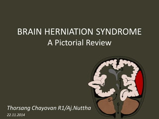
  • 1. BRAIN HERNIATION SYNDROME A Pictorial Review Thorsang Chayovan R1/Aj.Nuttha 22.11.2014
  • 2.
  • 3.
  • 4. BRAIN HERNIATION • most common types –Subfalcine herniation –descending transtentorial herniation •Others –Posterior fossa herniations •ascending transtentorial herniation •tonsillar herniation –Transalar herniation •Rare but important types –transdural/transcranial herniations –brain displacements across the sphenoid wing
  • 6. Subfalcine herniation •most common •supratentorial mass in one hemicranium •affected hemisphere pushes across the midline under the inferior "free" margin of the falx, extending into the contralateral hemicranium
  • 7.
  • 8.
  • 9. Subfalcine herniation: imaging Axial and coronal images show that •cingulate gyrus •anterior cerebral artery (ACA) •internal cerebral vein (ICV) are pushed from one side to the other under the falx cerebri. The ipsilateral ventricle appears compressed and displaced across the midline
  • 10.
  • 11. Complications •unilateral obstructive hydrocephalus –foramen of Monro occlusion •Periventricular hypodensity with "blurred" margins of the lateral ventricle –Fluid accumulates in the periventricular white matter
  • 12. Complications •When severe, the herniating ACA can be pinned against the inferior "free" margin of the falx cerebri  secondary infarction of the cingulate gyrus
  • 13.
  • 15. Transtentorial herniations descending herniations ascending herniations
  • 16. Descending transtentorial herniations •the second most common •a hemispheric mass •initially produces subfalcine herniation •As the mass effect increases, the uncus of the temporal lobe is pushed medially begins to encroach on the suprasellar cistern hippocampus follows hippocampus effaces the ipsilateral quadrigeminal cistern both the uncus and hippocampus herniate inferiorly through the tentorial incisura
  • 17. "Dysautonomia, Multisystem Atrophy and Parkinson's." Dysautonomia, Multisystem Atrophy and Parkinson's. N.p., n.d. Web. 18 Nov. 2014
  • 18.
  • 19. Descending transtentorial herniation •Unilateral •Bilateral ("central“) –Severe
  • 20. unilateral DTH: imaging early uncus is displaced medially Ipsilateral aspect of the suprasellar cistern effaced Ipsilateral prepontine + cerebellopontine angle cistern enlarged
  • 21.
  • 22. Descending transtentorial herniation As DTH increases hippocampus also herniates medially quadrigeminal cistern compression midbrain pushed toward the opposite side of the incisura
  • 23. Descending transtentorial herniation severe cases entire suprasellar and quadrigeminal cisterns are effaced. The temporal horn can even be displaced almost into the midline
  • 24.
  • 25. bilateral DTH both hemispheres become swollen the whole central brain is flattened against the skull base All the basal cisterns are obliterated hypothalamus and optic chiasm are crushed against the sella turcica
  • 26.
  • 27.
  • 28. Complete bilateral DTH both temporal lobes herniate medially into the tentorial hiatus midbrain and pons displaced inferiorly through the tentorial incisura The angle between the midbrain and pons is progressively reduced from 90° to almost 0°
  • 29.
  • 30. Complications •CN III (oculomotor) nerve compression –CN III palsy •PCA occlusion as it passes back up over the medial edge of the tentorium –secondary PCA (occipital) infarct
  • 31.
  • 32.
  • 33. Kernohan notch •As the herniating temporal lobe pushes the midbrain toward the opposite side of the incisura –contralateral cerebral peduncle is forced against the hard edge of the tentorium •Pressure ischemia  ipsilateral hemiplegia –the "false localizing" sign
  • 34.
  • 35.
  • 36.
  • 37. Duret hemorrhage "Top-down" mass effect displaces the midbrain inferiorly closes the midbrain-pontine angle Perforating arteries from basilar artery are compressed and buckled secondary hemorrhagic midbrain infarct
  • 38. Brainstem hemorrhage Brainstem hemorrhage Dorsolateral Primary injury Severe DAI Ventral paramedian Duret
  • 39. Hemorrhage in diffuse axonal injury •Gray-white junction •Corpus callosum •Brainstem
  • 40. hypothalamic and basal ganglia infarcts complete bilateral DTH perforating arteries from the circle of Willis compression against the central skull base hypothalamic and basal ganglia infarcts
  • 41.
  • 42.
  • 43.
  • 44. POSTERIOR FOSSA MASS: TONSILLAR HERNIATION ASCENDING TRANSTENTORIAL HERNIATION
  • 45.
  • 46. Tonsillar herniation •The cerebellar tonsils are displaced inferiorly and become impacted into the foramen magnum. •congenital (e.g., Chiari 1 malformation) – mismatch between size and content of the posterior fossa •Acquired –an expanding posterior fossa mass pushing the tonsils downward—more common –intracranial hypotension: abnormally low intraspinal CSF pressure •tonsils are pulled downward
  • 47. Tonsillar herniation: imaging •Diagnosing tonsillar herniation on NECT scans may be problematic. Cisterna magna obliteration
  • 48.
  • 49. Tonsillar herniation: imaging •MR: much more easily diagnosed •In the sagittal plane –the tonsillar folia become vertically oriented –the inferior aspect of the tonsils becomes pointed –Tonsils > 5 mm (or 7 mm in children) below the foramen magnum are generally abnormal •especially if they are peg-like or pointed (rather than rounded)
  • 50. Tonsillar herniation: imaging •In the axial plane, T2 scans show that the tonsils are impacted into the foramen magnum –obliterating CSF in the cisterna magna –displacing the medulla anteriorly
  • 51.
  • 54.
  • 55. Ascending transtentorial herniation •caused by any expanding posterior fossa mass –Neoplasms > trauma
  • 56.
  • 57.
  • 58. Complications •Acute intraventricular obstructive hydrocephalus –caused by compression of the cerebral aqueduct
  • 59.
  • 60.
  • 61. OTHER LESS COMMON HERNIATION: TRANSALAR TRANSDURAL/TRANSCRANIAL HERNIATIONS
  • 62. Transalar Herniation •brain herniates across the greater sphenoid wing (GSW) or "ala" •ascending > descending
  • 63. Ascending transalar herniation •caused by a large middle cranial fossa mass •An intratemporal or large extraaxial mass Temporal lobe + sylvian fissure + MCA up and over the greater sphenoid wing
  • 64.
  • 65.
  • 66.
  • 67. Descending transalar herniation •caused by a large anterior cranial fossa mass Gyrus rectus is forced posteroinferiorly over the GSW displacing the sylvian fissure and shifting the MCA backward
  • 68. Transdural/Transcranial Herniation •Rare •Sometimes called a "brain fungus" •can be life-threatening Lacerated dura + a skull defect + increased ICP
  • 69.
  • 70. Transdural/Transcranial Herniation •Traumatic –infants or young children with a comminuted inward skull fracture •Iatrogenic –a burr hole, craniotomy, or craniectomy
  • 71. Transdural/Transcranial Herniation •MR best depicts these unusual herniations. •The disrupted dura –discontinuous black line on T2WI –Brain tissue, blood vessels, and CSF, are extruded through the defects into the subgaleal space
  • 72.
  • 73.
  • 74. Kaewlai, R. Imaging of Traumatic Brain Injury. 2013.
  • 76.
  • 77.
  • 78.
  • 79.
  • 80. References •Osborn, Anne G. "Secondary Effects and Sequellae of CNS Trauma."Osborn's Brain: Imaging, Pathology, and Anatomy. Salt Lake City, UT: Amirsys Pub., 2013. N. pag. Print. •Osborn, Anne G. "Cerebral Vasculature: Normal Anatomy and Pathology."Diagnostic Neuroradiology. St. Louis: Mosby, 1994. N. pag. Print. •Kaewlai, R. Imaging of Traumatic Brain Injury. 2013. Web.