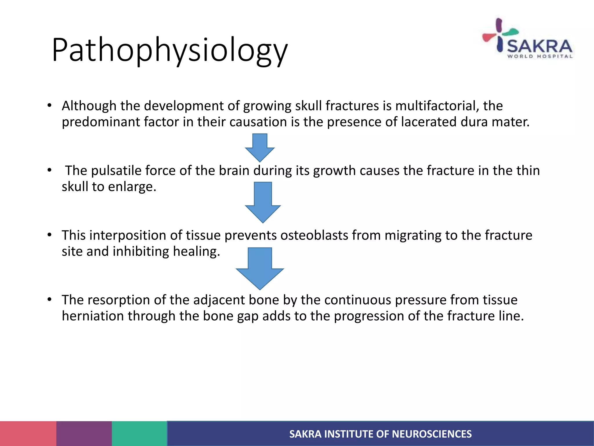
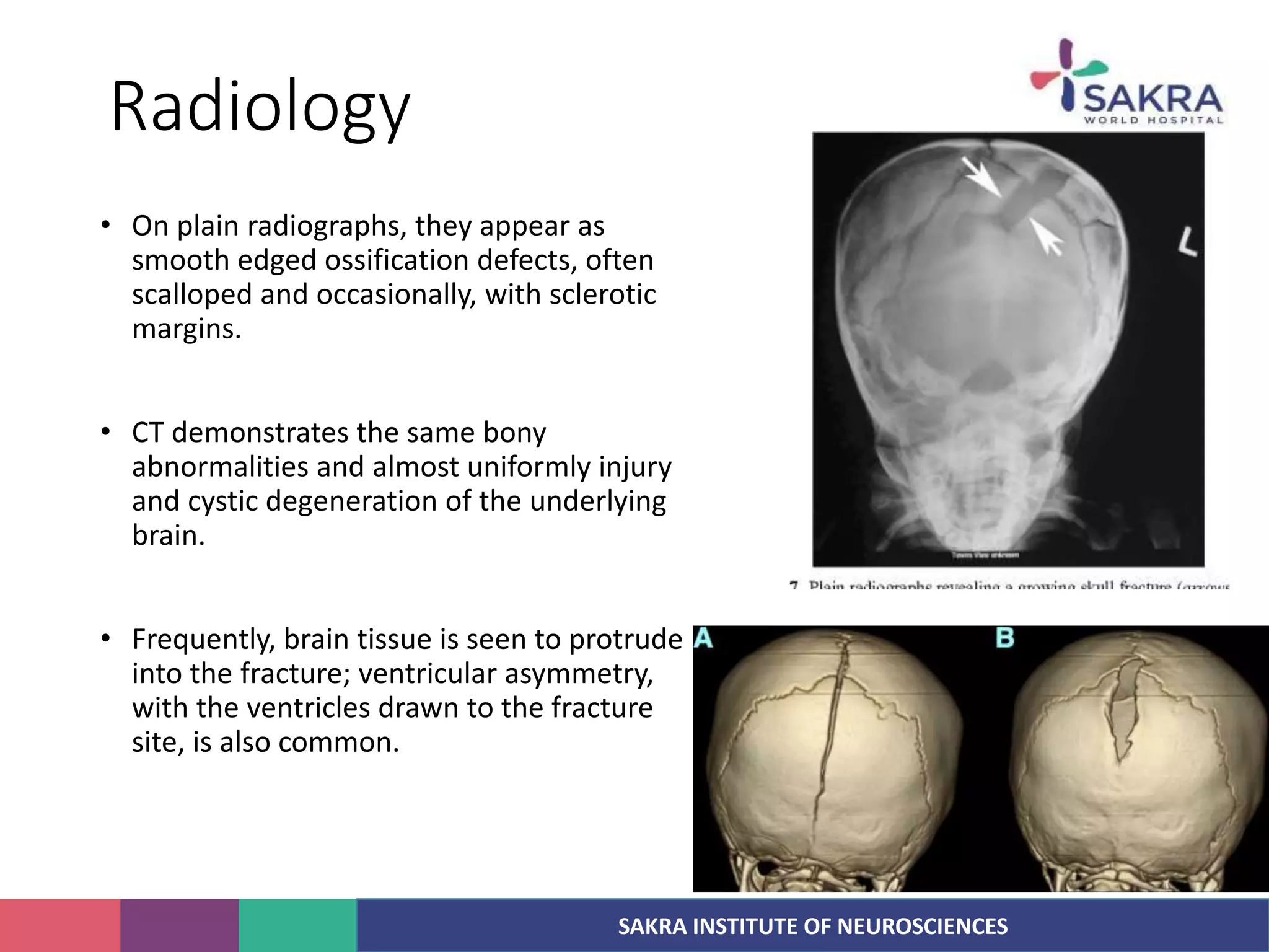
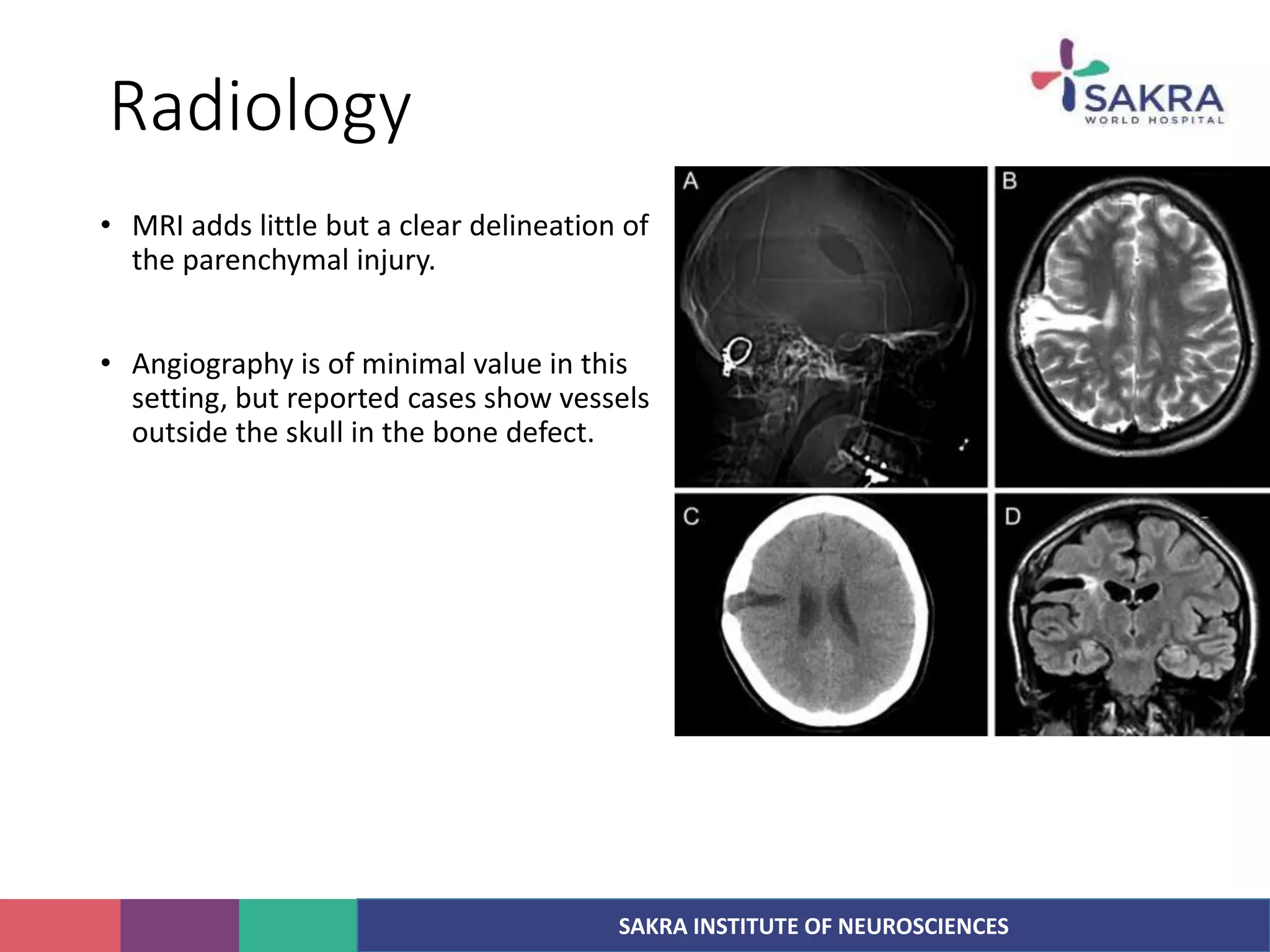
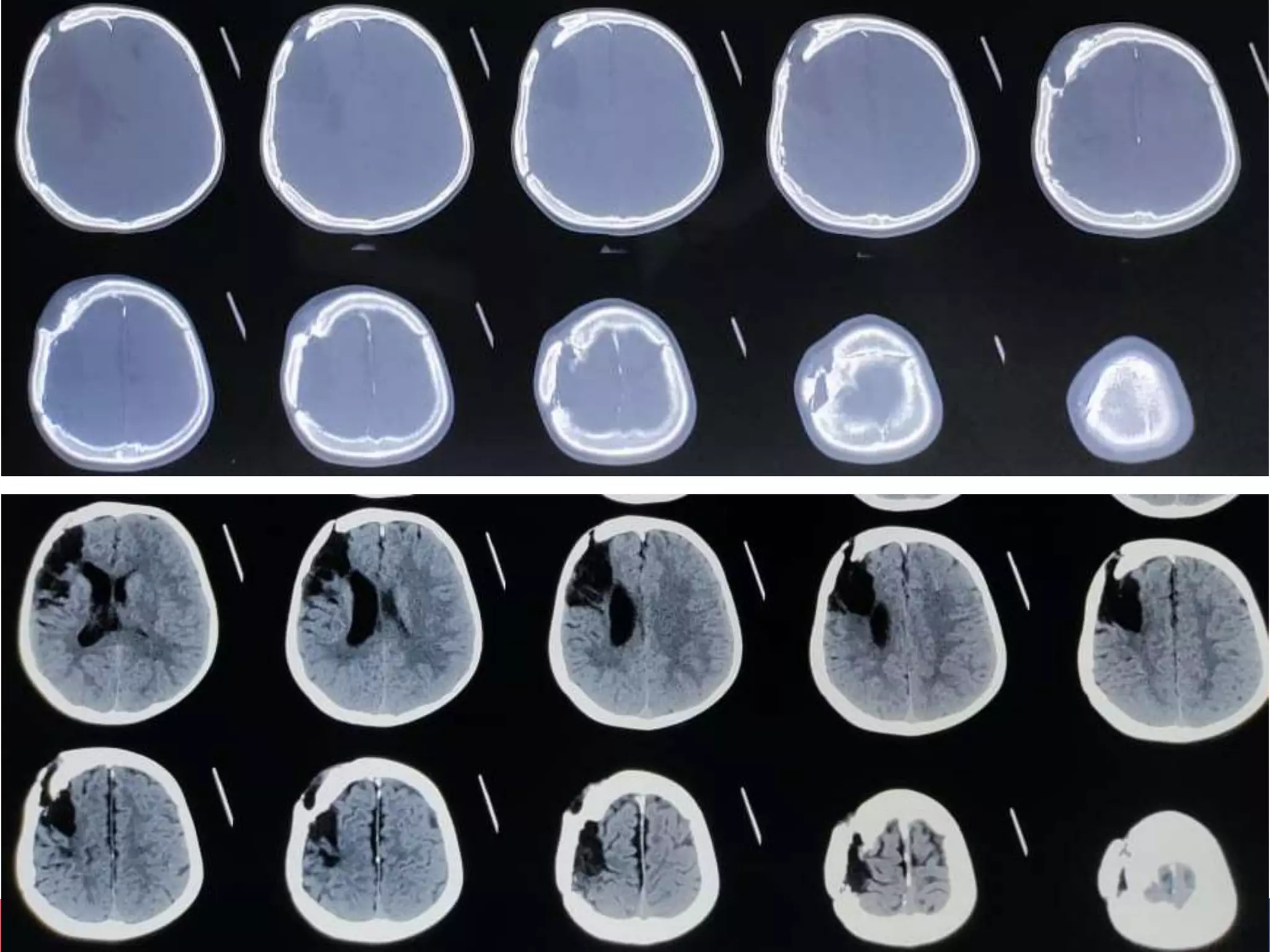
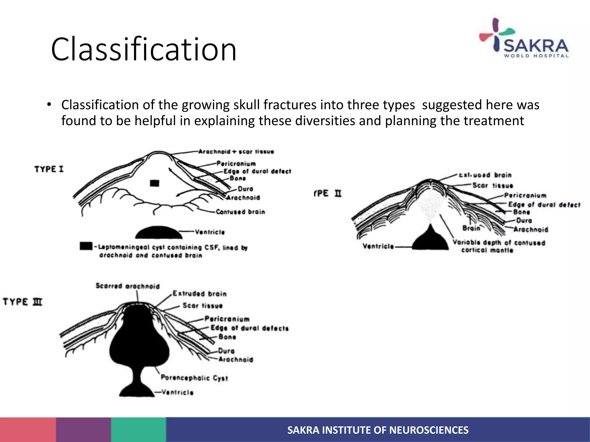
SAKRA INSTITUTE OF NEUROSCIENCES
Growing skull fracture
Dr Abhishek Rai
Resident neurosurgery
SAKRA INSTITUTE OF NEUROSCIENCES
Introduction
• A skull fracture is a break in one or more of the
eight bones that form the cranial portion of the
skull, usually occurring as a result of blunt force
trauma.
• Any significant blow to the head results in a
concussion, with or without loss of
consciousness.
SAKRA INSTITUTE OF NEUROSCIENCES
Compound fracture
• A fracture in conjunction with an overlying
laceration that tears the epidermis and the
meninges, or runs through the paranasal
sinuses and the middle ear structures,
bringing the outside environment into
contact with the cranial cavity is called a
compound fracture.
• Compound fractures can either be clean or
contaminated.
• Compound elevated fracture : Its rare type of
skull fracture where the fractured bone is
elevated above the intact outer table of the
skull.
SAKRA INSTITUTE OF NEUROSCIENCES
Types
• Cranial vault
1. Linear/ Fissure fracture
2. Depressed
3. Elevated
4. Comminuted
5. Pond fracture
6. Gutter fracture
7. Diastatic
8. Basilar
9. Growing skull fracture
10. Cranial burst fracture
• Fracture of skull base
1. Ring fracture
2. Hinge fracture
3. ACF fracture
4. MCF fracture
5. PCF fracture
SAKRA INSTITUTE OF NEUROSCIENCES
Linear/Fissure fracture
• Linear skull fractures are breaks in the bone that
transverse the full thickness of the skull from the
outer to inner table.
• They are usually with no bone displacement.
• The common cause of injury is blunt force trauma
where the impact energy transferred over a wide
area of the skull.
• Not easily detectable in radiology.
• Mc type(70%)
• Line of fracture runs parallel to axis of compression
SAKRA INSTITUTE OF NEUROSCIENCES
Linear fracture
• Linear skull fractures are usually of little
clinical significance unless they parallel
in close proximity or transverse a
suture, or they involve a venous sinus
groove or vascular channel.
• The resulting complications may include
suture diastasis, venous sinus
thrombosis, and epidural hematoma.
• In young children, although rare, the
possibility exists of developing a
growing skull fracture especially if the
fracture occurs in the parietal bone
SAKRA INSTITUTE OF NEUROSCIENCES
Depressed fracture
• A depressed skull fracture aka
Fracture a la signature, since it
reflects the type of weapon used.
• It occurs if the fractured bone is
driven inwards to distance
equivalent to thickness of skull
table. Sometime only involve outer
table.
• Risk of post traumatic epilepsy is
15%.
SAKRA INSTITUTE OF NEUROSCIENCES
Depressed fracture
• Depressed skull fractures present a high risk of increased pressure on the brain,
or a hemorrhage to the brain that crushes the delicate tissue.
• In complex depressed fractures, the dura mater is torn.
• Depressed skull fractures may require surgery to lift the bones off the brain if
they are pressing on it by making burr holes on the adjacent normal skull.
SAKRA INSTITUTE OF NEUROSCIENCES
Comminuted fracture
• Spider web/Mosaic fracture
• 2 or more intersecting lines of fracture
dividing the bone into 3 or more
fragments.
• Cause : Significant force striking over
broad surface area.
• Complications of linear and depressed
fracture
SAKRA INSTITUTE OF NEUROSCIENCES
Pond fracture
• Smooth concave dent resulting from in-
buckling of skull.
• Occurs in pliable skull (<4 years)
• Inner table not fractured.
• Brain and meninges are not damaged.
• Cause :
1. Obstetric fracture
2. Forcible delivery
SAKRA INSTITUTE OF NEUROSCIENCES
Gutter fracture
• Part of thickness of skull is removed
to form gutter.
• Cause: Oblique bullet wound
• Associated with irregular depressed
fracture of outer table of skull.
• Dura and brain may be torn.
SAKRA INSTITUTE OF NEUROSCIENCES
Diastatic fracture
• Separation of sutures due to blow on the
head with blunt weapon.
• Young children
• Mc Sagittal suture
• Widening of the sutures may be
appreciated on plain radiograph and is
better still appreciated on CT
• The following widths are considered to be
diagnostic of sutural diastasis
a. >10 mm at birth
b. >3 mm at two years
c. >2 mm at three years
SAKRA INSTITUTE OF NEUROSCIENCES
Growing skull fracture
• Linear or non-linear skull fractures in children
that enlarge with time are termed growing skull
fractures.
• Growing skull fractures are rare and occur,
almost exclusively, during the first years of life.
• Also described as traumatic ventricular
cysts, craniocerebral erosions, cranial
malacia, and leptomeningeal cysts,
among others.
• They appear most frequently as soft, subgaleal
collections overlying an enlarging bony defect at
the site of a previous diastatic fracture.
SAKRA INSTITUTE OF NEUROSCIENCES
History
• Howship was the first to report a patient with a growing skull fracture. In 1816,
he described a 9-month-old child in whom an enlarging defect in his parietal
bone developed after an injury. The defect was apparent within 2 weeks after
the injury and never resolved.
• The first account of the pathology of this condition was by Rokitansky in 1856.
He reported 5 month old who struck his head during a seizure and subsequently
an enlarging scalp mass developed.
• Therapeutic puncture of the mass was performed, after which meningitis
developed and the child died. At autopsy a large bone defect was found along
with an associated dural tear and injury to the underlying brain.
SAKRA INSTITUTE OF NEUROSCIENCES
History
• Dyke introduced the term leptomeningeal cyst in 1937.
• Other observations have failed to confirm the central role of leptomeningeal
cysts in the pathogenesis of growing fractures.
• Penfield and Erickson described a patient with an enlarging defect at the site of
a childhood fracture. At surgery, brain, but no cyst was found in the defect.
• Tenner and Stein also described herniated brain within the ossification defects in
children with growing fractures and highlighted the importance of local brain
injury and swelling in their development.
• Rosenthal and associates found that both the dura and arachnoid had to be
opened to produce an enlarging fracture and that concomitant injury to the
brain did not increase the likelihood that a growing fracture.
SAKRA INSTITUTE OF NEUROSCIENCES
History
• Many of the children described have had significant neurological deficits from
the time of their injury, there are several reports of progressive dysfunction that
develops months or years after the event.
• This has been attributed to brain injury caused by
(1) local brain herniation and consequent ischemia
(2) repetitive trauma to the exposed brain on the bone edge or by direct
concussion,
(3) physical distortion of the brain related to its displacement
• Growing fractures occur only in settings in which there is rapid brain growth or,
much less commonly, increased intracranial pressure.
SAKRA INSTITUTE OF NEUROSCIENCES
Epidemiology
• They account for less than 1% of skull fractures in large pediatric series and, for
practical purposes, are never seen in adults.
• Almost all enlarging fractures are found in children younger than 3 years; two
thirds of them occur in children younger than 1 year.
• Neurological deficit more than 50% in most case series, and for the incidence of
posttraumatic seizures, which is also around 50%
SAKRA INSTITUTE OF NEUROSCIENCES
Natural history
• 3 stages of development of GSF:
• Stage I : Aka Prephase : Skull fracture (linear or comminuted) with dural
laceration and herniation of brain tissue or arachnoid membrane through torn
dura.
• Stage II : Early phase. It last approx. 2 months, Bone defect is small, deformity
relatively limited, mild neurologic deficit, Entrapped tissue prevents fracture
healing.
• Stage III : Late stage, after 2 months, bone defect significantly enlarges, brain
tissue and CSF extend between bony edge of fracture through torn dura and
arachnoid
SAKRA INSTITUTE OF NEUROSCIENCES
Pathophysiology
• Although the development of growing skull fractures is multifactorial, the
predominant factor in their causation is the presence of lacerated dura mater.
• The pulsatile force of the brain during its growth causes the fracture in the thin
skull to enlarge.
• This interposition of tissue prevents osteoblasts from migrating to the fracture
site and inhibiting healing.
• The resorption of the adjacent bone by the continuous pressure from tissue
herniation through the bone gap adds to the progression of the fracture line.
SAKRA INSTITUTE OF NEUROSCIENCES
Pathophysiology
• The brain extrusion may be present shortly after diastatic linear fracture in
neonates and young infants resulting in focal dilation of the lateral ventricle near
the growing fracture.
SAKRA INSTITUTE OF NEUROSCIENCES
Evaluation
• The typical patient with a growing fracture is
1. Being younger than 3 years
2. Has a subgaleal fluid collection overlying the fracture, (although the collection
may resolve soon after injury)
3. Has a neurological deficit caused by the injury
4. Has skull radiographs that demonstrate a diastatic fracture with at least 3.5-mm
separation of the bone edges
• Some authors have advised obtaining skull radiographs 6 weeks and again
several months after the injury while others asked to see these patients in the
clinic after 8 weeks.
SAKRA INSTITUTE OF NEUROSCIENCES
Evaluation
• Common initial complaints are
1. pain at the fracture site
2. Epilepsy
3. progressive neurological deficit.
• On examination :
1. Palpable ossification defect, larger than the one initially present, that is usually
but not always overlain by a soft, often pulsatile subgaleal collection. -
Operative
2. The ossification defect is small or only minimally different than it was at initial
evaluation, and no overlying collection is present. - re-examine these children
several weeks later.
SAKRA INSTITUTE OF NEUROSCIENCES
Radiology
• On plain radiographs, they appear as
smooth edged ossification defects, often
scalloped and occasionally, with sclerotic
margins.
• CT demonstrates the same bony
abnormalities and almost uniformly injury
and cystic degeneration of the underlying
brain.
• Frequently, brain tissue is seen to protrude
into the fracture; ventricular asymmetry,
with the ventricles drawn to the fracture
site, is also common.
SAKRA INSTITUTE OF NEUROSCIENCES
Radiology
• MRI adds little but a clear delineation of
the parenchymal injury.
• Angiography is of minimal value in this
setting, but reported cases show vessels
outside the skull in the bone defect.
SAKRA INSTITUTE OF NEUROSCIENCES
Classification
• Classification of the growing skull fractures into three types suggested here was
found to be helpful in explaining these diversities and planning the treatment
SAKRA INSTITUTE OF NEUROSCIENCES
Classification
• Classification of the growing skull fractures into three types suggested here was
found to be helpful in explaining these diversities and planning the treatment
SAKRA INSTITUTE OF NEUROSCIENCES
Classification
• Duro-cranioplasty was the correct treatment in type 1.
• A shunting procedure required as an initial or definitive management for type II
and III fractures with raised intracranial pressure.
SAKRA INSTITUTE OF NEUROSCIENCES
Treatment
• Successful repair requires identification of the dural margin, repair of the dural
defect, and coverage of the missing bone.
• A large incision is needed; it must extend well beyond the margins of the skull
defect.
• The skin flap is reflected in the subgaleal plane and the bony defect is defined.
• The dural edge has a tendency to recede and is often located well back from the
edge of the bone.
1. It can be found by progressively removing bone from the margins of the defect
until the dura is circumferentially exposed.
2. A burr hole can be placed a few centimeters from the bone edge.
SAKRA INSTITUTE OF NEUROSCIENCES
Treatment
• Circumferential craniotomy
• Once the dural defect is exposed, the underlying brain is carefully dissected. If a
large area of cortex is herniating through the dural defect, an attempt to reduce
it should be made.
• Resection of herniated gliotic tissue
• Closure of the dural defect is necessary to prevent recurrence f the growing
fracture.
• Primary closure rarely possible, so some graft material must be used.
SAKRA INSTITUTE OF NEUROSCIENCES
Treatment
• Several options are available for bone coverage, the most frequently used of
which are split-thickness calvarial grafts, full thickness calvarial autografts,
synthetic materials, and split rib grafts.
• VP Shunt
SAKRA INSTITUTE OF NEUROSCIENCES

Growing skull fracture

  • 1.
    SAKRA INSTITUTE OFNEUROSCIENCES Growing skull fracture Dr Abhishek Rai Resident neurosurgery
  • 2.
    SAKRA INSTITUTE OFNEUROSCIENCES Introduction • A skull fracture is a break in one or more of the eight bones that form the cranial portion of the skull, usually occurring as a result of blunt force trauma. • Any significant blow to the head results in a concussion, with or without loss of consciousness.
  • 3.
    SAKRA INSTITUTE OFNEUROSCIENCES Compound fracture • A fracture in conjunction with an overlying laceration that tears the epidermis and the meninges, or runs through the paranasal sinuses and the middle ear structures, bringing the outside environment into contact with the cranial cavity is called a compound fracture. • Compound fractures can either be clean or contaminated. • Compound elevated fracture : Its rare type of skull fracture where the fractured bone is elevated above the intact outer table of the skull.
  • 4.
    SAKRA INSTITUTE OFNEUROSCIENCES Types • Cranial vault 1. Linear/ Fissure fracture 2. Depressed 3. Elevated 4. Comminuted 5. Pond fracture 6. Gutter fracture 7. Diastatic 8. Basilar 9. Growing skull fracture 10. Cranial burst fracture • Fracture of skull base 1. Ring fracture 2. Hinge fracture 3. ACF fracture 4. MCF fracture 5. PCF fracture
  • 5.
    SAKRA INSTITUTE OFNEUROSCIENCES Linear/Fissure fracture • Linear skull fractures are breaks in the bone that transverse the full thickness of the skull from the outer to inner table. • They are usually with no bone displacement. • The common cause of injury is blunt force trauma where the impact energy transferred over a wide area of the skull. • Not easily detectable in radiology. • Mc type(70%) • Line of fracture runs parallel to axis of compression
  • 6.
    SAKRA INSTITUTE OFNEUROSCIENCES Linear fracture • Linear skull fractures are usually of little clinical significance unless they parallel in close proximity or transverse a suture, or they involve a venous sinus groove or vascular channel. • The resulting complications may include suture diastasis, venous sinus thrombosis, and epidural hematoma. • In young children, although rare, the possibility exists of developing a growing skull fracture especially if the fracture occurs in the parietal bone
  • 7.
    SAKRA INSTITUTE OFNEUROSCIENCES Depressed fracture • A depressed skull fracture aka Fracture a la signature, since it reflects the type of weapon used. • It occurs if the fractured bone is driven inwards to distance equivalent to thickness of skull table. Sometime only involve outer table. • Risk of post traumatic epilepsy is 15%.
  • 8.
    SAKRA INSTITUTE OFNEUROSCIENCES Depressed fracture • Depressed skull fractures present a high risk of increased pressure on the brain, or a hemorrhage to the brain that crushes the delicate tissue. • In complex depressed fractures, the dura mater is torn. • Depressed skull fractures may require surgery to lift the bones off the brain if they are pressing on it by making burr holes on the adjacent normal skull.
  • 9.
    SAKRA INSTITUTE OFNEUROSCIENCES Comminuted fracture • Spider web/Mosaic fracture • 2 or more intersecting lines of fracture dividing the bone into 3 or more fragments. • Cause : Significant force striking over broad surface area. • Complications of linear and depressed fracture
  • 10.
    SAKRA INSTITUTE OFNEUROSCIENCES Pond fracture • Smooth concave dent resulting from in- buckling of skull. • Occurs in pliable skull (<4 years) • Inner table not fractured. • Brain and meninges are not damaged. • Cause : 1. Obstetric fracture 2. Forcible delivery
  • 11.
    SAKRA INSTITUTE OFNEUROSCIENCES Gutter fracture • Part of thickness of skull is removed to form gutter. • Cause: Oblique bullet wound • Associated with irregular depressed fracture of outer table of skull. • Dura and brain may be torn.
  • 12.
    SAKRA INSTITUTE OFNEUROSCIENCES Diastatic fracture • Separation of sutures due to blow on the head with blunt weapon. • Young children • Mc Sagittal suture • Widening of the sutures may be appreciated on plain radiograph and is better still appreciated on CT • The following widths are considered to be diagnostic of sutural diastasis a. >10 mm at birth b. >3 mm at two years c. >2 mm at three years
  • 13.
    SAKRA INSTITUTE OFNEUROSCIENCES Growing skull fracture • Linear or non-linear skull fractures in children that enlarge with time are termed growing skull fractures. • Growing skull fractures are rare and occur, almost exclusively, during the first years of life. • Also described as traumatic ventricular cysts, craniocerebral erosions, cranial malacia, and leptomeningeal cysts, among others. • They appear most frequently as soft, subgaleal collections overlying an enlarging bony defect at the site of a previous diastatic fracture.
  • 14.
    SAKRA INSTITUTE OFNEUROSCIENCES History • Howship was the first to report a patient with a growing skull fracture. In 1816, he described a 9-month-old child in whom an enlarging defect in his parietal bone developed after an injury. The defect was apparent within 2 weeks after the injury and never resolved. • The first account of the pathology of this condition was by Rokitansky in 1856. He reported 5 month old who struck his head during a seizure and subsequently an enlarging scalp mass developed. • Therapeutic puncture of the mass was performed, after which meningitis developed and the child died. At autopsy a large bone defect was found along with an associated dural tear and injury to the underlying brain.
  • 15.
    SAKRA INSTITUTE OFNEUROSCIENCES History • Dyke introduced the term leptomeningeal cyst in 1937. • Other observations have failed to confirm the central role of leptomeningeal cysts in the pathogenesis of growing fractures. • Penfield and Erickson described a patient with an enlarging defect at the site of a childhood fracture. At surgery, brain, but no cyst was found in the defect. • Tenner and Stein also described herniated brain within the ossification defects in children with growing fractures and highlighted the importance of local brain injury and swelling in their development. • Rosenthal and associates found that both the dura and arachnoid had to be opened to produce an enlarging fracture and that concomitant injury to the brain did not increase the likelihood that a growing fracture.
  • 16.
    SAKRA INSTITUTE OFNEUROSCIENCES History • Many of the children described have had significant neurological deficits from the time of their injury, there are several reports of progressive dysfunction that develops months or years after the event. • This has been attributed to brain injury caused by (1) local brain herniation and consequent ischemia (2) repetitive trauma to the exposed brain on the bone edge or by direct concussion, (3) physical distortion of the brain related to its displacement • Growing fractures occur only in settings in which there is rapid brain growth or, much less commonly, increased intracranial pressure.
  • 17.
    SAKRA INSTITUTE OFNEUROSCIENCES Epidemiology • They account for less than 1% of skull fractures in large pediatric series and, for practical purposes, are never seen in adults. • Almost all enlarging fractures are found in children younger than 3 years; two thirds of them occur in children younger than 1 year. • Neurological deficit more than 50% in most case series, and for the incidence of posttraumatic seizures, which is also around 50%
  • 18.
    SAKRA INSTITUTE OFNEUROSCIENCES Natural history • 3 stages of development of GSF: • Stage I : Aka Prephase : Skull fracture (linear or comminuted) with dural laceration and herniation of brain tissue or arachnoid membrane through torn dura. • Stage II : Early phase. It last approx. 2 months, Bone defect is small, deformity relatively limited, mild neurologic deficit, Entrapped tissue prevents fracture healing. • Stage III : Late stage, after 2 months, bone defect significantly enlarges, brain tissue and CSF extend between bony edge of fracture through torn dura and arachnoid
  • 19.
    SAKRA INSTITUTE OFNEUROSCIENCES Pathophysiology • Although the development of growing skull fractures is multifactorial, the predominant factor in their causation is the presence of lacerated dura mater. • The pulsatile force of the brain during its growth causes the fracture in the thin skull to enlarge. • This interposition of tissue prevents osteoblasts from migrating to the fracture site and inhibiting healing. • The resorption of the adjacent bone by the continuous pressure from tissue herniation through the bone gap adds to the progression of the fracture line.
  • 20.
    SAKRA INSTITUTE OFNEUROSCIENCES Pathophysiology • The brain extrusion may be present shortly after diastatic linear fracture in neonates and young infants resulting in focal dilation of the lateral ventricle near the growing fracture.
  • 21.
    SAKRA INSTITUTE OFNEUROSCIENCES Evaluation • The typical patient with a growing fracture is 1. Being younger than 3 years 2. Has a subgaleal fluid collection overlying the fracture, (although the collection may resolve soon after injury) 3. Has a neurological deficit caused by the injury 4. Has skull radiographs that demonstrate a diastatic fracture with at least 3.5-mm separation of the bone edges • Some authors have advised obtaining skull radiographs 6 weeks and again several months after the injury while others asked to see these patients in the clinic after 8 weeks.
  • 22.
    SAKRA INSTITUTE OFNEUROSCIENCES Evaluation • Common initial complaints are 1. pain at the fracture site 2. Epilepsy 3. progressive neurological deficit. • On examination : 1. Palpable ossification defect, larger than the one initially present, that is usually but not always overlain by a soft, often pulsatile subgaleal collection. - Operative 2. The ossification defect is small or only minimally different than it was at initial evaluation, and no overlying collection is present. - re-examine these children several weeks later.
  • 23.
    SAKRA INSTITUTE OFNEUROSCIENCES Radiology • On plain radiographs, they appear as smooth edged ossification defects, often scalloped and occasionally, with sclerotic margins. • CT demonstrates the same bony abnormalities and almost uniformly injury and cystic degeneration of the underlying brain. • Frequently, brain tissue is seen to protrude into the fracture; ventricular asymmetry, with the ventricles drawn to the fracture site, is also common.
  • 24.
    SAKRA INSTITUTE OFNEUROSCIENCES Radiology • MRI adds little but a clear delineation of the parenchymal injury. • Angiography is of minimal value in this setting, but reported cases show vessels outside the skull in the bone defect.
  • 25.
    SAKRA INSTITUTE OFNEUROSCIENCES
  • 26.
    SAKRA INSTITUTE OFNEUROSCIENCES
  • 27.
    SAKRA INSTITUTE OFNEUROSCIENCES Classification • Classification of the growing skull fractures into three types suggested here was found to be helpful in explaining these diversities and planning the treatment
  • 28.
    SAKRA INSTITUTE OFNEUROSCIENCES Classification • Classification of the growing skull fractures into three types suggested here was found to be helpful in explaining these diversities and planning the treatment
  • 29.
    SAKRA INSTITUTE OFNEUROSCIENCES Classification • Duro-cranioplasty was the correct treatment in type 1. • A shunting procedure required as an initial or definitive management for type II and III fractures with raised intracranial pressure.
  • 30.
    SAKRA INSTITUTE OFNEUROSCIENCES Treatment • Successful repair requires identification of the dural margin, repair of the dural defect, and coverage of the missing bone. • A large incision is needed; it must extend well beyond the margins of the skull defect. • The skin flap is reflected in the subgaleal plane and the bony defect is defined. • The dural edge has a tendency to recede and is often located well back from the edge of the bone. 1. It can be found by progressively removing bone from the margins of the defect until the dura is circumferentially exposed. 2. A burr hole can be placed a few centimeters from the bone edge.
  • 31.
    SAKRA INSTITUTE OFNEUROSCIENCES Treatment • Circumferential craniotomy • Once the dural defect is exposed, the underlying brain is carefully dissected. If a large area of cortex is herniating through the dural defect, an attempt to reduce it should be made. • Resection of herniated gliotic tissue • Closure of the dural defect is necessary to prevent recurrence f the growing fracture. • Primary closure rarely possible, so some graft material must be used.
  • 32.
    SAKRA INSTITUTE OFNEUROSCIENCES Treatment • Several options are available for bone coverage, the most frequently used of which are split-thickness calvarial grafts, full thickness calvarial autografts, synthetic materials, and split rib grafts. • VP Shunt
  • 33.
    SAKRA INSTITUTE OFNEUROSCIENCES

Editor's Notes

  • #3 If the force of the impact is excessive, the bone may fracture at or near the site of the impact and cause damage to the underlying structures within the skull such as the membranes, blood vessels, and brain.
  • #4 The most serious complication of compound skull fractures is infection. Increased risk factors for infection include visible contamination, meningeal tear, loose bone fragments and presenting for treatment more than eight hours after initial injury. Compound elevated : initial blow penetrates the skull and the underlying meninges and, on withdrawal, the weapon lifts the fractured portion of the skull outward. It can also be caused by the skull rotating while being struck in a case of blunt force trauma, the skull rotating while striking an inanimate object as in a fall, or it may occur during transfer of a patient after an initial compound head injury.
  • #5 Cervical myelopathy may manifest with subtle to severe neurological deficits.
  • #7 Linear fractures exteds into SSS, causing diastasis of sagittal suture diastasis
  • #10 When no displacement of fragment it resemble a spider’s web or mosaic pattern.
  • #11 Aka ping pong Fracture
  • #13 Aka Sutural fracture Causes : Trauma or Raised ICP
  • #15 The boy was re-examined when he was 4 years old and “the opening in the cranium remained undiminished. Upon laying the hand on the part, the pulsations of the brain were felt strong and distinct.”
  • #16 Additionally, these authors made the point that porencephalic cysts can be seen in the parenchyma adjacent to the fracture site. Rosenthal findings confirmed by Winston and coworkers
  • #18 No specific or unique mechanism of injury predisposes patients to the development of a growing fracture, but in many cases the trauma suffered seems to have been relatively severe.
  • #19 St 1 from TOI to to just before fracture enlarges
  • #20 laceration of the dura and the brain pulsations may herniate a pouch of arachnoid
  • #21 The brain extrusion may be present shortly after diastatic linear fracture in neonates and young infants resulting in focal dilation of the lateral ventricle near the growing fracture, This dilatation is said to be reversible and may normalize after surgical repair.
  • #23 There have been several reports of patients more than 10 years after injury who have had enlarging bone defects andworsening neurological conditions. pseudogrowing” fractures. No skull defect after 2 months require no intervention
  • #24 enlargement of cerebral ventricles and subarachnoid spaces, caused by encephalic volume loss
  • #26 ex vacio dilatation, right ventricle enlargement, rt frontal subdural and interhemispheric calcification, chronic infarct with gliosis,
  • #27 Coronal Sutural diastasis ,
  • #29 An active form with evidence of raised intracranial pressure (ICE’), mass effect on CT, progressive separation of bone edges with a tense bulge between them; and an arrested or ‘burnt out’ form with a normal ICP, and a slack/sunken gap between the bone edges often showing some evidence of bone regrowth and healing at the fracture site.
  • #30 An active form with evidence of raised intracranial pressure (ICE’), mass effect on CT, progressive separation of bone edges with a tense bulge between them; and an arrested or ‘burnt out’ form with a normal ICP, and a slack/sunken gap between the bone edges often showing some evidence of bone regrowth and healing at the fracture site.
  • #31 There is no place for minimally invasive techniques with this condition.
  • #32 circumferential craniotomy is then performed to allow identification of the dura around the entire lesion
  • #33 Finally, placement of a shunt is best reserved for patients with clear evidence of hydrocephalus, but it does not replace or alter the need for operative repair of these lesions