SlideShare a Scribd company logo
cka
BASIC PRINCIPLES IN
MENINGIOMAS
DR. SURESH BISHOKARMA
MCH NEUROSURGERY ®
UPENDRA DEVKOTA MEMORIAL NATIONAL INSTITUTE OF
NEUROLOGICAL AND ALLIED SCIENCES
BANSBARI, NEPAL
 1922: Harvey Cushing coined the term meningioma to describe a benign
globoid tumor arising from the leptomeninges.
 Eisenhardt believed meningioma arises from arachnoid cap cells that are
particularly abundant in the arachnoid granulation.
 Virchow was the first to describe the classic pathologic feature of the
meningioma with the term psammoma body (sand like, rightly describing
the granules noted within the tumor).
 1957: Donald Simpson's 1957 paper described the correlation between
surgical resection of meningiomas and the rates of symptomatic
recurrence:
HISTORY
 Slow growing, extra-axial tumor, usually benign neoplasms thought to
arise from meningothelial cells found within arachnoid granulations (not
dura).
 Concentrated in the walls of the major venous sinuses, these structures,
which contain “arachnoid cap cells,” account for the dural localization of
most meningiomas within the cranium and spinal cord.
 Although most are benign (70-75%), a few are classified as atypical (20-
24%) or anaplastic (4%).
 Metastases are uncommon and may be seen with benign or malignant
meningiomas.
INTRODUCTION
 Meningiomas: 13 to 26% of all intracranial tumors.
 Skull base meningiomas comprise approximately 44% of all skull base
tumors.
 98% of all CNS meningiomas (spinal 2%).
 An annual estimated incidence of 2.3 cases per 100,000 persons.
 Autopsy: 3% of the population over 60 years of age.
 Female preponderance (1.8)
 Peaks age: fourth decade (45yrs)
 Meningiomas are less often seen in the younger age groups.
 Less than 2% occur in childhood and adolescence.
 About 20% of cases seen in adolescents are associated with NF-I.
 Approximately 1% of meningioma patients have neurofibromatosis 2 (NF2).
 May be multiple in up to 8% of cases.
EPIDEMIOLOGY
 Ionisation radiation: DNA damage resulting for SS or DS DNA break.
 Atomic bomb (epicenter); low dose scalp IR for ringworm; 10x; Israel (1948-60); Full mouth dental
IR; XRT for IC tumors.
 Hormones:
 Est, Prog, Androgens; Size (+): Luteal/pregnancy; 22q12 related with progesterone expression.
 Progesterone alone: Good : Absence of Prog or Est recep or Presence of estrogen alone: Bad.
 Three Chicago areas hospital: 1987-1992: Protective effect of OCP/HRT.
 OCP/HRT: Interphone group: 1993-2002: Increases risk (OR 1.7 HRT/2.7X OCP)
 Mayo Clinic jacksonville patient database; OR 3X
 Infer Limited statistical evidence of increaed risk.
 BMI: Increased BMI association.
 Head Trauma: Harvey Cushing; Danish study: 228,055 Cohort of head injury: F/u 8yrs; No evidence.
Proinflammatory enzyme COX-2 is upregulated after head trauma: Celecoxib# decreases progression.
 Cell Phone use: 10 studies including Interphone Study ; inconclusive; Small sample size; short f/u.
 Association with breast cancer: Common hormonal risk factor and genetic predisposition; BRCA1
interacting protein 1 (BR1P1) is associated with meningioma risk.*
 Industry/Occupation/Diet/Allergy: Chemical (inconclusive); Diet (no): International Case control study;
Allergic diseases like asthma/eczema associated with glial tumor not evident with meningioma.
 Positive familial history:2X: 1st degree.
 Genetic polymorphism: BR1P1; ATM gene. LOH of chromosome 22 and NF2 gene mutation are
implicated in ~50% of sporadic and 100% of NF2-associated meningiomas.
RISK FACTORS
# Ragel Btet al. Celecoxib inhibits meningioma tumor growth in a mouse xenograft model. Cancer 2007.
*Bethke L et al. Comprehensive analysis of DNA repair gene variants and risk of meningioma. J Natl Cancer Inst 2008.
MENINGIOMA TUMORIGENESIS
MENINGIOMA TUMOR PROGRESSION
 The loss of the long arm of chromosome 22 occurs in 40 to 70% of
meningiomas and is associated with loss of the tumor suppressor gene
(MERLIN) for neurofibromatosis type 2 (NF2), located at 22q12.
 The product of this gene, merlin, is thought to be critical for meningioma
tumorigenesis.
 Merlin belongs to the family of structural proteins that link the
cytoskeleton to several proteins of the cytoplasmic membrane, and some
authors have suggested that merlin may act as a tumor suppressor via its
interactions with the cellular cytoskeleton.
Merlin and Meningioma
 It has also been demonstrated that MRI hypointensity on T2-weighted images
is associated with slowed tumor growth, and that MRI hyperintensity on T2-
weighted images is associated significantly with faster tumor growth.
 An annual growth rate of > 1 cm3/year or volume increases > 15% were
considered tumor growth.
 Growth has been defined as a change in tumor size of at least 2 mm,3 5
mm,12 or any measurable change
 Many authors have pointed out that meningiomas without calcification on
imaging are more likely to progress than are calcified meningiomas and no
negative correlation between calcification and slow growth has been
reported.
 Presence of peritumoral edema, ambiguous brain- tumor borders, and
irregular tumor shape are predictor of tumor growth: needs further study.
 Location don’t affects tumor growth.
Tumor growth
LOCATION OF MENIGIOMA
GREENBERG 8TH ED
LOCATION OF MENINGIOMA PERCENTAGE
Parasagittal 20.80%
Convexity 15%
Tuberculum sella 13%
Sphenoid wing 12%
Olfactory 10%
Falcine 8%
CPA 6%
Intraventricular 4%
Tentorial 4%
Suprasellar 3%
Middle fossa 3%
Foramen magnum 2%
Orbital 1.20%
Spinal 1.20%
Multiple 0.90%
Pineal 0.50%
Intrasylvian 0.30%
Extracalvarian 0.30%
Others 5%
Artist's Composite of Different Potential Locations
of Meningiomas
1. Convexity; 2. olfactory groove: 3. sphenoid ridge; 4. Tuberculum sella: 5. Dorsum sellae; 6. Meckel cave. cranial nerve V:
7. Clivus: 8. Petrosal : 9. Tentorial: 10. Sigmoid sinus: 11. Straight sinus: 12. Transverse sinus: 13. Torcular-supra-infra
tentorial meningioma: 14.Supratentorial interhemispheric meningioma: 15. Falx; 16. superior sagittal sinus.
Nader Tricks and Trade of cranial Neurosurgery
 The grading has been done on the basis of histological markers, which
includes,
1. Presence and number of mitotic figures,
2. Overall cellularity,
3. Nuclear to cytoplasmic ratio,
4. Nuclear prominence,
5. The presence of necrosis
 Though the majority of meningiomas are benign Grade I tumors, 23-24% of
the tumors are atypical (Grade II) and 1-3% are found to be anaplastic (Grade
III) under the new WHO classification system.
 Meningiomas with a low risk of recurrence and nonaggressive growth are
classified as grade I, whereas those with a higher likelihood of recurrence and
more aggressive behavior are classified as either grade II or grade III
meningiomas.
CLASSIFICATION
 In general, cellular proliferation increases in proportion to grade.
 The mitotic index and Ki-67 proliferation index correlate
approximately with volume growth rate.
 Meningiomas with an KI-67 index of:
 > 4% have an increased risk of recurrence similar to that of atypical
meningioma
 > 20% are associated with death rates analogous to those associated
with anaplastic meningioma.
 Mitotic index: > 4 mitoses per 10 high-power fields: Malignant.
PROLIFERATION
 The symptoms and signs are consistent with;
 Localization
 Mass effect
 Increased intracranial pressure due to tumor size.
 Massive intra- cranial hemorrhage and
 Hypoglycemia from tumors that release insulin-like growth factor:
rare.
CLINICAL FEATURE OF MENINGIOMA
 The most common clinical features are the following:
• Headaches
• Weakness/paresis
• Altered mental status
 Less commonly, other features noted due to the location of the meningioma are as follows:
• Seizures : Parasagittal/sphenoid wing/convexity
• Hemiplegia : Parasagittal/convexity
• Behavioral changes : Olfactoty groove
• Anosmia : Olfactory groove/ planum sphenoidale
• Foster Kennedy syndrome : Sub-frontal/olfactory groove/Anterior clinoidal
• Visual field defects : Suprasellar/TSM/OGM/Clinoidal
• Proptosis : Orbital/sphenoid wing
• Palpable mass : Convexity/intraosseous
• Stalk effect : TSM, Sellar
• Dizziness, Vertigo, Tinnitus : Cerebellopontine angle meningiomas
• Cranial neuropathies : Suprasellar/medial sphenoid wing/infratentorial
• Hydrocephalus : Intraventricular/pineal region/Infratentorial/TSM
• Parinaud syndrome : Pineal region
CLINICAL FEATURE OF MENINGIOMA
WHO grading: 2007
Meningioma variants grouped by WHO grade and
biological behavior
2016
Donald Simpson's grade:
 Grade I: Macroscopically complete resection of tumor,with excision of dural
attachment and removal of abnormal bone (involved venous sinus is also excised in
relevant cases)
 Grade II: Macroscopically complete resection of tumor with coagulation of dural
attachment
 Grade III: Resection of tumor without coagulation or excision of dural attachment
 Grade IV: Partial debulking of tumor
 Grade V: Biopsy or decompression only
The risks of eventual recurrence after Simpson grades
I, II, III, IV and V were 9%, 16%, 29%, 39%, and 99%.
when the patients survived for 6 months or longer after surgery.
 More recent studies*, however, suggest that recurrence-free survival is not statistically
different between patients undergoing Simpson grade I, II, III, or IV resections.
 Despite this, the Simpson grade of surgical resection should be part of the routine
reporting of surgical results, as it resects more subtle involvement of the dura,
arachnoid, arteries, veins, and nerves that only the surgeon can observe at open
operation and that is not always obvious on routine postoperative magnetic resonance
imaging (MRI).
SIMPSON’S GRADING
*Sughrue ME et al. The relevance of Simpson Grade I and II resection in modern neurosurgical treatment of WHO Grade I
meningiomas. J Neurosurg. 2010.
Simpson Classification of Surgical Resection
 Lang and coworkers classified them as follows:
Type 1: Purely extracranial
Type II: Purely calvarial
Type III: Calvarial with extracalvarial extension.
Convexity meningioma
 Parasagittal meningiomas are classified based on their location along the
superior sagittal sinus into
o Anterior: Between the crista galli and the coronal suture
o Middle: Coronal suture to the lambdoid suture
o Posterior: From the lambdoid suture to the torcula.
They could invade the superior sagittal sinus.
o The sagittal sinus could be ligated when dealing with an anterior
parasagittal meningioma; however, doing the same when dealing with a
more posteriorly placed meningioma could lead to venous infarction as a
result of the substantial number of venous tributaries.
OLFACTORY GROOVE MENINGIOMA (OGM)
Type I Tumor is attached to the outer surface of the sinus
Type II Tumor enters the lateral recess of the SSS
Type III Tumor invades one wall of the SSS
Type IV Tumor invades two walls of a still patent sinus
Type V
Tumor spreads over the midline, invades the three
walls, and occludes the SSS
CLASSIFICATION OF PARASAGITAL MENIGNIOMA
Hancq S, Baleriaux D, Brotchi J. Surgical treatment of parasagittal meningiomas. Semin Neurosurg 2003.
Sindou MP, Alvernia JE. Results of attempted radical tumor removal and venous repair in 100 consecutive meningiomas
involving the major dural sinuses. J Neurosurg 2006.
Type I Tumor attaches to the outer surface of the sinus wall
Type II Tumor fragment inside the lateral recess
Type III Tumor invades the ipsilateral wall
Type IV Tumor invades the lateral wall and roof
Type V Complete sinus occlusion with one free wall
Type VI Complete sinus occlusion without any free walls
BROTCHI CLASSIFICATION: 2003 SINDOU CLASSIFICATION: 2006
SINDOU’S CLASS
 Classification of meningiomas
according to sinus invasion.
1. Type 1: meningioma attached to
the outer layer of the sinus wall.
2. Type II: Lateral recess Invaded.
3. Type Ill: Ipsilateral wall Invaded
4. Type IV: both Ipsilateral wall
and roof invaded.
5. Type V: sinus totally occluded,
but contralateral wall free of
invasion
6. Type VI: sinus totally invaded
including three walls
Classification of meningioma according to sinus
invasion
SINDOU CLASSIFICATION: 2006
 Due to their slow compressive effect on the frontal lobes, they usually
cause behavioral changes or psychiatric symptoms before the onset of
neurologic deficits.
 Foster-Kennedy syndrome, a triad of optic atrophy in the ipsilateral eye,
papilledema in the contralateral eye, and anosmia.
OLFACTORY GROOVE MENINGIOMA
COMPARING OGM VS TSM
 Patients often present with insidious, progressive visual loss, which is
usually asymmetric. Frequently these tumors are mistaken for pituitary
adenomas, but they are centered on the chiasmatic sulcus of the
tuberculum and grow down the front face of the sella as well as over the
planum
 Pterional, cranioorbital, or unilateral subfrontal: Small to medium-sized
tumors
 Bifrontal extended frontal craniotomy : Larger tumors.
TUBERCULUM SELLAE MENINGIOMA
Origin: region of the chiasmatic sulcus and
tuberculum, usually with a point of origin at the
junction of the optic canal and lateral aspect of the
chiasmatic sulcus.
C.F: Most patients with TSM present with visual
loss; bitemporal visual field loss; Frontal lobe
syndrome.
Displacement:
The optic nerves are usually displaced laterally and
superiorly and the optic chiasm superiorly or
posteriorly. Frequently there is extension down the
medial aspect of one or both optic canals
Significant involvement of the internal carotid, anterior cerebral, or
anterior communicating arteries largely contraindicates the endonasal
approaches.
 Optic nerve sheath tumors are a rare form of meningioma that no longer
should be approached surgically unless the entire tumor is confined to the
orbit and the patient has no useful vision.
 Most often these tumors are diagnosed by imaging studies and treated
with fractionated external beam radiotherapy.
 Combined neurosurgical and ophthalmologic approach: FTOZ.
OPTIC NERVE SHEATH MENINGIOMAS
 Challenge: They often involve the supraclinoid internal carotid artery and its branches, the
cavernous sinus (CS), and its associated oculomotor nerves and the anterior visual
pathways.
 Middle third meningiomas present with headaches, seizures, and altered mental status.
SPHENOID WING MENIGIOMA
 A, En-plaque: Carpet-like dural growth especially located on the sphenoid ridge invades the haversian
canals along the pterion. orbital walls, and zygoma, creating a hyperostotic reaction and the orbital roof,
B, Nodular or Globoid: This variety has been sub-classified into three types:
 Cushing and Eisenhardt distinction: (1) Deep, inner, or clinoidal; (2) Middle or alar; and (3) Lateral,
outer, or pterional.
SPHENOID WING MENIGIOMA:
CLASSIFICATION
PIRROTE AND BROTCHI
CLASSIFICATION OF SWM
Roser’s sub- division
A Deep or Clinoidal or sphenocavernous
B Invading en plaque of sphenoid wing
C Invading en masse of sphenoid wing Roser et al further
sub- divided C tumors
into meningiomas en
plaque with and
without cavernous
sinus infiltration, and
purely intraosseous
tumors.
D Middle ridge meningioma
E Pterional or sylvian point
meningiomas.
CLASSIFICATION OF SWM
Group C tumors are thought to combine
features of both group A and B tumors (i.e.,
globular and invasive growth en plaque).
1. Group I arising from the inferior
aspect of the anterior clinoid process
(ACP) proximal to end of carotid
cistern: Anterior clinoidal
meningioma (aka medial SWM) :
Tends to encase ICA without
intervening arachnoid.
2. Group II arising from the superior
and /or lateral aspect of the ACP
separated from the ICA and cranial
nerves by an arachnoidal membrane
of the carotid and sylvian cisterns.
3. Group Ill arising at the level of the
optic foramen extending into canal:
separate from ICA.
CLINOIDAL MENINGIOMAS
Clinoidal meningiomas (CMs) are divided into three groups: Al-Mefty
Some clinoidal meningiomas, however, may grow
into the region of the mesial sphenoid wing and be
confused with medial sphenoid wing meningiomas
Other school of thoughts/Fallacies: Group I tumors are probably rare, and the lack of an arachnoidal dissection plane between the tumor
and the carotid artery may be related to other factors such as repeat surgery. Group III tumors may be more appropriately referred to as
optic foramen, optic sheath, or optic canal meningiomas.
ANTERIOR CLINOIDAL MENINGIOMA
6.5% of all meningioma and 25% of Ant fossa Meningiomas
Types of anterior clinoidal meningioma.
Preoperative contrast-enhanced MR images show that the sizes of the tumor on coronal dimensions are <2 cm, 2–
4 cm, and >4 cm, respectively.
Tumors without extradural growth and tumors with extradural growth into the cavernous sinus.
Visual deterioration with ipsilateral nasal hemianopsia, Foster-Kennedy syndrome
Meningothelial >> Transitional are the common variant in ACM.
Pamir combined
coronal diameter in
Al-mefty’s class
SPHENOCAVERNOUS MENINGIOMA
“Hirsch criteria for sinus involvement”
Grade Tumor Description
1
Touch or partially encircle the cavernous internal carotid
artery
2
Completely encircle but do not narrow the lumen of the
internal carotid artery
3
Encircle and narrow the lumen of the cavernous internal
carotid artery
Radiological Grade of Cavernous Sinus Tumors as Determined by Magnetic Resonance
Imaging and Angiography According to the Criteria of Hirsch et al
 With anterior clinoidal meningiomas, the epicenter of the tumor base is on the anterior clinoid
process and the tumor typically grows upward, forming a small pedicle toward the suprasellar area
and the sylvian fissure. (Fig B upper arrow)
 In contrast, meningiomas of the medial third of the sphenoid wing grow in the direction of the
anterior aspect of the medial temporal lobe. (Fig A and B lower arrow)
 Another characteristic feature of anterior clinoidal meningiomas is the presence of hyperostosis of
the anterior clinoidal process on coronal CT. (Fig C)
CLUE TO DIFFERENTIATE ACM FROM
OTHER SWM
Fig A: Medial sphenoidal wing meningiomas grow in the direction of the anterior aspect of the
medial temporal lobe (lower arrow on A), whereas
Fig B: Anterior clinoidal meningiomas grow upward with a small pedicle (upper arrow on B)
C
 Challenging tumors
 Occurs in relatively young patient (life expectancy of at least 15 years),
 Surgery is indicated at the time of detection, regardless of the size or the
presence of symptoms.
 Various skull-base approaches with or without intra- or extradural removal of
anterior clinoid.
 Pterional and subfrontal approaches
 Al-Mefty exclusively used the orbitocranial approach: shortest distance to
tumor, suitability for surgical attack via multiple routes, and early
interception of the tumor’s blood supply through the sphenoid ridge.
 Lee and colleagues 2011: Modified Dolenc cranial base approach: involves
extradural clinoidectomy, removal of the roof of the optic canal, and
opening of the optic nerve sheath.
 Oculomotor morbidity is extremely high after a direct approach to this region
ANTERIOR CLINOIDAL MENINGIOMA:
SURGERY
Surgical approach
Extent of Tumor Removal: A Grading System According to De Monte et al.
Grade Tumor Description
1 Complete microscopic removal of tumor and its dural attachment with any abnormal bone
2
Complete microscopic removal of tumor with diathermy coagulation of its dural
attachment
3a
Complete microscopic removal of intra- and extradural tumor without resection or
coagulation of its dural attachment
3b
Complete microscopic removal of intradural tumor without resection or coagulation of its
dural attachment or any extradural tumor
4a
Intentional subtotal removal to preserve cranial nerves or blood vessels; complete
microscopic removal of tumor dural attachment
4b Partial removal leaving tumor <10% in volume
5 Partial removal leaving >10% in volume or decompression with or without biopsy
CAVERNOUS SINUS MENINGIOMA
Series (Ref. No.) No. of Cases
No. of Total Resections
(%)
Follow-up (mo)
Cioffi et al., 1987 (6) 12 1 (8.3) 24
De Monte et al., 1994 (9)a 41 31 (76) 45
Dolenc et al., 1987 (12) 40 Unknown Unknown
Hakuba et al., 1989 (16) 4 Unknown Unknown
Kawase et al., 1987 (20) 9 7 (77.8) Unknown
Lesoin and Jomin, 1987 (25) 16 0 Unknown
Risi et al., 1994 (31) 15 4 (26.7) Unknown
Sekhar et al., 1992 (36)a 70 61 (87.1) 36
Sepehrnia et al., 1991 (38) 36 18 (50)
Von Wild and Eskinja, 1989 (44) 9 4 (44.4) 10
O'Sullivan et al. (present study) 39 8 (20.5) 24
Reported Cases of Meningiomas of the Cavernous
Sinus
 The resectability of meningiomas of the cavernous sinus depends on the
degree of internal carotid artery involvement;
 Total excision of cavernous sinus meningiomas is possible but rarely
achieved in holocavernous meningiomas;
 Cranial nerve morbidity is significant; and
 Subtotal excision with or without postoperative radiotherapy is an
effective short-term oncological strategy.
SURGERY
 Optic nerve sheath meningiomas (ONSMs) involve the optic nerve and the anterior visual pathways.
 They usually arise from the arachnoidal membrane of the intraorbital nerve and extend through the optic
canal to the anterior fossa.
 Without treatment, slowly but progressive growth often results in unremitting visual loss.
 Axial CECT: Tram track sign: Hyperdense encasement of optic sheath surrounding the hypodense optic
nerve.
 XRT: EBRT: 50-55Gy: 75% improvement in VA, 33% complication; while SRS or SCRT: Visual
improvement 80%, 4% complication.
OPTIC NERVE SHEATH MENINGIOMA
SCHICK AND COLLEAGUES CLASSIFIICATION Treatment (SCHICK)
Type I: Intraorbital
lesions
IA Flat extension around the optic nerve XRT without biopsy
IB Bulbiform mass around the optic nerve XRT, if painful with no useful vision: surgery
IC Exophytic tumor around the optic nerve Surgery
Type II: : Intraorbital
tumors with intracranial
extension through the
optic canal or superior
orbital fissure
IIA Intraorbital growth through the optic canal Intradural exploration with Optic canal and SOF
decompression = STR ff XRT.
IIB Growth through the superior orbital fissure or
cavernous sinus)
Cavernous sinus involvement be treated with XRT
Type III: Intraorbital
tumors with widespread
intracranial tumor
extension
IIIA Extension to chiasm Preventive surgery to avoid involvement of
chiasma and contralateral optic nerve.
IIIB Extension to chiasm, contralateral optic nerve, and
planum sphenoidale
 Apart from the cavernous sinus, they could also arise from the posterior
aspect of the sphenoid wing, clivus, petrous bone or from the middle
cranial fossa floor.
 These meningiomas are usually closely associated with the cavernous
sinus and present a challenge to the neurosurgeon as surgical resection
could result in a cranial neuropathy.
 Approaches: sub-temporal, middle fossa or supra petrosal approaches.
MIDDLE FOSSA MENINGIOMA
 1% of all intracranial meningiomas.
 10-15% of CP tumor (80-90% of VS in CPA region)
 Desgeorges and colleagues classified these meningiomas into three groups based on their
proximity to the internal acoustic canal (lAC):
 I : Meningiomas anterior to the lAC.
II : Meningiomas centered at the lAC, (Sometime not used this class)
III : Meningiomas posterior to the lAC.
 CPA meningioma vs Vestibular Schawannoma
 Widening of the lAC when there is a vestibular schwannoma.
 Hyperostosis, intratumoral calcification, broad tumor base on the tentorium, and a dural
tail all favor the diagnosis of a meningioma.
 T2-weighted gradient echo sequences: microhemorrhages of VS
 Angle with petrous bone: 81% vestibular schwannoma will form an acute angle, while
a meningioma will form an obtuse angle.
 Retrosigmoid and trans-labyrinthine approaches
CEREBELLOPONTINE ANGLE (CPA)
MENINGIOMAS
 Foramen magnum meningiomas present with variable symptoms.
 Most patients have a history of neck or sub-occipital pain, and motor sensory
symptoms develop, usually in one arm, and then in the contralateral leg.
 Because of the lower cranial nerve morbidity associated with surgical resection of
these tumors, incidentally discovered tumors in this location can initially be followed.
 Class: Anterior, Lateral and Posterior
 The standard approach for these tumors is the far lateral sub-occipital approach with
the patient in the three-quarter prone position.
 The posterior one third of the occipital condyle is removed to improve access to the
ventral dura.
 Once the dura is opened, the vertebral artery can be identified and is usually not
directly invaded by meningioma.
 With Meninigoma below VA, Ther lower CNs are displaced superiorly while tumor
above VA, the position is unpredictable. avid
 Sharp dissection of the arachnoid plane is key to preserving the rootlets of cranial
nerves IX, X, XI, and XII.
 The patient should be warned that for the larger tumors such dissections could result in
transient and possibly permanent problems with dysphasia and dysphonia.
FORAMEN MAGNUM MENINGIOMAS
 Arise from arachnoidal cap cells in the region of the torcular Herophili.
 Meningiomas invading the torcular region can be categorized into two main subtypes based on predominant site of
invasion:
 1. Torcular (T)
 2. Transverse sinus (TS): Cerebellar convexity ( medial, Lateral or superior) and tentorial origin
(Medial, Lateral and falcotentorial)
 Torcular Meningiomas
 • Frequently, torcular meningiomas present with neurologic signs indicative of intracranial
hypertension.
 • All of the patients in the Cushing and Eisenhardt series exhibited papilledema, while in half of them a
homonymous field deficit was round Infratentorial extension may result in cerebellar signs, including
ataxia, dysmetria, hypotonia, and nystagmus.
 Transverse Sinus Meningiomas
 These are typically placed into two categories: those arising from the cerebellar convexity or from the
tentorium
 Cerebellar Convexity/Superior origin
 These are divided into medial, lateral, or superior, with the superior type most likely to invade the TS.
Symptoms are relatively few and nonspecific: headache, cerebellar signs, symptoms of increased
intracranial pressure, and hydrocephalus.
 Tentorial Origin
 These are divided into medial, lateral, or falcotentorial types based on assessment of tumor origin at
surgery.
 Medial and lateral types are further classified as anterior, middle, or posterior.
 Most tumors of tentorial origin affecting the sinus grow infratentorially and produce characteristic
symptoms of headache and truncal ataxia.
TORCULAR MENINGIOMA Diagnosis and Imaging
Patients must undergo CT
scaning and MRI with
gadolinium including an MR
venogram (MRV) to establish
the dimensions, degree of
bone invasion, presence of
surrounding edema,
relationships of the tumor
with the surrounding
structures, and degree of
venous sinus involvement.
Digital subtraction
angiography (DSA) to
determine variants of
circulation in the torcular and
lateral sinuses is critical
before surgical intervention
can be established.
Nader’s classification about
degree of the meningioma's sinus
invasion:
As parasagital menigioma
Type I - attachment to outer
surface of a sinus wall,
Type II - fragment inside a lateral
recess,
Type III - invasion of one wall,
Type IV - invasion of two walls,
Types V and VI - complete sinus
occlusion, with or without one
wall free,
 Surgery usually is the preferred treatment modality for patients with torcular
and peritorcular meningiomas.
 The debate lies in the decision of the surgeon to restore venous circulation by
way of bypass or to rely on collateral circulation after tumor re- section.
 Various surgical interventions have been established depending on the type of
sinus invasion as well as patient-specific factors of the case, such as age and
preoperative symptoms of the patient.
 Anatomic variants of the dural sinuses should be anticipated through imaging
as they will affect the surgeon's decision to restore venous.
 the external jugular vein for short grafting (< 10 em) or the internal
saphenous vein for larger grafts.
 In the eventuality of "inoperable" tumors, radiosurgery such as gamma knife
treatment and hypo fractionated stereotactic radiotherapy (hFSRT) have
emerged as means of effectively reducing neurologic deficits with relatively
limited morbidity.
Treatment
 Yasargil’s classification of tentorial meningiomas (TMs)
defines eight types of tumors according to their location
on the cerebellar tentorium.
 They were subsequently regrouped as follows: 5
TENTORIAL MENINGIOMA
3-6%
Group Characteristics
I Anteromedial, arising from the apex of the tentorial margin
II Anterolateral, arising from the lateral aspect of the tentorial incisural
margin
III Intermediate, arising from the intermediate aspect of the tentorium remote
from the incisura and the dural sinuses
IV Posteromedial, arising from posteromedial aspect of the tentorium close
to straight sinus or venous confluence at the torcula; this group also
includes the falcotentorial and torcular meningiomas.
V Posterolateral, arising from the posterolateral aspect of the tentorium
close to the sigmoid sinus
The most frequent benign meningomas in these locations are fibroblastic and meningothelial
AL-MEFTY’S MENINGIOMA
 Occipital Interhemispheric Approach: I II, III
 Transtentorial access : I, II, and IV.
 Subtentorial: II
 Bioccipital-Suboccipital Approach: IV
 Midline Supracerebellar Infratentorial Approach: I, IV
 Paramedian Supracerebellar Infratentorial Retrosigmoid Approach: II, III,
and V meningiomas
APPROACH
Transzygomatic approach: Medial
tentorial meningiomas
 5% of all intracranial meningiomas.
 The most common location is the atrium of the lateral ventricle. (80%), 3rd (15%), 4th (5%)
 They may arise either from the stroma of the choroid plexus or from rests of arachnoid tissue inside it.
 A patient usually presents with symptoms related to obstructive hydrocephalus such as headaches,
nausea, and vomiting.
 Intraventricular and subarachnoid hemorrhage
 Weakness of upper limb progressing to contralateral leg.
 Seizures can occur with large tumors in either hemisphere, and speech disturbance can occur for left-
sided lesions.
 Approach
 Lateral ventricle tumors: anterior transcallosal or parietooccipital approach.
 Trigone: Non-dominant: Temporal parietal craniotomy; dominant hemisphere: superior parietal
lobule approach..
 Anterior third ventricle : anterior transcallosal approach.
 Posterior third ventricle: Posterior transcallosal or supra- cerebellar infratentorial trajectory.
 Fourth ventricle : Medial sub-occipital approach.
INTRAVENTRICULAR MENINGIOMAS
2%
GROUP CHARACTERISTICS
A Pure convexity meningiomas arising from the dura over the posterior convexity of the
cerebellum
B Inferior peritorcular meningiomas arising from or invading the inferior wall of the torcular
herophili or the medial transverse sinus
C Parasinus meningiomas arising in the angle between petrous and convexity dura, including the
wall of the sigmoid and transverse sinuses
D Meningiomas with secondary invasion of cerebellar convexity/fossa.
CEREBELLAR CONVEXITY MENINGIOMA
Midline or paramedial suboccipital infratentorial trajectory.
 SMs arise at the junction of the spinal arachnoids and the dura of the
nerve root sheath.
 Approximately 83% to 94% have an intradural component, 5% to 14%
are extradural, and 10% may grow in both compartments.
 Thoracic spine: 73%, cervical: 16% and lumbar region : 5%.
 C.F: sensitive motor deficit, Myelopathy, BSS, autonomic symptoms.
 MC: psammomatous subtype
 Somatosensory and motor evoked potentials are generally used.
SPINAL MENINGIOMAS
7.5%-12%
Review: Sandalcioglu and co-workers: 137 SM
WORK-UP
 Appear as homogeneous, densely enhancing mass with broad base of attachment
along dural border.
 60-70HU : calcifications.
 Cerebral edema, Mild or marked: white matter of the entire hemisphere.
 Intraventricular meningiomas: 50% produce extra ventricular edema. On
angiogram, these may falsely appear malignant.
CT SCAN
 Occasionally may be isointense with brain on T1WI and T2WI, but most
enhance with gadolinium.
 Brain edema may or may not be present.
 Calcifications appear as signal voids on MRI.
 Gives information regarding patency of dural venous sinuses (accuracy in
predicting sinus involvement is ≈ 90%).
 Dural tail is a common finding. (60%).
MRI
 DWI/ADC: atypical and malignant subtypes may show greater than expected
restricted diffusion although recent work suggests that this is not useful in
prospectively predicting histological grade.
 MR spectroscopy: usually it does not play a significant role in diagnosis but
can help distinguish meningiomas from mimics.
 Features include:
 Increase in alanine (1.3-1.5 ppm)
 Increased gluatamine/glutamate
 Increased choline (CHO): cellular tumour
 Absent or significantly reduced N-acetylaspartate (NAA): non-
neuronal origin
 absent or significantly reduced creatine (Cr)
 MR perfusion: good correlation between volume transfer constatn (K-
trans) and histological grade
Typical V/S Atypical/Malignant
 CSF vascular cleft sign : extra-axial; invasion: grade II and grade III
 Dural tail is seen in 60-72% (note that a dural tail is also seen in other
processes)
 Sunbrust or spokewheel appearance of the vessels.
 Arterial narrowing
 Typically seen in meningiomas which encase arteries.
 Useful sign in parasellar tumours, in distinguishing a meningioma
from a pituitary macroadenoma; the latter typically does not narrow
vessels.
 Meningiomas characteristically have external carotid artery feeders.
 Exceptions: OGM: ICA (ethmoidal branches of the ophthalmic
artery), Suprasellar meningiomas :ophthalmic arteries, Parasellar
meningiomas : ICA.
 Secondary vascular supply may be derived from pial branches of the
anterior, middle, and posterior cerebral arteries.
 Artery of Bernasconi & Cassinari AKA artery of tentorium (a branch of
the meningohypophyseal trunk) AKA the “Italian” artery: enlarged in
lesions involving tentorium (e.g. tentorial meningiomas).
 Angiography also gives information about occlusion of dural venous
sinuses, especially for para-sagittal/falx meningiomas.
 Oblique views are often best for evaluating patency of the superior
sagittal sinus (SSS).
CT Angiogram
 The "Mother-in-Law" sign is commonly seen when there is a
meningioma, as the tumor contrast blush "comes early (in the arterial
phase), stays late (beyond the venous phase), and is very dense."
 Angiography can also help confirm diagnosis by the distinctive
prolonged homogeneous tumor blush.
 Angiography also provides an opportunity for pre-op embolization.
DIGITAL SUBTRACTION ANGIOGRAMS (DSA)
LOCATION EXTRADURAL VESSELS INTRADURAL VESSELS
Convexity MMA -
Parasagittal MMA -
Falcine MMA, Ant falx artery -
Sphenoid Wing/ACM MMA -
OGM Ant br. MMA Cavernous ICA, Ethmoid br. of ophthalmic a.
TSM Post Ethmodal br of ophthalmic a.
Supra sellar/ Parasellar Ophthalmic a./ ICA respectively
Tentorial MMA Marginal tentorial br of SCA, Bernasconi &
Cassinari artery
Tentorial/ Clivus MMA Cavernous ICA, Davidoff & Schecter
Petroclival MMA, IMA, APA MHT, ILT
Post. Fossa (PM) M br of VA, PMA -
Post Fossa (Lateral) - Transmastoid br OA
Intraventricular - Choroidal a.
Foramen magnum Mening. br APA, OA, VA, PICA, and PSA
Abbreviation: OGM: Olfactory groove meningioma; MMA: Middle meningeal artery; OA: Occipital artery; MHT: meningohypophyseal trunk;
ILT: the anterior branch of the inferolateral trunk; APA: Asc. Pharyngeal a; PSA: Post spinal a.
Note secondary supply may be derived from pial br of ACA, MCA or PCA.
ARTERIAL FEEDER OF MENINGIOMAS
 Calcifications within the tumor (in ≈10%).
 Hyperostosis or blistering of the skull (including floor of frontal fossa
with olfactory groove meningiomas).
 Enlargement of vascular grooves (especially middle meningeal artery).
PLAIN X-RAYS
 Manelfe and associates, in 1973, first described the microcatheter
technique of meningioma embolization.
 Preoperative embolization can be an important adjuvant treatment
modality.
 Embolizing materials could be liquid or particulate agents.
 The liquid agents :
1. N-butyl cyanoacrylate (NBCA),
2. ONYX (ethylene vinyl alcohol polymer dissolved in dimethyl
sulfoxide), and
3. Fibrin glue
 Particulate agents: PVA particles and microspheres .
PRE-OP PLANNING
EMBOLIZATION
Indications:
1. Surgeons’ preference
institutional practices,
2. Large tumor size,
3. Extensive tumor vascularity,
4. Skull base meningiomas with
difficult-to-access arterial
supply
EMBOLIZATION
Benefits:
1. Reduced operative blood loss,
2. Easier tumor resection, and
3. Shortened surgical time).
1. Hemorrhage ( Intratumoral and SAH)
2. Cranial nerve deficits (usually transient)
3. Stroke from embolization through ICA or VA anastomoses
4. Scalp necrosis
5. Retinal embolus, and
6. Potentially dangerous tumor swelling.
7. Some meningiomas (e.g. olfactory groove) are less amenable to
embolization due to risk of blindness.
Complication of embolisation
 Pre-op embolization: Reduces the vascularity of these often bloody
tumors, facilitating surgical removal.
 Timing of subsequent surgery is controversial. Some advocate waiting 7–
10 days to permit tumor necrosis which simplifies resection.
 Antiepileptic medication should naturally be continued during the
perioperative period in patients with a known history of seizures. For
meningioma patients with no history of seizures.
 However, a meta-analysis of five randomized controlled trials in patients
with primary brain neoplasms found no effect of prophylactic
antiepileptic medication on seizure control.
 The American Academy of Neurology does not recommend the use of
antiepileptic medications in brain tumor patients with no history of
seizures.
PREOPERATIVE SEIZURE PROPHYLAXIS
Sirven JI et al. Seizure prophylaxis in patients with brain tumors: a meta- analysis. Mayo Clin Proc 2004.
 Meningioma resection improves seizure control in patients with
preoperative epilepsy.
 ~5 to 20% of patients may develop new postoperative seizures.1,2
 The use of postoperative antiepileptic medications such as phenytoin
after intracranial neurosurgical procedures has been shown to reduce
early postoperative seizures by ~40 to 50%.
 Recent studies comparing levitiracetam to phenytoin have also shown no
difference in postoperative seizure rates in glioma patients.
 The American Academy of Neurology currently recommends
discontinuing postoperative seizure prophylaxis after 1 week in patients
with no history of seizures. 3
POSTOPERATIVE SEIZURE PROPHYLAXIS
1. Chozick BS et al. Incidence of seizures after surgery for supratentorial meningiomas: a modern analysis. J Neurosurg 1996.
2. Lieu AS et al. Intracranial meningiomas and epilepsy: incidence, prognosis and influencing factors. Epilepsy Res 2000.
3. Glantz MJ et al; Anticonvulsant prophylaxis in patients with newly diagnosed brain tumors. Neurology 2000.
 Intraoperative monitoring including,
 Brainstem Evoked Potentials
 Somatosensory Evoked Potentials (SSEPS)
 Neuronavigation
 Microscopic Magnification
 Ultrasonic Aspirator
 Microsurgical instruments:
 A lumbar drain may be necessary to reduce brain turgor intra- operatively.
 The abdomen and/or thigh should be prepared for possible fat or fascial
graft harvesting.
 Perioperative antibiotics, steroids, and anticonvulsants are given.
 Central line and arterial lines are placed.
Preoperative Planning and Special Equipment
 Every meningioma patient is unique and deserves a tailored approach.
 Surgeons approaching these lesions should have mastered all cranial
approaches including the ones to the skull base. as the choice of the
proper approach should always be based on what is best for the patient
and what carries the least amount of risk leading to deficits and
complications while providing a high chance of success.
 Four critical HPE variables
1. Grade: WHO I, II, III
2. Histological subtype: Meningothelial, clear cell, rhabdoid, etc.
3. Proliferative indices: Mean KI-67 index
4. Brain invasion: Increases the likelihood of recurrence to level
similar to atypical meningioma (not anaplastic).
 But it is not an indicator of malignant grade.
PATHOLOGY
 Multiple menigioma: suggest NF2
 Pleomorphic xanthoastrocytoma: Peripherally located/dural tail.
 Rosai-Dorfman disease: Connective tissue Dz, Sinus histocytosis and
massive pain less lymphadenopathy/ Dural tail. CD68 and S-100 (+):
XRT.
DIFFERENTIAL DIAGNOSIS
 Immunohistochemical markers of proliferation potential such as Ki-67,
MIB-1, and proliferating cell nuclear antigen (PCNA) indices have also
been correlated with risk of recurrence.
 Oya and colleagues indicated that an MIB-1value of 3% or higher was
associated with a greater recurrence rate of World Health Organization
(WHO) grade I meningiomas surgically treated by Simpson grade II or
III resection.
 Ideally, pathological grade and histology, Simpson surgical grade,
and a marker of proliferation index should be included in the
reporting/analysis of operative meningioma specimens.
RECURRENCE
Meningioma Recurrence Remarks
OGM 5-41% Obeid and Almefty’s series
Parasagittal 9,19,29,40% Simpson’s 10 year F/U
Recurrence
Description and WHO
grade
Mean KI-67 index Recurrence rate
WHO I 0.7% 9%
WHO II 2.1% 29%
WHO III 11% 50%
TREATMENT ALGORITHM FOR
MANAGEMENT OF MENINGIOMA
 Subfrontal approach: large and giant olfactory groove meningiomas.
 FTOZ: Large OGM
 Supraorbital craniotomy : small unilateral olfactory groove meningiomas.
 Pterional approach : sphenoid wing, OGM or temporal meningiomas.
 Middle fossa, retrosigmoid, translabyrinthine, transcochlear, infratemporal fossa
and retrolabyrinthine approaches
 For skull base meningiomas (petroclival/lpetrous ridge, tentorium, sigmoid,
internal auditory canal and jugular foramen regions).
 TSM: Bifrontal, FT, FL, Expanded endonasal.
 Endoscopic sellar surgery: pioneered (Jankowski in 1992) : Planum sphenoidale
meningiomas and other meningiomas in the sellar/parasellar region.
 FM meningiomas: Anterior and Lateral: Posterolateral/transcondylar app
*Posterior: Posterior suboccipital approach.
Surgical approach
 Radiotherapy treatment has proved to be effective in controlling tumor growth,
particularly for residual tumor following resection of skull base meningiomas.
 Value of XRT:
1. Partially resected meningiomas.
2. Inaccessible meningiomas,
3. Inoperable meningiomas,
4. Post-resection cavity for grade iii meningiomas.
5. Remnants that have demonstrated confirmed growth..
 High dose of about 55-60 Gy could be beneficial.
 Alternatively, one can follow these patients with CT or MRI and use XRT for
documented progression.
XRT
Barbaro NM et al. Radiation Therapy in the Treatment of Partially Resected Meningiomas. Neurosurgery.
1987.
Barbaro et al. Option Recurrence
UCSF series of 135 non malignant
meningoma F/u 5-15 years
Total resection 4%
PR with XRT 32%
PR without XRT 60%
PR: Partial resection: Extracted from Greenberg 8th
Mean time to recurrence was longer in the XRT group (125 mos) than in the non-XRT group (66 mos).
RADIOTHERAPY
For small to medium-size tumors less than 30 mm, or less than 8 cc, at sites
not associated with critical structures such as optic nerves, chiasm, or
brainstem, radiosurgery is a powerful technique as either a primary form of
therapy, for residual
disease, or for treatment of recurrence
Case report of malignant Astrocytoma developing after XRT used to treat
meningioma.
 Lars Leksell: 1960s,
 It is considered most effective for tumors less than 3 cm in diameter or 10
cm3 in volume.
 Influence factor: clear tumor-brain interphase, proximity to areas of
functionally important brain or nerves, and other critical structures.
 Excellent tumor control for WHO; grade I meningiomas is usually
achieved with 12 to 16 Gy. (Ganz et al)
 No improvement with marginal doses greater than 15 Gy (Kondziolka et)
STEREOTACTIC RADIOSURGERY
 PBT delivers protons instead of radiotherapy photons.
 Protons are more conformal and homogeneous than photons and their use
decreases the dose in surrounding tissue compared to photon beam
therapy.
 However, the treatment results of PBT appear to be similar to those for
IMRT.
PROTON BEAM THERAPY
 Many modalities have been tested including cytotoxic drugs,
immunomodulation, molecular agents, and hormonal therapy.
 No effective chemotherapy till date
 Promise: Interferon alpha-2b and hydroxyurea.
 Hydroxyurea suggested for treatment of unresectable and recurrent
meningiomas: arrests meningioma cell growth in the S phase DNA
synthesis inhibitionapoptosis
 Conventional combined chemotherapy— cyclophosphamide, adriamycin,
and vincristine—showed a modest activity against malignant
meningiomas and may improve the median survival time.
 Treatment with interferon alpha-2b has presented some success in
preventing meningioma growth.
CHEMOTHERAPY
 Ki-67, MIB-1, and proliferating cell nuclear antigen (PCNA)
are predictors increased recurrence, while CDKN2A
deletion, along with a 9p21 deletion, is a predictor of
malignant progression, increased recurrence, and poor
survival.
PROGNOSTIC MARKERS
Criteria:
Radiation-Induced Meningioma
1. Tumor must arise in the irradiated field
2. Histological features must differ from those of any previous neoplasm
3. A sufficient latency or induction period following radiation must elapse before
meningioma is diagnosed (usually > 5 years)
4. No family history of phacomatosis.
5. Tumor must not be recurrent or metastatic
6. Tumor must not be present before radiation therapy
Thank you

MENINGIOMA
DR. SURESH BISHOKARMA
MCH NEUROSURGERY ®
UPENDRA DEVKOTA MEMORIAL NATIONAL INSTITUTE OF
NEUROLOGICAL AND ALLIED SCIENCES
BANSBARI, NEPAL

More Related Content

What's hot

Ependymoma
EpendymomaEpendymoma
Ependymoma
Inga Nalivaiko
 
Cerebellopontine Angle Tumors
Cerebellopontine Angle Tumors Cerebellopontine Angle Tumors
Cerebellopontine Angle Tumors
Ade Wijaya
 
Meningioma final
Meningioma finalMeningioma final
Meningioma final
DrAyush Garg
 
Brain abscess
Brain abscessBrain abscess
Brain abscess
joemdas
 
Brain metastasis - Simplified
Brain metastasis - SimplifiedBrain metastasis - Simplified
Brain metastasis - Simplified
suresh Bishokarma
 
APPROACH TO PINEAL TUMOR
APPROACH TO PINEAL TUMORAPPROACH TO PINEAL TUMOR
APPROACH TO PINEAL TUMOR
suresh Bishokarma
 
Brain tumours
Brain tumoursBrain tumours
Brain tumours
Dr Fakir Mohan Sahu
 
Intraventricular tumors
Intraventricular tumorsIntraventricular tumors
Intraventricular tumors
mestetyibeltal
 
Epidural haematoma extradural haemorrhage
Epidural haematoma extradural haemorrhageEpidural haematoma extradural haemorrhage
Epidural haematoma extradural haemorrhage
Karan Rawat
 
Cp angle tumors
Cp angle tumorsCp angle tumors
Brain stem gliomas
Brain stem gliomasBrain stem gliomas
Brain stem gliomas
Dr Himanshu Soni
 
Supratentorial brain tumours
Supratentorial brain tumoursSupratentorial brain tumours
Supratentorial brain tumours
trial4neha
 
Parasagittal Meningioma
Parasagittal MeningiomaParasagittal Meningioma
Parasagittal Meningioma
Farrukh Javeed
 
2021 WHO Classification of brain tumours.pptx
2021 WHO Classification of brain tumours.pptx2021 WHO Classification of brain tumours.pptx
2021 WHO Classification of brain tumours.pptx
RejoyceAnto
 
Pterional craniotomy
Pterional craniotomyPterional craniotomy
Pterional craniotomy
Dr Fakir Mohan Sahu
 
Primary CNS lymphoma
Primary CNS lymphomaPrimary CNS lymphoma
Primary CNS lymphoma
NeurologyKota
 
BRAIN ARTERIOVENOUS MALFORMATION
BRAIN ARTERIOVENOUS MALFORMATIONBRAIN ARTERIOVENOUS MALFORMATION
BRAIN ARTERIOVENOUS MALFORMATION
suresh Bishokarma
 
Chiari malformation
Chiari malformationChiari malformation
Chiari malformation
Sourabh Jain
 
Sphenoid wing meningioma
Sphenoid wing meningiomaSphenoid wing meningioma
Sphenoid wing meningioma
Neurosurgeon Mumtaz Ali Narejo
 

What's hot (20)

Ependymoma
EpendymomaEpendymoma
Ependymoma
 
Cerebellopontine Angle Tumors
Cerebellopontine Angle Tumors Cerebellopontine Angle Tumors
Cerebellopontine Angle Tumors
 
Meningioma final
Meningioma finalMeningioma final
Meningioma final
 
Brain abscess
Brain abscessBrain abscess
Brain abscess
 
Brain metastasis - Simplified
Brain metastasis - SimplifiedBrain metastasis - Simplified
Brain metastasis - Simplified
 
APPROACH TO PINEAL TUMOR
APPROACH TO PINEAL TUMORAPPROACH TO PINEAL TUMOR
APPROACH TO PINEAL TUMOR
 
Brain tumours
Brain tumoursBrain tumours
Brain tumours
 
Intraventricular tumors
Intraventricular tumorsIntraventricular tumors
Intraventricular tumors
 
Epidural haematoma extradural haemorrhage
Epidural haematoma extradural haemorrhageEpidural haematoma extradural haemorrhage
Epidural haematoma extradural haemorrhage
 
Cp angle tumors
Cp angle tumorsCp angle tumors
Cp angle tumors
 
Brain stem gliomas
Brain stem gliomasBrain stem gliomas
Brain stem gliomas
 
Supratentorial brain tumours
Supratentorial brain tumoursSupratentorial brain tumours
Supratentorial brain tumours
 
Parasagittal Meningioma
Parasagittal MeningiomaParasagittal Meningioma
Parasagittal Meningioma
 
2021 WHO Classification of brain tumours.pptx
2021 WHO Classification of brain tumours.pptx2021 WHO Classification of brain tumours.pptx
2021 WHO Classification of brain tumours.pptx
 
Pterional craniotomy
Pterional craniotomyPterional craniotomy
Pterional craniotomy
 
Primary CNS lymphoma
Primary CNS lymphomaPrimary CNS lymphoma
Primary CNS lymphoma
 
BRAIN ARTERIOVENOUS MALFORMATION
BRAIN ARTERIOVENOUS MALFORMATIONBRAIN ARTERIOVENOUS MALFORMATION
BRAIN ARTERIOVENOUS MALFORMATION
 
Chiari malformation
Chiari malformationChiari malformation
Chiari malformation
 
Sphenoid wing meningioma
Sphenoid wing meningiomaSphenoid wing meningioma
Sphenoid wing meningioma
 
Glioblastoma
GlioblastomaGlioblastoma
Glioblastoma
 

Similar to Meningioma of brain

Ocular surface squamous neoplasia
Ocular surface squamous neoplasiaOcular surface squamous neoplasia
Ocular surface squamous neoplasia
Nikita Jaiswal
 
pharyngeal Tumors
pharyngeal Tumorspharyngeal Tumors
pharyngeal Tumors
AmeenaAjam1
 
Benign brain tumours
Benign brain tumoursBenign brain tumours
O.O.Bogomolets National Medical University's Achivement
O.O.Bogomolets National Medical University's AchivementO.O.Bogomolets National Medical University's Achivement
O.O.Bogomolets National Medical University's Achivement
Mevar Nirav
 
Meningioma
MeningiomaMeningioma
Meningioma
TengkuRizkyAditya
 
Unusual Nonepithelial cancers of H&N
Unusual Nonepithelial cancers of H&NUnusual Nonepithelial cancers of H&N
Unusual Nonepithelial cancers of H&N
bernard_biro
 
Tumours of the head & neck in the childhood
Tumours of the head & neck in the childhoodTumours of the head & neck in the childhood
Tumours of the head & neck in the childhoodShekhar Krishna Debnath
 
Salivary Gland Cytology case of adenoid cyst carcinoma
Salivary Gland Cytology case of adenoid cyst carcinoma Salivary Gland Cytology case of adenoid cyst carcinoma
Salivary Gland Cytology case of adenoid cyst carcinoma
SEJOJO PHAAROE
 
Retinoblastoma - Diagnosis and Management Presentation
Retinoblastoma - Diagnosis and Management PresentationRetinoblastoma - Diagnosis and Management Presentation
Retinoblastoma - Diagnosis and Management Presentation
documesh
 
I LOVE NEUROSURGERY INITIATIVE: INTRACRANIAL TUMORS.ppt
I LOVE NEUROSURGERY INITIATIVE: INTRACRANIAL TUMORS.pptI LOVE NEUROSURGERY INITIATIVE: INTRACRANIAL TUMORS.ppt
I LOVE NEUROSURGERY INITIATIVE: INTRACRANIAL TUMORS.ppt
walid maani
 
Meningioma- Dr Kiran
Meningioma- Dr KiranMeningioma- Dr Kiran
Meningioma- Dr Kiran
Kiran Ramakrishna
 
Neuroblastoma
NeuroblastomaNeuroblastoma
Malignant tumours of the salivary glands
Malignant tumours of the salivary glandsMalignant tumours of the salivary glands
Malignant tumours of the salivary glandsShekhar Krishna Debnath
 
DR OLATUNYA NEPHROBLASTOMA & NEUROBLASTOMA LECTURE.pptx
DR OLATUNYA  NEPHROBLASTOMA & NEUROBLASTOMA LECTURE.pptxDR OLATUNYA  NEPHROBLASTOMA & NEUROBLASTOMA LECTURE.pptx
DR OLATUNYA NEPHROBLASTOMA & NEUROBLASTOMA LECTURE.pptx
FeniksRetails
 
MENINGIOMA
MENINGIOMAMENINGIOMA
MENINGIOMA
KIST Surgery
 
Brain tumours
Brain tumoursBrain tumours
CNS tumors and Neuroblastomas
CNS tumors and NeuroblastomasCNS tumors and Neuroblastomas
CNS tumors and NeuroblastomasSariu Ali
 
Neuroblastoma
Neuroblastoma Neuroblastoma
Neuroblastoma
drksreenath
 

Similar to Meningioma of brain (20)

MENINGIOMA.pptx
MENINGIOMA.pptxMENINGIOMA.pptx
MENINGIOMA.pptx
 
Ocular surface squamous neoplasia
Ocular surface squamous neoplasiaOcular surface squamous neoplasia
Ocular surface squamous neoplasia
 
pharyngeal Tumors
pharyngeal Tumorspharyngeal Tumors
pharyngeal Tumors
 
Benign brain tumours
Benign brain tumoursBenign brain tumours
Benign brain tumours
 
O.O.Bogomolets National Medical University's Achivement
O.O.Bogomolets National Medical University's AchivementO.O.Bogomolets National Medical University's Achivement
O.O.Bogomolets National Medical University's Achivement
 
Meningioma
MeningiomaMeningioma
Meningioma
 
Unusual Nonepithelial cancers of H&N
Unusual Nonepithelial cancers of H&NUnusual Nonepithelial cancers of H&N
Unusual Nonepithelial cancers of H&N
 
Tumours of the head & neck in the childhood
Tumours of the head & neck in the childhoodTumours of the head & neck in the childhood
Tumours of the head & neck in the childhood
 
Cases 12 fna 7
Cases 12 fna  7Cases 12 fna  7
Cases 12 fna 7
 
Salivary Gland Cytology case of adenoid cyst carcinoma
Salivary Gland Cytology case of adenoid cyst carcinoma Salivary Gland Cytology case of adenoid cyst carcinoma
Salivary Gland Cytology case of adenoid cyst carcinoma
 
Retinoblastoma - Diagnosis and Management Presentation
Retinoblastoma - Diagnosis and Management PresentationRetinoblastoma - Diagnosis and Management Presentation
Retinoblastoma - Diagnosis and Management Presentation
 
I LOVE NEUROSURGERY INITIATIVE: INTRACRANIAL TUMORS.ppt
I LOVE NEUROSURGERY INITIATIVE: INTRACRANIAL TUMORS.pptI LOVE NEUROSURGERY INITIATIVE: INTRACRANIAL TUMORS.ppt
I LOVE NEUROSURGERY INITIATIVE: INTRACRANIAL TUMORS.ppt
 
Meningioma- Dr Kiran
Meningioma- Dr KiranMeningioma- Dr Kiran
Meningioma- Dr Kiran
 
Neuroblastoma
NeuroblastomaNeuroblastoma
Neuroblastoma
 
Malignant tumours of the salivary glands
Malignant tumours of the salivary glandsMalignant tumours of the salivary glands
Malignant tumours of the salivary glands
 
DR OLATUNYA NEPHROBLASTOMA & NEUROBLASTOMA LECTURE.pptx
DR OLATUNYA  NEPHROBLASTOMA & NEUROBLASTOMA LECTURE.pptxDR OLATUNYA  NEPHROBLASTOMA & NEUROBLASTOMA LECTURE.pptx
DR OLATUNYA NEPHROBLASTOMA & NEUROBLASTOMA LECTURE.pptx
 
MENINGIOMA
MENINGIOMAMENINGIOMA
MENINGIOMA
 
Brain tumours
Brain tumoursBrain tumours
Brain tumours
 
CNS tumors and Neuroblastomas
CNS tumors and NeuroblastomasCNS tumors and Neuroblastomas
CNS tumors and Neuroblastomas
 
Neuroblastoma
Neuroblastoma Neuroblastoma
Neuroblastoma
 

More from suresh Bishokarma

Water dynamic of UBE Unilateral Biportal Endoscopy.pptx
Water dynamic of UBE Unilateral Biportal Endoscopy.pptxWater dynamic of UBE Unilateral Biportal Endoscopy.pptx
Water dynamic of UBE Unilateral Biportal Endoscopy.pptx
suresh Bishokarma
 
Posterior lumbar fusion vs Lumbar interbody fusion Evidence based.pptx
Posterior lumbar fusion vs Lumbar interbody fusion Evidence based.pptxPosterior lumbar fusion vs Lumbar interbody fusion Evidence based.pptx
Posterior lumbar fusion vs Lumbar interbody fusion Evidence based.pptx
suresh Bishokarma
 
Brain Abscess - Algorithmic approach
Brain Abscess - Algorithmic approachBrain Abscess - Algorithmic approach
Brain Abscess - Algorithmic approach
suresh Bishokarma
 
NEUROSURGICAL TENETS OF PITUITARY GLAND
NEUROSURGICAL TENETS OF PITUITARY GLANDNEUROSURGICAL TENETS OF PITUITARY GLAND
NEUROSURGICAL TENETS OF PITUITARY GLAND
suresh Bishokarma
 
Third ventricular surgical approaches
Third ventricular surgical approachesThird ventricular surgical approaches
Third ventricular surgical approaches
suresh Bishokarma
 
BRAIN ABSCESS: ALGORITHM SIMPLIFIED
BRAIN ABSCESS: ALGORITHM SIMPLIFIEDBRAIN ABSCESS: ALGORITHM SIMPLIFIED
BRAIN ABSCESS: ALGORITHM SIMPLIFIED
suresh Bishokarma
 
Screw trajectories and ergonomics in spine surgery
Screw trajectories and ergonomics in spine surgeryScrew trajectories and ergonomics in spine surgery
Screw trajectories and ergonomics in spine surgery
suresh Bishokarma
 
Stereotactic Radiosurgery (SRS)
Stereotactic Radiosurgery (SRS)Stereotactic Radiosurgery (SRS)
Stereotactic Radiosurgery (SRS)
suresh Bishokarma
 
Foramen Magnum Meningioma
Foramen Magnum MeningiomaForamen Magnum Meningioma
Foramen Magnum Meningioma
suresh Bishokarma
 
Dandy Walker syndrome
Dandy Walker syndromeDandy Walker syndrome
Dandy Walker syndrome
suresh Bishokarma
 
Cranioplasty complications
Cranioplasty complicationsCranioplasty complications
Cranioplasty complications
suresh Bishokarma
 
Caroticocavernous fistula CCF
Caroticocavernous fistula CCFCaroticocavernous fistula CCF
Caroticocavernous fistula CCF
suresh Bishokarma
 
Choroid plexus
Choroid plexus Choroid plexus
Choroid plexus
suresh Bishokarma
 
Brain stem glioma
Brain stem gliomaBrain stem glioma
Brain stem glioma
suresh Bishokarma
 
Potts spine- TB spine.
Potts spine- TB spine.Potts spine- TB spine.
Potts spine- TB spine.
suresh Bishokarma
 
Lilliquist Membrane
Lilliquist MembraneLilliquist Membrane
Lilliquist Membrane
suresh Bishokarma
 
Head Injury AKA Traumatic Brain Injury
Head Injury AKA Traumatic Brain InjuryHead Injury AKA Traumatic Brain Injury
Head Injury AKA Traumatic Brain Injury
suresh Bishokarma
 
Cerebellar haematoma
Cerebellar haematomaCerebellar haematoma
Cerebellar haematoma
suresh Bishokarma
 
Cerebellum surgical anatomy
Cerebellum surgical anatomyCerebellum surgical anatomy
Cerebellum surgical anatomy
suresh Bishokarma
 
Empty sella syndrome
Empty sella syndromeEmpty sella syndrome
Empty sella syndrome
suresh Bishokarma
 

More from suresh Bishokarma (20)

Water dynamic of UBE Unilateral Biportal Endoscopy.pptx
Water dynamic of UBE Unilateral Biportal Endoscopy.pptxWater dynamic of UBE Unilateral Biportal Endoscopy.pptx
Water dynamic of UBE Unilateral Biportal Endoscopy.pptx
 
Posterior lumbar fusion vs Lumbar interbody fusion Evidence based.pptx
Posterior lumbar fusion vs Lumbar interbody fusion Evidence based.pptxPosterior lumbar fusion vs Lumbar interbody fusion Evidence based.pptx
Posterior lumbar fusion vs Lumbar interbody fusion Evidence based.pptx
 
Brain Abscess - Algorithmic approach
Brain Abscess - Algorithmic approachBrain Abscess - Algorithmic approach
Brain Abscess - Algorithmic approach
 
NEUROSURGICAL TENETS OF PITUITARY GLAND
NEUROSURGICAL TENETS OF PITUITARY GLANDNEUROSURGICAL TENETS OF PITUITARY GLAND
NEUROSURGICAL TENETS OF PITUITARY GLAND
 
Third ventricular surgical approaches
Third ventricular surgical approachesThird ventricular surgical approaches
Third ventricular surgical approaches
 
BRAIN ABSCESS: ALGORITHM SIMPLIFIED
BRAIN ABSCESS: ALGORITHM SIMPLIFIEDBRAIN ABSCESS: ALGORITHM SIMPLIFIED
BRAIN ABSCESS: ALGORITHM SIMPLIFIED
 
Screw trajectories and ergonomics in spine surgery
Screw trajectories and ergonomics in spine surgeryScrew trajectories and ergonomics in spine surgery
Screw trajectories and ergonomics in spine surgery
 
Stereotactic Radiosurgery (SRS)
Stereotactic Radiosurgery (SRS)Stereotactic Radiosurgery (SRS)
Stereotactic Radiosurgery (SRS)
 
Foramen Magnum Meningioma
Foramen Magnum MeningiomaForamen Magnum Meningioma
Foramen Magnum Meningioma
 
Dandy Walker syndrome
Dandy Walker syndromeDandy Walker syndrome
Dandy Walker syndrome
 
Cranioplasty complications
Cranioplasty complicationsCranioplasty complications
Cranioplasty complications
 
Caroticocavernous fistula CCF
Caroticocavernous fistula CCFCaroticocavernous fistula CCF
Caroticocavernous fistula CCF
 
Choroid plexus
Choroid plexus Choroid plexus
Choroid plexus
 
Brain stem glioma
Brain stem gliomaBrain stem glioma
Brain stem glioma
 
Potts spine- TB spine.
Potts spine- TB spine.Potts spine- TB spine.
Potts spine- TB spine.
 
Lilliquist Membrane
Lilliquist MembraneLilliquist Membrane
Lilliquist Membrane
 
Head Injury AKA Traumatic Brain Injury
Head Injury AKA Traumatic Brain InjuryHead Injury AKA Traumatic Brain Injury
Head Injury AKA Traumatic Brain Injury
 
Cerebellar haematoma
Cerebellar haematomaCerebellar haematoma
Cerebellar haematoma
 
Cerebellum surgical anatomy
Cerebellum surgical anatomyCerebellum surgical anatomy
Cerebellum surgical anatomy
 
Empty sella syndrome
Empty sella syndromeEmpty sella syndrome
Empty sella syndrome
 

Recently uploaded

Surat @ℂall @Girls ꧁❤8527049040❤꧂@ℂall @Girls Service Vip Top Model Safe
Surat @ℂall @Girls ꧁❤8527049040❤꧂@ℂall @Girls Service Vip Top Model SafeSurat @ℂall @Girls ꧁❤8527049040❤꧂@ℂall @Girls Service Vip Top Model Safe
Surat @ℂall @Girls ꧁❤8527049040❤꧂@ℂall @Girls Service Vip Top Model Safe
Savita Shen $i11
 
Flu Vaccine Alert in Bangalore Karnataka
Flu Vaccine Alert in Bangalore KarnatakaFlu Vaccine Alert in Bangalore Karnataka
Flu Vaccine Alert in Bangalore Karnataka
addon Scans
 
Couples presenting to the infertility clinic- Do they really have infertility...
Couples presenting to the infertility clinic- Do they really have infertility...Couples presenting to the infertility clinic- Do they really have infertility...
Couples presenting to the infertility clinic- Do they really have infertility...
Sujoy Dasgupta
 
HOT NEW PRODUCT! BIG SALES FAST SHIPPING NOW FROM CHINA!! EU KU DB BK substit...
HOT NEW PRODUCT! BIG SALES FAST SHIPPING NOW FROM CHINA!! EU KU DB BK substit...HOT NEW PRODUCT! BIG SALES FAST SHIPPING NOW FROM CHINA!! EU KU DB BK substit...
HOT NEW PRODUCT! BIG SALES FAST SHIPPING NOW FROM CHINA!! EU KU DB BK substit...
GL Anaacs
 
Alcohol_Dr. Jeenal Mistry MD Pharmacology.pdf
Alcohol_Dr. Jeenal Mistry MD Pharmacology.pdfAlcohol_Dr. Jeenal Mistry MD Pharmacology.pdf
Alcohol_Dr. Jeenal Mistry MD Pharmacology.pdf
Dr Jeenal Mistry
 
Tom Selleck Health: A Comprehensive Look at the Iconic Actor’s Wellness Journey
Tom Selleck Health: A Comprehensive Look at the Iconic Actor’s Wellness JourneyTom Selleck Health: A Comprehensive Look at the Iconic Actor’s Wellness Journey
Tom Selleck Health: A Comprehensive Look at the Iconic Actor’s Wellness Journey
greendigital
 
Phone Us ❤85270-49040❤ #ℂall #gIRLS In Surat By Surat @ℂall @Girls Hotel With...
Phone Us ❤85270-49040❤ #ℂall #gIRLS In Surat By Surat @ℂall @Girls Hotel With...Phone Us ❤85270-49040❤ #ℂall #gIRLS In Surat By Surat @ℂall @Girls Hotel With...
Phone Us ❤85270-49040❤ #ℂall #gIRLS In Surat By Surat @ℂall @Girls Hotel With...
Savita Shen $i11
 
Evaluation of antidepressant activity of clitoris ternatea in animals
Evaluation of antidepressant activity of clitoris ternatea in animalsEvaluation of antidepressant activity of clitoris ternatea in animals
Evaluation of antidepressant activity of clitoris ternatea in animals
Shweta
 
Lung Cancer: Artificial Intelligence, Synergetics, Complex System Analysis, S...
Lung Cancer: Artificial Intelligence, Synergetics, Complex System Analysis, S...Lung Cancer: Artificial Intelligence, Synergetics, Complex System Analysis, S...
Lung Cancer: Artificial Intelligence, Synergetics, Complex System Analysis, S...
Oleg Kshivets
 
The hemodynamic and autonomic determinants of elevated blood pressure in obes...
The hemodynamic and autonomic determinants of elevated blood pressure in obes...The hemodynamic and autonomic determinants of elevated blood pressure in obes...
The hemodynamic and autonomic determinants of elevated blood pressure in obes...
Catherine Liao
 
BRACHYTHERAPY OVERVIEW AND APPLICATORS
BRACHYTHERAPY OVERVIEW  AND  APPLICATORSBRACHYTHERAPY OVERVIEW  AND  APPLICATORS
BRACHYTHERAPY OVERVIEW AND APPLICATORS
Krishan Murari
 
ANATOMY AND PHYSIOLOGY OF URINARY SYSTEM.pptx
ANATOMY AND PHYSIOLOGY OF URINARY SYSTEM.pptxANATOMY AND PHYSIOLOGY OF URINARY SYSTEM.pptx
ANATOMY AND PHYSIOLOGY OF URINARY SYSTEM.pptx
Swetaba Besh
 
THOA 2.ppt Human Organ Transplantation Act
THOA 2.ppt Human Organ Transplantation ActTHOA 2.ppt Human Organ Transplantation Act
THOA 2.ppt Human Organ Transplantation Act
DrSathishMS1
 
Prix Galien International 2024 Forum Program
Prix Galien International 2024 Forum ProgramPrix Galien International 2024 Forum Program
Prix Galien International 2024 Forum Program
Levi Shapiro
 
Triangles of Neck and Clinical Correlation by Dr. RIG.pptx
Triangles of Neck and Clinical Correlation by Dr. RIG.pptxTriangles of Neck and Clinical Correlation by Dr. RIG.pptx
Triangles of Neck and Clinical Correlation by Dr. RIG.pptx
Dr. Rabia Inam Gandapore
 
Report Back from SGO 2024: What’s the Latest in Cervical Cancer?
Report Back from SGO 2024: What’s the Latest in Cervical Cancer?Report Back from SGO 2024: What’s the Latest in Cervical Cancer?
Report Back from SGO 2024: What’s the Latest in Cervical Cancer?
bkling
 
Surgical Site Infections, pathophysiology, and prevention.pptx
Surgical Site Infections, pathophysiology, and prevention.pptxSurgical Site Infections, pathophysiology, and prevention.pptx
Surgical Site Infections, pathophysiology, and prevention.pptx
jval Landero
 
POST OPERATIVE OLIGURIA and its management
POST OPERATIVE OLIGURIA and its managementPOST OPERATIVE OLIGURIA and its management
POST OPERATIVE OLIGURIA and its management
touseefaziz1
 
Antiulcer drugs Advance Pharmacology .pptx
Antiulcer drugs Advance Pharmacology .pptxAntiulcer drugs Advance Pharmacology .pptx
Antiulcer drugs Advance Pharmacology .pptx
Rohit chaurpagar
 
Are There Any Natural Remedies To Treat Syphilis.pdf
Are There Any Natural Remedies To Treat Syphilis.pdfAre There Any Natural Remedies To Treat Syphilis.pdf
Are There Any Natural Remedies To Treat Syphilis.pdf
Little Cross Family Clinic
 

Recently uploaded (20)

Surat @ℂall @Girls ꧁❤8527049040❤꧂@ℂall @Girls Service Vip Top Model Safe
Surat @ℂall @Girls ꧁❤8527049040❤꧂@ℂall @Girls Service Vip Top Model SafeSurat @ℂall @Girls ꧁❤8527049040❤꧂@ℂall @Girls Service Vip Top Model Safe
Surat @ℂall @Girls ꧁❤8527049040❤꧂@ℂall @Girls Service Vip Top Model Safe
 
Flu Vaccine Alert in Bangalore Karnataka
Flu Vaccine Alert in Bangalore KarnatakaFlu Vaccine Alert in Bangalore Karnataka
Flu Vaccine Alert in Bangalore Karnataka
 
Couples presenting to the infertility clinic- Do they really have infertility...
Couples presenting to the infertility clinic- Do they really have infertility...Couples presenting to the infertility clinic- Do they really have infertility...
Couples presenting to the infertility clinic- Do they really have infertility...
 
HOT NEW PRODUCT! BIG SALES FAST SHIPPING NOW FROM CHINA!! EU KU DB BK substit...
HOT NEW PRODUCT! BIG SALES FAST SHIPPING NOW FROM CHINA!! EU KU DB BK substit...HOT NEW PRODUCT! BIG SALES FAST SHIPPING NOW FROM CHINA!! EU KU DB BK substit...
HOT NEW PRODUCT! BIG SALES FAST SHIPPING NOW FROM CHINA!! EU KU DB BK substit...
 
Alcohol_Dr. Jeenal Mistry MD Pharmacology.pdf
Alcohol_Dr. Jeenal Mistry MD Pharmacology.pdfAlcohol_Dr. Jeenal Mistry MD Pharmacology.pdf
Alcohol_Dr. Jeenal Mistry MD Pharmacology.pdf
 
Tom Selleck Health: A Comprehensive Look at the Iconic Actor’s Wellness Journey
Tom Selleck Health: A Comprehensive Look at the Iconic Actor’s Wellness JourneyTom Selleck Health: A Comprehensive Look at the Iconic Actor’s Wellness Journey
Tom Selleck Health: A Comprehensive Look at the Iconic Actor’s Wellness Journey
 
Phone Us ❤85270-49040❤ #ℂall #gIRLS In Surat By Surat @ℂall @Girls Hotel With...
Phone Us ❤85270-49040❤ #ℂall #gIRLS In Surat By Surat @ℂall @Girls Hotel With...Phone Us ❤85270-49040❤ #ℂall #gIRLS In Surat By Surat @ℂall @Girls Hotel With...
Phone Us ❤85270-49040❤ #ℂall #gIRLS In Surat By Surat @ℂall @Girls Hotel With...
 
Evaluation of antidepressant activity of clitoris ternatea in animals
Evaluation of antidepressant activity of clitoris ternatea in animalsEvaluation of antidepressant activity of clitoris ternatea in animals
Evaluation of antidepressant activity of clitoris ternatea in animals
 
Lung Cancer: Artificial Intelligence, Synergetics, Complex System Analysis, S...
Lung Cancer: Artificial Intelligence, Synergetics, Complex System Analysis, S...Lung Cancer: Artificial Intelligence, Synergetics, Complex System Analysis, S...
Lung Cancer: Artificial Intelligence, Synergetics, Complex System Analysis, S...
 
The hemodynamic and autonomic determinants of elevated blood pressure in obes...
The hemodynamic and autonomic determinants of elevated blood pressure in obes...The hemodynamic and autonomic determinants of elevated blood pressure in obes...
The hemodynamic and autonomic determinants of elevated blood pressure in obes...
 
BRACHYTHERAPY OVERVIEW AND APPLICATORS
BRACHYTHERAPY OVERVIEW  AND  APPLICATORSBRACHYTHERAPY OVERVIEW  AND  APPLICATORS
BRACHYTHERAPY OVERVIEW AND APPLICATORS
 
ANATOMY AND PHYSIOLOGY OF URINARY SYSTEM.pptx
ANATOMY AND PHYSIOLOGY OF URINARY SYSTEM.pptxANATOMY AND PHYSIOLOGY OF URINARY SYSTEM.pptx
ANATOMY AND PHYSIOLOGY OF URINARY SYSTEM.pptx
 
THOA 2.ppt Human Organ Transplantation Act
THOA 2.ppt Human Organ Transplantation ActTHOA 2.ppt Human Organ Transplantation Act
THOA 2.ppt Human Organ Transplantation Act
 
Prix Galien International 2024 Forum Program
Prix Galien International 2024 Forum ProgramPrix Galien International 2024 Forum Program
Prix Galien International 2024 Forum Program
 
Triangles of Neck and Clinical Correlation by Dr. RIG.pptx
Triangles of Neck and Clinical Correlation by Dr. RIG.pptxTriangles of Neck and Clinical Correlation by Dr. RIG.pptx
Triangles of Neck and Clinical Correlation by Dr. RIG.pptx
 
Report Back from SGO 2024: What’s the Latest in Cervical Cancer?
Report Back from SGO 2024: What’s the Latest in Cervical Cancer?Report Back from SGO 2024: What’s the Latest in Cervical Cancer?
Report Back from SGO 2024: What’s the Latest in Cervical Cancer?
 
Surgical Site Infections, pathophysiology, and prevention.pptx
Surgical Site Infections, pathophysiology, and prevention.pptxSurgical Site Infections, pathophysiology, and prevention.pptx
Surgical Site Infections, pathophysiology, and prevention.pptx
 
POST OPERATIVE OLIGURIA and its management
POST OPERATIVE OLIGURIA and its managementPOST OPERATIVE OLIGURIA and its management
POST OPERATIVE OLIGURIA and its management
 
Antiulcer drugs Advance Pharmacology .pptx
Antiulcer drugs Advance Pharmacology .pptxAntiulcer drugs Advance Pharmacology .pptx
Antiulcer drugs Advance Pharmacology .pptx
 
Are There Any Natural Remedies To Treat Syphilis.pdf
Are There Any Natural Remedies To Treat Syphilis.pdfAre There Any Natural Remedies To Treat Syphilis.pdf
Are There Any Natural Remedies To Treat Syphilis.pdf
 

Meningioma of brain

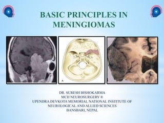
  • 1. cka BASIC PRINCIPLES IN MENINGIOMAS DR. SURESH BISHOKARMA MCH NEUROSURGERY ® UPENDRA DEVKOTA MEMORIAL NATIONAL INSTITUTE OF NEUROLOGICAL AND ALLIED SCIENCES BANSBARI, NEPAL
  • 2.  1922: Harvey Cushing coined the term meningioma to describe a benign globoid tumor arising from the leptomeninges.  Eisenhardt believed meningioma arises from arachnoid cap cells that are particularly abundant in the arachnoid granulation.  Virchow was the first to describe the classic pathologic feature of the meningioma with the term psammoma body (sand like, rightly describing the granules noted within the tumor).  1957: Donald Simpson's 1957 paper described the correlation between surgical resection of meningiomas and the rates of symptomatic recurrence: HISTORY
  • 3.  Slow growing, extra-axial tumor, usually benign neoplasms thought to arise from meningothelial cells found within arachnoid granulations (not dura).  Concentrated in the walls of the major venous sinuses, these structures, which contain “arachnoid cap cells,” account for the dural localization of most meningiomas within the cranium and spinal cord.  Although most are benign (70-75%), a few are classified as atypical (20- 24%) or anaplastic (4%).  Metastases are uncommon and may be seen with benign or malignant meningiomas. INTRODUCTION
  • 4.  Meningiomas: 13 to 26% of all intracranial tumors.  Skull base meningiomas comprise approximately 44% of all skull base tumors.  98% of all CNS meningiomas (spinal 2%).  An annual estimated incidence of 2.3 cases per 100,000 persons.  Autopsy: 3% of the population over 60 years of age.  Female preponderance (1.8)  Peaks age: fourth decade (45yrs)  Meningiomas are less often seen in the younger age groups.  Less than 2% occur in childhood and adolescence.  About 20% of cases seen in adolescents are associated with NF-I.  Approximately 1% of meningioma patients have neurofibromatosis 2 (NF2).  May be multiple in up to 8% of cases. EPIDEMIOLOGY
  • 5.  Ionisation radiation: DNA damage resulting for SS or DS DNA break.  Atomic bomb (epicenter); low dose scalp IR for ringworm; 10x; Israel (1948-60); Full mouth dental IR; XRT for IC tumors.  Hormones:  Est, Prog, Androgens; Size (+): Luteal/pregnancy; 22q12 related with progesterone expression.  Progesterone alone: Good : Absence of Prog or Est recep or Presence of estrogen alone: Bad.  Three Chicago areas hospital: 1987-1992: Protective effect of OCP/HRT.  OCP/HRT: Interphone group: 1993-2002: Increases risk (OR 1.7 HRT/2.7X OCP)  Mayo Clinic jacksonville patient database; OR 3X  Infer Limited statistical evidence of increaed risk.  BMI: Increased BMI association.  Head Trauma: Harvey Cushing; Danish study: 228,055 Cohort of head injury: F/u 8yrs; No evidence. Proinflammatory enzyme COX-2 is upregulated after head trauma: Celecoxib# decreases progression.  Cell Phone use: 10 studies including Interphone Study ; inconclusive; Small sample size; short f/u.  Association with breast cancer: Common hormonal risk factor and genetic predisposition; BRCA1 interacting protein 1 (BR1P1) is associated with meningioma risk.*  Industry/Occupation/Diet/Allergy: Chemical (inconclusive); Diet (no): International Case control study; Allergic diseases like asthma/eczema associated with glial tumor not evident with meningioma.  Positive familial history:2X: 1st degree.  Genetic polymorphism: BR1P1; ATM gene. LOH of chromosome 22 and NF2 gene mutation are implicated in ~50% of sporadic and 100% of NF2-associated meningiomas. RISK FACTORS # Ragel Btet al. Celecoxib inhibits meningioma tumor growth in a mouse xenograft model. Cancer 2007. *Bethke L et al. Comprehensive analysis of DNA repair gene variants and risk of meningioma. J Natl Cancer Inst 2008.
  • 8.  The loss of the long arm of chromosome 22 occurs in 40 to 70% of meningiomas and is associated with loss of the tumor suppressor gene (MERLIN) for neurofibromatosis type 2 (NF2), located at 22q12.  The product of this gene, merlin, is thought to be critical for meningioma tumorigenesis.  Merlin belongs to the family of structural proteins that link the cytoskeleton to several proteins of the cytoplasmic membrane, and some authors have suggested that merlin may act as a tumor suppressor via its interactions with the cellular cytoskeleton. Merlin and Meningioma
  • 9.  It has also been demonstrated that MRI hypointensity on T2-weighted images is associated with slowed tumor growth, and that MRI hyperintensity on T2- weighted images is associated significantly with faster tumor growth.  An annual growth rate of > 1 cm3/year or volume increases > 15% were considered tumor growth.  Growth has been defined as a change in tumor size of at least 2 mm,3 5 mm,12 or any measurable change  Many authors have pointed out that meningiomas without calcification on imaging are more likely to progress than are calcified meningiomas and no negative correlation between calcification and slow growth has been reported.  Presence of peritumoral edema, ambiguous brain- tumor borders, and irregular tumor shape are predictor of tumor growth: needs further study.  Location don’t affects tumor growth. Tumor growth
  • 10. LOCATION OF MENIGIOMA GREENBERG 8TH ED LOCATION OF MENINGIOMA PERCENTAGE Parasagittal 20.80% Convexity 15% Tuberculum sella 13% Sphenoid wing 12% Olfactory 10% Falcine 8% CPA 6% Intraventricular 4% Tentorial 4% Suprasellar 3% Middle fossa 3% Foramen magnum 2% Orbital 1.20% Spinal 1.20% Multiple 0.90% Pineal 0.50% Intrasylvian 0.30% Extracalvarian 0.30% Others 5%
  • 11. Artist's Composite of Different Potential Locations of Meningiomas 1. Convexity; 2. olfactory groove: 3. sphenoid ridge; 4. Tuberculum sella: 5. Dorsum sellae; 6. Meckel cave. cranial nerve V: 7. Clivus: 8. Petrosal : 9. Tentorial: 10. Sigmoid sinus: 11. Straight sinus: 12. Transverse sinus: 13. Torcular-supra-infra tentorial meningioma: 14.Supratentorial interhemispheric meningioma: 15. Falx; 16. superior sagittal sinus. Nader Tricks and Trade of cranial Neurosurgery
  • 12.  The grading has been done on the basis of histological markers, which includes, 1. Presence and number of mitotic figures, 2. Overall cellularity, 3. Nuclear to cytoplasmic ratio, 4. Nuclear prominence, 5. The presence of necrosis  Though the majority of meningiomas are benign Grade I tumors, 23-24% of the tumors are atypical (Grade II) and 1-3% are found to be anaplastic (Grade III) under the new WHO classification system.  Meningiomas with a low risk of recurrence and nonaggressive growth are classified as grade I, whereas those with a higher likelihood of recurrence and more aggressive behavior are classified as either grade II or grade III meningiomas. CLASSIFICATION
  • 13.  In general, cellular proliferation increases in proportion to grade.  The mitotic index and Ki-67 proliferation index correlate approximately with volume growth rate.  Meningiomas with an KI-67 index of:  > 4% have an increased risk of recurrence similar to that of atypical meningioma  > 20% are associated with death rates analogous to those associated with anaplastic meningioma.  Mitotic index: > 4 mitoses per 10 high-power fields: Malignant. PROLIFERATION
  • 14.  The symptoms and signs are consistent with;  Localization  Mass effect  Increased intracranial pressure due to tumor size.  Massive intra- cranial hemorrhage and  Hypoglycemia from tumors that release insulin-like growth factor: rare. CLINICAL FEATURE OF MENINGIOMA
  • 15.  The most common clinical features are the following: • Headaches • Weakness/paresis • Altered mental status  Less commonly, other features noted due to the location of the meningioma are as follows: • Seizures : Parasagittal/sphenoid wing/convexity • Hemiplegia : Parasagittal/convexity • Behavioral changes : Olfactoty groove • Anosmia : Olfactory groove/ planum sphenoidale • Foster Kennedy syndrome : Sub-frontal/olfactory groove/Anterior clinoidal • Visual field defects : Suprasellar/TSM/OGM/Clinoidal • Proptosis : Orbital/sphenoid wing • Palpable mass : Convexity/intraosseous • Stalk effect : TSM, Sellar • Dizziness, Vertigo, Tinnitus : Cerebellopontine angle meningiomas • Cranial neuropathies : Suprasellar/medial sphenoid wing/infratentorial • Hydrocephalus : Intraventricular/pineal region/Infratentorial/TSM • Parinaud syndrome : Pineal region CLINICAL FEATURE OF MENINGIOMA
  • 17. Meningioma variants grouped by WHO grade and biological behavior 2016
  • 18. Donald Simpson's grade:  Grade I: Macroscopically complete resection of tumor,with excision of dural attachment and removal of abnormal bone (involved venous sinus is also excised in relevant cases)  Grade II: Macroscopically complete resection of tumor with coagulation of dural attachment  Grade III: Resection of tumor without coagulation or excision of dural attachment  Grade IV: Partial debulking of tumor  Grade V: Biopsy or decompression only The risks of eventual recurrence after Simpson grades I, II, III, IV and V were 9%, 16%, 29%, 39%, and 99%. when the patients survived for 6 months or longer after surgery.  More recent studies*, however, suggest that recurrence-free survival is not statistically different between patients undergoing Simpson grade I, II, III, or IV resections.  Despite this, the Simpson grade of surgical resection should be part of the routine reporting of surgical results, as it resects more subtle involvement of the dura, arachnoid, arteries, veins, and nerves that only the surgeon can observe at open operation and that is not always obvious on routine postoperative magnetic resonance imaging (MRI). SIMPSON’S GRADING *Sughrue ME et al. The relevance of Simpson Grade I and II resection in modern neurosurgical treatment of WHO Grade I meningiomas. J Neurosurg. 2010.
  • 19. Simpson Classification of Surgical Resection
  • 20.  Lang and coworkers classified them as follows: Type 1: Purely extracranial Type II: Purely calvarial Type III: Calvarial with extracalvarial extension. Convexity meningioma
  • 21.  Parasagittal meningiomas are classified based on their location along the superior sagittal sinus into o Anterior: Between the crista galli and the coronal suture o Middle: Coronal suture to the lambdoid suture o Posterior: From the lambdoid suture to the torcula. They could invade the superior sagittal sinus. o The sagittal sinus could be ligated when dealing with an anterior parasagittal meningioma; however, doing the same when dealing with a more posteriorly placed meningioma could lead to venous infarction as a result of the substantial number of venous tributaries. OLFACTORY GROOVE MENINGIOMA (OGM)
  • 22. Type I Tumor is attached to the outer surface of the sinus Type II Tumor enters the lateral recess of the SSS Type III Tumor invades one wall of the SSS Type IV Tumor invades two walls of a still patent sinus Type V Tumor spreads over the midline, invades the three walls, and occludes the SSS CLASSIFICATION OF PARASAGITAL MENIGNIOMA Hancq S, Baleriaux D, Brotchi J. Surgical treatment of parasagittal meningiomas. Semin Neurosurg 2003. Sindou MP, Alvernia JE. Results of attempted radical tumor removal and venous repair in 100 consecutive meningiomas involving the major dural sinuses. J Neurosurg 2006. Type I Tumor attaches to the outer surface of the sinus wall Type II Tumor fragment inside the lateral recess Type III Tumor invades the ipsilateral wall Type IV Tumor invades the lateral wall and roof Type V Complete sinus occlusion with one free wall Type VI Complete sinus occlusion without any free walls BROTCHI CLASSIFICATION: 2003 SINDOU CLASSIFICATION: 2006 SINDOU’S CLASS
  • 23.  Classification of meningiomas according to sinus invasion. 1. Type 1: meningioma attached to the outer layer of the sinus wall. 2. Type II: Lateral recess Invaded. 3. Type Ill: Ipsilateral wall Invaded 4. Type IV: both Ipsilateral wall and roof invaded. 5. Type V: sinus totally occluded, but contralateral wall free of invasion 6. Type VI: sinus totally invaded including three walls Classification of meningioma according to sinus invasion SINDOU CLASSIFICATION: 2006
  • 24.  Due to their slow compressive effect on the frontal lobes, they usually cause behavioral changes or psychiatric symptoms before the onset of neurologic deficits.  Foster-Kennedy syndrome, a triad of optic atrophy in the ipsilateral eye, papilledema in the contralateral eye, and anosmia. OLFACTORY GROOVE MENINGIOMA
  • 26.  Patients often present with insidious, progressive visual loss, which is usually asymmetric. Frequently these tumors are mistaken for pituitary adenomas, but they are centered on the chiasmatic sulcus of the tuberculum and grow down the front face of the sella as well as over the planum  Pterional, cranioorbital, or unilateral subfrontal: Small to medium-sized tumors  Bifrontal extended frontal craniotomy : Larger tumors.
  • 27.
  • 28. TUBERCULUM SELLAE MENINGIOMA Origin: region of the chiasmatic sulcus and tuberculum, usually with a point of origin at the junction of the optic canal and lateral aspect of the chiasmatic sulcus. C.F: Most patients with TSM present with visual loss; bitemporal visual field loss; Frontal lobe syndrome. Displacement: The optic nerves are usually displaced laterally and superiorly and the optic chiasm superiorly or posteriorly. Frequently there is extension down the medial aspect of one or both optic canals Significant involvement of the internal carotid, anterior cerebral, or anterior communicating arteries largely contraindicates the endonasal approaches.
  • 29.  Optic nerve sheath tumors are a rare form of meningioma that no longer should be approached surgically unless the entire tumor is confined to the orbit and the patient has no useful vision.  Most often these tumors are diagnosed by imaging studies and treated with fractionated external beam radiotherapy.  Combined neurosurgical and ophthalmologic approach: FTOZ. OPTIC NERVE SHEATH MENINGIOMAS
  • 30.  Challenge: They often involve the supraclinoid internal carotid artery and its branches, the cavernous sinus (CS), and its associated oculomotor nerves and the anterior visual pathways.  Middle third meningiomas present with headaches, seizures, and altered mental status. SPHENOID WING MENIGIOMA
  • 31.  A, En-plaque: Carpet-like dural growth especially located on the sphenoid ridge invades the haversian canals along the pterion. orbital walls, and zygoma, creating a hyperostotic reaction and the orbital roof, B, Nodular or Globoid: This variety has been sub-classified into three types:  Cushing and Eisenhardt distinction: (1) Deep, inner, or clinoidal; (2) Middle or alar; and (3) Lateral, outer, or pterional. SPHENOID WING MENIGIOMA: CLASSIFICATION
  • 32. PIRROTE AND BROTCHI CLASSIFICATION OF SWM Roser’s sub- division A Deep or Clinoidal or sphenocavernous B Invading en plaque of sphenoid wing C Invading en masse of sphenoid wing Roser et al further sub- divided C tumors into meningiomas en plaque with and without cavernous sinus infiltration, and purely intraosseous tumors. D Middle ridge meningioma E Pterional or sylvian point meningiomas. CLASSIFICATION OF SWM Group C tumors are thought to combine features of both group A and B tumors (i.e., globular and invasive growth en plaque).
  • 33. 1. Group I arising from the inferior aspect of the anterior clinoid process (ACP) proximal to end of carotid cistern: Anterior clinoidal meningioma (aka medial SWM) : Tends to encase ICA without intervening arachnoid. 2. Group II arising from the superior and /or lateral aspect of the ACP separated from the ICA and cranial nerves by an arachnoidal membrane of the carotid and sylvian cisterns. 3. Group Ill arising at the level of the optic foramen extending into canal: separate from ICA. CLINOIDAL MENINGIOMAS Clinoidal meningiomas (CMs) are divided into three groups: Al-Mefty Some clinoidal meningiomas, however, may grow into the region of the mesial sphenoid wing and be confused with medial sphenoid wing meningiomas Other school of thoughts/Fallacies: Group I tumors are probably rare, and the lack of an arachnoidal dissection plane between the tumor and the carotid artery may be related to other factors such as repeat surgery. Group III tumors may be more appropriately referred to as optic foramen, optic sheath, or optic canal meningiomas.
  • 34. ANTERIOR CLINOIDAL MENINGIOMA 6.5% of all meningioma and 25% of Ant fossa Meningiomas Types of anterior clinoidal meningioma. Preoperative contrast-enhanced MR images show that the sizes of the tumor on coronal dimensions are <2 cm, 2– 4 cm, and >4 cm, respectively. Tumors without extradural growth and tumors with extradural growth into the cavernous sinus. Visual deterioration with ipsilateral nasal hemianopsia, Foster-Kennedy syndrome Meningothelial >> Transitional are the common variant in ACM. Pamir combined coronal diameter in Al-mefty’s class
  • 35. SPHENOCAVERNOUS MENINGIOMA “Hirsch criteria for sinus involvement” Grade Tumor Description 1 Touch or partially encircle the cavernous internal carotid artery 2 Completely encircle but do not narrow the lumen of the internal carotid artery 3 Encircle and narrow the lumen of the cavernous internal carotid artery Radiological Grade of Cavernous Sinus Tumors as Determined by Magnetic Resonance Imaging and Angiography According to the Criteria of Hirsch et al
  • 36.  With anterior clinoidal meningiomas, the epicenter of the tumor base is on the anterior clinoid process and the tumor typically grows upward, forming a small pedicle toward the suprasellar area and the sylvian fissure. (Fig B upper arrow)  In contrast, meningiomas of the medial third of the sphenoid wing grow in the direction of the anterior aspect of the medial temporal lobe. (Fig A and B lower arrow)  Another characteristic feature of anterior clinoidal meningiomas is the presence of hyperostosis of the anterior clinoidal process on coronal CT. (Fig C) CLUE TO DIFFERENTIATE ACM FROM OTHER SWM Fig A: Medial sphenoidal wing meningiomas grow in the direction of the anterior aspect of the medial temporal lobe (lower arrow on A), whereas Fig B: Anterior clinoidal meningiomas grow upward with a small pedicle (upper arrow on B) C
  • 37.  Challenging tumors  Occurs in relatively young patient (life expectancy of at least 15 years),  Surgery is indicated at the time of detection, regardless of the size or the presence of symptoms.  Various skull-base approaches with or without intra- or extradural removal of anterior clinoid.  Pterional and subfrontal approaches  Al-Mefty exclusively used the orbitocranial approach: shortest distance to tumor, suitability for surgical attack via multiple routes, and early interception of the tumor’s blood supply through the sphenoid ridge.  Lee and colleagues 2011: Modified Dolenc cranial base approach: involves extradural clinoidectomy, removal of the roof of the optic canal, and opening of the optic nerve sheath.  Oculomotor morbidity is extremely high after a direct approach to this region ANTERIOR CLINOIDAL MENINGIOMA: SURGERY
  • 39. Extent of Tumor Removal: A Grading System According to De Monte et al. Grade Tumor Description 1 Complete microscopic removal of tumor and its dural attachment with any abnormal bone 2 Complete microscopic removal of tumor with diathermy coagulation of its dural attachment 3a Complete microscopic removal of intra- and extradural tumor without resection or coagulation of its dural attachment 3b Complete microscopic removal of intradural tumor without resection or coagulation of its dural attachment or any extradural tumor 4a Intentional subtotal removal to preserve cranial nerves or blood vessels; complete microscopic removal of tumor dural attachment 4b Partial removal leaving tumor <10% in volume 5 Partial removal leaving >10% in volume or decompression with or without biopsy CAVERNOUS SINUS MENINGIOMA
  • 40. Series (Ref. No.) No. of Cases No. of Total Resections (%) Follow-up (mo) Cioffi et al., 1987 (6) 12 1 (8.3) 24 De Monte et al., 1994 (9)a 41 31 (76) 45 Dolenc et al., 1987 (12) 40 Unknown Unknown Hakuba et al., 1989 (16) 4 Unknown Unknown Kawase et al., 1987 (20) 9 7 (77.8) Unknown Lesoin and Jomin, 1987 (25) 16 0 Unknown Risi et al., 1994 (31) 15 4 (26.7) Unknown Sekhar et al., 1992 (36)a 70 61 (87.1) 36 Sepehrnia et al., 1991 (38) 36 18 (50) Von Wild and Eskinja, 1989 (44) 9 4 (44.4) 10 O'Sullivan et al. (present study) 39 8 (20.5) 24 Reported Cases of Meningiomas of the Cavernous Sinus
  • 41.  The resectability of meningiomas of the cavernous sinus depends on the degree of internal carotid artery involvement;  Total excision of cavernous sinus meningiomas is possible but rarely achieved in holocavernous meningiomas;  Cranial nerve morbidity is significant; and  Subtotal excision with or without postoperative radiotherapy is an effective short-term oncological strategy. SURGERY
  • 42.  Optic nerve sheath meningiomas (ONSMs) involve the optic nerve and the anterior visual pathways.  They usually arise from the arachnoidal membrane of the intraorbital nerve and extend through the optic canal to the anterior fossa.  Without treatment, slowly but progressive growth often results in unremitting visual loss.  Axial CECT: Tram track sign: Hyperdense encasement of optic sheath surrounding the hypodense optic nerve.  XRT: EBRT: 50-55Gy: 75% improvement in VA, 33% complication; while SRS or SCRT: Visual improvement 80%, 4% complication. OPTIC NERVE SHEATH MENINGIOMA SCHICK AND COLLEAGUES CLASSIFIICATION Treatment (SCHICK) Type I: Intraorbital lesions IA Flat extension around the optic nerve XRT without biopsy IB Bulbiform mass around the optic nerve XRT, if painful with no useful vision: surgery IC Exophytic tumor around the optic nerve Surgery Type II: : Intraorbital tumors with intracranial extension through the optic canal or superior orbital fissure IIA Intraorbital growth through the optic canal Intradural exploration with Optic canal and SOF decompression = STR ff XRT. IIB Growth through the superior orbital fissure or cavernous sinus) Cavernous sinus involvement be treated with XRT Type III: Intraorbital tumors with widespread intracranial tumor extension IIIA Extension to chiasm Preventive surgery to avoid involvement of chiasma and contralateral optic nerve. IIIB Extension to chiasm, contralateral optic nerve, and planum sphenoidale
  • 43.  Apart from the cavernous sinus, they could also arise from the posterior aspect of the sphenoid wing, clivus, petrous bone or from the middle cranial fossa floor.  These meningiomas are usually closely associated with the cavernous sinus and present a challenge to the neurosurgeon as surgical resection could result in a cranial neuropathy.  Approaches: sub-temporal, middle fossa or supra petrosal approaches. MIDDLE FOSSA MENINGIOMA
  • 44.  1% of all intracranial meningiomas.  10-15% of CP tumor (80-90% of VS in CPA region)  Desgeorges and colleagues classified these meningiomas into three groups based on their proximity to the internal acoustic canal (lAC):  I : Meningiomas anterior to the lAC. II : Meningiomas centered at the lAC, (Sometime not used this class) III : Meningiomas posterior to the lAC.  CPA meningioma vs Vestibular Schawannoma  Widening of the lAC when there is a vestibular schwannoma.  Hyperostosis, intratumoral calcification, broad tumor base on the tentorium, and a dural tail all favor the diagnosis of a meningioma.  T2-weighted gradient echo sequences: microhemorrhages of VS  Angle with petrous bone: 81% vestibular schwannoma will form an acute angle, while a meningioma will form an obtuse angle.  Retrosigmoid and trans-labyrinthine approaches CEREBELLOPONTINE ANGLE (CPA) MENINGIOMAS
  • 45.  Foramen magnum meningiomas present with variable symptoms.  Most patients have a history of neck or sub-occipital pain, and motor sensory symptoms develop, usually in one arm, and then in the contralateral leg.  Because of the lower cranial nerve morbidity associated with surgical resection of these tumors, incidentally discovered tumors in this location can initially be followed.  Class: Anterior, Lateral and Posterior  The standard approach for these tumors is the far lateral sub-occipital approach with the patient in the three-quarter prone position.  The posterior one third of the occipital condyle is removed to improve access to the ventral dura.  Once the dura is opened, the vertebral artery can be identified and is usually not directly invaded by meningioma.  With Meninigoma below VA, Ther lower CNs are displaced superiorly while tumor above VA, the position is unpredictable. avid  Sharp dissection of the arachnoid plane is key to preserving the rootlets of cranial nerves IX, X, XI, and XII.  The patient should be warned that for the larger tumors such dissections could result in transient and possibly permanent problems with dysphasia and dysphonia. FORAMEN MAGNUM MENINGIOMAS
  • 46.  Arise from arachnoidal cap cells in the region of the torcular Herophili.  Meningiomas invading the torcular region can be categorized into two main subtypes based on predominant site of invasion:  1. Torcular (T)  2. Transverse sinus (TS): Cerebellar convexity ( medial, Lateral or superior) and tentorial origin (Medial, Lateral and falcotentorial)  Torcular Meningiomas  • Frequently, torcular meningiomas present with neurologic signs indicative of intracranial hypertension.  • All of the patients in the Cushing and Eisenhardt series exhibited papilledema, while in half of them a homonymous field deficit was round Infratentorial extension may result in cerebellar signs, including ataxia, dysmetria, hypotonia, and nystagmus.  Transverse Sinus Meningiomas  These are typically placed into two categories: those arising from the cerebellar convexity or from the tentorium  Cerebellar Convexity/Superior origin  These are divided into medial, lateral, or superior, with the superior type most likely to invade the TS. Symptoms are relatively few and nonspecific: headache, cerebellar signs, symptoms of increased intracranial pressure, and hydrocephalus.  Tentorial Origin  These are divided into medial, lateral, or falcotentorial types based on assessment of tumor origin at surgery.  Medial and lateral types are further classified as anterior, middle, or posterior.  Most tumors of tentorial origin affecting the sinus grow infratentorially and produce characteristic symptoms of headache and truncal ataxia. TORCULAR MENINGIOMA Diagnosis and Imaging Patients must undergo CT scaning and MRI with gadolinium including an MR venogram (MRV) to establish the dimensions, degree of bone invasion, presence of surrounding edema, relationships of the tumor with the surrounding structures, and degree of venous sinus involvement. Digital subtraction angiography (DSA) to determine variants of circulation in the torcular and lateral sinuses is critical before surgical intervention can be established. Nader’s classification about degree of the meningioma's sinus invasion: As parasagital menigioma Type I - attachment to outer surface of a sinus wall, Type II - fragment inside a lateral recess, Type III - invasion of one wall, Type IV - invasion of two walls, Types V and VI - complete sinus occlusion, with or without one wall free,
  • 47.  Surgery usually is the preferred treatment modality for patients with torcular and peritorcular meningiomas.  The debate lies in the decision of the surgeon to restore venous circulation by way of bypass or to rely on collateral circulation after tumor re- section.  Various surgical interventions have been established depending on the type of sinus invasion as well as patient-specific factors of the case, such as age and preoperative symptoms of the patient.  Anatomic variants of the dural sinuses should be anticipated through imaging as they will affect the surgeon's decision to restore venous.  the external jugular vein for short grafting (< 10 em) or the internal saphenous vein for larger grafts.  In the eventuality of "inoperable" tumors, radiosurgery such as gamma knife treatment and hypo fractionated stereotactic radiotherapy (hFSRT) have emerged as means of effectively reducing neurologic deficits with relatively limited morbidity. Treatment
  • 48.  Yasargil’s classification of tentorial meningiomas (TMs) defines eight types of tumors according to their location on the cerebellar tentorium.  They were subsequently regrouped as follows: 5 TENTORIAL MENINGIOMA 3-6% Group Characteristics I Anteromedial, arising from the apex of the tentorial margin II Anterolateral, arising from the lateral aspect of the tentorial incisural margin III Intermediate, arising from the intermediate aspect of the tentorium remote from the incisura and the dural sinuses IV Posteromedial, arising from posteromedial aspect of the tentorium close to straight sinus or venous confluence at the torcula; this group also includes the falcotentorial and torcular meningiomas. V Posterolateral, arising from the posterolateral aspect of the tentorium close to the sigmoid sinus The most frequent benign meningomas in these locations are fibroblastic and meningothelial
  • 50.  Occipital Interhemispheric Approach: I II, III  Transtentorial access : I, II, and IV.  Subtentorial: II  Bioccipital-Suboccipital Approach: IV  Midline Supracerebellar Infratentorial Approach: I, IV  Paramedian Supracerebellar Infratentorial Retrosigmoid Approach: II, III, and V meningiomas APPROACH Transzygomatic approach: Medial tentorial meningiomas
  • 51.  5% of all intracranial meningiomas.  The most common location is the atrium of the lateral ventricle. (80%), 3rd (15%), 4th (5%)  They may arise either from the stroma of the choroid plexus or from rests of arachnoid tissue inside it.  A patient usually presents with symptoms related to obstructive hydrocephalus such as headaches, nausea, and vomiting.  Intraventricular and subarachnoid hemorrhage  Weakness of upper limb progressing to contralateral leg.  Seizures can occur with large tumors in either hemisphere, and speech disturbance can occur for left- sided lesions.  Approach  Lateral ventricle tumors: anterior transcallosal or parietooccipital approach.  Trigone: Non-dominant: Temporal parietal craniotomy; dominant hemisphere: superior parietal lobule approach..  Anterior third ventricle : anterior transcallosal approach.  Posterior third ventricle: Posterior transcallosal or supra- cerebellar infratentorial trajectory.  Fourth ventricle : Medial sub-occipital approach. INTRAVENTRICULAR MENINGIOMAS 2%
  • 52. GROUP CHARACTERISTICS A Pure convexity meningiomas arising from the dura over the posterior convexity of the cerebellum B Inferior peritorcular meningiomas arising from or invading the inferior wall of the torcular herophili or the medial transverse sinus C Parasinus meningiomas arising in the angle between petrous and convexity dura, including the wall of the sigmoid and transverse sinuses D Meningiomas with secondary invasion of cerebellar convexity/fossa. CEREBELLAR CONVEXITY MENINGIOMA Midline or paramedial suboccipital infratentorial trajectory.
  • 53.  SMs arise at the junction of the spinal arachnoids and the dura of the nerve root sheath.  Approximately 83% to 94% have an intradural component, 5% to 14% are extradural, and 10% may grow in both compartments.  Thoracic spine: 73%, cervical: 16% and lumbar region : 5%.  C.F: sensitive motor deficit, Myelopathy, BSS, autonomic symptoms.  MC: psammomatous subtype  Somatosensory and motor evoked potentials are generally used. SPINAL MENINGIOMAS 7.5%-12% Review: Sandalcioglu and co-workers: 137 SM
  • 55.  Appear as homogeneous, densely enhancing mass with broad base of attachment along dural border.  60-70HU : calcifications.  Cerebral edema, Mild or marked: white matter of the entire hemisphere.  Intraventricular meningiomas: 50% produce extra ventricular edema. On angiogram, these may falsely appear malignant. CT SCAN
  • 56.  Occasionally may be isointense with brain on T1WI and T2WI, but most enhance with gadolinium.  Brain edema may or may not be present.  Calcifications appear as signal voids on MRI.  Gives information regarding patency of dural venous sinuses (accuracy in predicting sinus involvement is ≈ 90%).  Dural tail is a common finding. (60%). MRI
  • 57.  DWI/ADC: atypical and malignant subtypes may show greater than expected restricted diffusion although recent work suggests that this is not useful in prospectively predicting histological grade.  MR spectroscopy: usually it does not play a significant role in diagnosis but can help distinguish meningiomas from mimics.  Features include:  Increase in alanine (1.3-1.5 ppm)  Increased gluatamine/glutamate  Increased choline (CHO): cellular tumour  Absent or significantly reduced N-acetylaspartate (NAA): non- neuronal origin  absent or significantly reduced creatine (Cr)  MR perfusion: good correlation between volume transfer constatn (K- trans) and histological grade Typical V/S Atypical/Malignant
  • 58.  CSF vascular cleft sign : extra-axial; invasion: grade II and grade III  Dural tail is seen in 60-72% (note that a dural tail is also seen in other processes)  Sunbrust or spokewheel appearance of the vessels.  Arterial narrowing  Typically seen in meningiomas which encase arteries.  Useful sign in parasellar tumours, in distinguishing a meningioma from a pituitary macroadenoma; the latter typically does not narrow vessels.
  • 59.  Meningiomas characteristically have external carotid artery feeders.  Exceptions: OGM: ICA (ethmoidal branches of the ophthalmic artery), Suprasellar meningiomas :ophthalmic arteries, Parasellar meningiomas : ICA.  Secondary vascular supply may be derived from pial branches of the anterior, middle, and posterior cerebral arteries.  Artery of Bernasconi & Cassinari AKA artery of tentorium (a branch of the meningohypophyseal trunk) AKA the “Italian” artery: enlarged in lesions involving tentorium (e.g. tentorial meningiomas).  Angiography also gives information about occlusion of dural venous sinuses, especially for para-sagittal/falx meningiomas.  Oblique views are often best for evaluating patency of the superior sagittal sinus (SSS). CT Angiogram
  • 60.  The "Mother-in-Law" sign is commonly seen when there is a meningioma, as the tumor contrast blush "comes early (in the arterial phase), stays late (beyond the venous phase), and is very dense."  Angiography can also help confirm diagnosis by the distinctive prolonged homogeneous tumor blush.  Angiography also provides an opportunity for pre-op embolization. DIGITAL SUBTRACTION ANGIOGRAMS (DSA)
  • 61. LOCATION EXTRADURAL VESSELS INTRADURAL VESSELS Convexity MMA - Parasagittal MMA - Falcine MMA, Ant falx artery - Sphenoid Wing/ACM MMA - OGM Ant br. MMA Cavernous ICA, Ethmoid br. of ophthalmic a. TSM Post Ethmodal br of ophthalmic a. Supra sellar/ Parasellar Ophthalmic a./ ICA respectively Tentorial MMA Marginal tentorial br of SCA, Bernasconi & Cassinari artery Tentorial/ Clivus MMA Cavernous ICA, Davidoff & Schecter Petroclival MMA, IMA, APA MHT, ILT Post. Fossa (PM) M br of VA, PMA - Post Fossa (Lateral) - Transmastoid br OA Intraventricular - Choroidal a. Foramen magnum Mening. br APA, OA, VA, PICA, and PSA Abbreviation: OGM: Olfactory groove meningioma; MMA: Middle meningeal artery; OA: Occipital artery; MHT: meningohypophyseal trunk; ILT: the anterior branch of the inferolateral trunk; APA: Asc. Pharyngeal a; PSA: Post spinal a. Note secondary supply may be derived from pial br of ACA, MCA or PCA. ARTERIAL FEEDER OF MENINGIOMAS
  • 62.  Calcifications within the tumor (in ≈10%).  Hyperostosis or blistering of the skull (including floor of frontal fossa with olfactory groove meningiomas).  Enlargement of vascular grooves (especially middle meningeal artery). PLAIN X-RAYS
  • 63.  Manelfe and associates, in 1973, first described the microcatheter technique of meningioma embolization.  Preoperative embolization can be an important adjuvant treatment modality.  Embolizing materials could be liquid or particulate agents.  The liquid agents : 1. N-butyl cyanoacrylate (NBCA), 2. ONYX (ethylene vinyl alcohol polymer dissolved in dimethyl sulfoxide), and 3. Fibrin glue  Particulate agents: PVA particles and microspheres . PRE-OP PLANNING EMBOLIZATION
  • 64. Indications: 1. Surgeons’ preference institutional practices, 2. Large tumor size, 3. Extensive tumor vascularity, 4. Skull base meningiomas with difficult-to-access arterial supply EMBOLIZATION Benefits: 1. Reduced operative blood loss, 2. Easier tumor resection, and 3. Shortened surgical time).
  • 65. 1. Hemorrhage ( Intratumoral and SAH) 2. Cranial nerve deficits (usually transient) 3. Stroke from embolization through ICA or VA anastomoses 4. Scalp necrosis 5. Retinal embolus, and 6. Potentially dangerous tumor swelling. 7. Some meningiomas (e.g. olfactory groove) are less amenable to embolization due to risk of blindness. Complication of embolisation
  • 66.  Pre-op embolization: Reduces the vascularity of these often bloody tumors, facilitating surgical removal.  Timing of subsequent surgery is controversial. Some advocate waiting 7– 10 days to permit tumor necrosis which simplifies resection.
  • 67.  Antiepileptic medication should naturally be continued during the perioperative period in patients with a known history of seizures. For meningioma patients with no history of seizures.  However, a meta-analysis of five randomized controlled trials in patients with primary brain neoplasms found no effect of prophylactic antiepileptic medication on seizure control.  The American Academy of Neurology does not recommend the use of antiepileptic medications in brain tumor patients with no history of seizures. PREOPERATIVE SEIZURE PROPHYLAXIS Sirven JI et al. Seizure prophylaxis in patients with brain tumors: a meta- analysis. Mayo Clin Proc 2004.
  • 68.  Meningioma resection improves seizure control in patients with preoperative epilepsy.  ~5 to 20% of patients may develop new postoperative seizures.1,2  The use of postoperative antiepileptic medications such as phenytoin after intracranial neurosurgical procedures has been shown to reduce early postoperative seizures by ~40 to 50%.  Recent studies comparing levitiracetam to phenytoin have also shown no difference in postoperative seizure rates in glioma patients.  The American Academy of Neurology currently recommends discontinuing postoperative seizure prophylaxis after 1 week in patients with no history of seizures. 3 POSTOPERATIVE SEIZURE PROPHYLAXIS 1. Chozick BS et al. Incidence of seizures after surgery for supratentorial meningiomas: a modern analysis. J Neurosurg 1996. 2. Lieu AS et al. Intracranial meningiomas and epilepsy: incidence, prognosis and influencing factors. Epilepsy Res 2000. 3. Glantz MJ et al; Anticonvulsant prophylaxis in patients with newly diagnosed brain tumors. Neurology 2000.
  • 69.  Intraoperative monitoring including,  Brainstem Evoked Potentials  Somatosensory Evoked Potentials (SSEPS)  Neuronavigation  Microscopic Magnification  Ultrasonic Aspirator  Microsurgical instruments:  A lumbar drain may be necessary to reduce brain turgor intra- operatively.  The abdomen and/or thigh should be prepared for possible fat or fascial graft harvesting.  Perioperative antibiotics, steroids, and anticonvulsants are given.  Central line and arterial lines are placed. Preoperative Planning and Special Equipment
  • 70.  Every meningioma patient is unique and deserves a tailored approach.  Surgeons approaching these lesions should have mastered all cranial approaches including the ones to the skull base. as the choice of the proper approach should always be based on what is best for the patient and what carries the least amount of risk leading to deficits and complications while providing a high chance of success.
  • 71.  Four critical HPE variables 1. Grade: WHO I, II, III 2. Histological subtype: Meningothelial, clear cell, rhabdoid, etc. 3. Proliferative indices: Mean KI-67 index 4. Brain invasion: Increases the likelihood of recurrence to level similar to atypical meningioma (not anaplastic).  But it is not an indicator of malignant grade. PATHOLOGY
  • 72.  Multiple menigioma: suggest NF2  Pleomorphic xanthoastrocytoma: Peripherally located/dural tail.  Rosai-Dorfman disease: Connective tissue Dz, Sinus histocytosis and massive pain less lymphadenopathy/ Dural tail. CD68 and S-100 (+): XRT. DIFFERENTIAL DIAGNOSIS
  • 73.  Immunohistochemical markers of proliferation potential such as Ki-67, MIB-1, and proliferating cell nuclear antigen (PCNA) indices have also been correlated with risk of recurrence.  Oya and colleagues indicated that an MIB-1value of 3% or higher was associated with a greater recurrence rate of World Health Organization (WHO) grade I meningiomas surgically treated by Simpson grade II or III resection.  Ideally, pathological grade and histology, Simpson surgical grade, and a marker of proliferation index should be included in the reporting/analysis of operative meningioma specimens. RECURRENCE
  • 74. Meningioma Recurrence Remarks OGM 5-41% Obeid and Almefty’s series Parasagittal 9,19,29,40% Simpson’s 10 year F/U Recurrence Description and WHO grade Mean KI-67 index Recurrence rate WHO I 0.7% 9% WHO II 2.1% 29% WHO III 11% 50%
  • 76.  Subfrontal approach: large and giant olfactory groove meningiomas.  FTOZ: Large OGM  Supraorbital craniotomy : small unilateral olfactory groove meningiomas.  Pterional approach : sphenoid wing, OGM or temporal meningiomas.  Middle fossa, retrosigmoid, translabyrinthine, transcochlear, infratemporal fossa and retrolabyrinthine approaches  For skull base meningiomas (petroclival/lpetrous ridge, tentorium, sigmoid, internal auditory canal and jugular foramen regions).  TSM: Bifrontal, FT, FL, Expanded endonasal.  Endoscopic sellar surgery: pioneered (Jankowski in 1992) : Planum sphenoidale meningiomas and other meningiomas in the sellar/parasellar region.  FM meningiomas: Anterior and Lateral: Posterolateral/transcondylar app *Posterior: Posterior suboccipital approach. Surgical approach
  • 77.  Radiotherapy treatment has proved to be effective in controlling tumor growth, particularly for residual tumor following resection of skull base meningiomas.  Value of XRT: 1. Partially resected meningiomas. 2. Inaccessible meningiomas, 3. Inoperable meningiomas, 4. Post-resection cavity for grade iii meningiomas. 5. Remnants that have demonstrated confirmed growth..  High dose of about 55-60 Gy could be beneficial.  Alternatively, one can follow these patients with CT or MRI and use XRT for documented progression. XRT Barbaro NM et al. Radiation Therapy in the Treatment of Partially Resected Meningiomas. Neurosurgery. 1987. Barbaro et al. Option Recurrence UCSF series of 135 non malignant meningoma F/u 5-15 years Total resection 4% PR with XRT 32% PR without XRT 60% PR: Partial resection: Extracted from Greenberg 8th Mean time to recurrence was longer in the XRT group (125 mos) than in the non-XRT group (66 mos).
  • 78. RADIOTHERAPY For small to medium-size tumors less than 30 mm, or less than 8 cc, at sites not associated with critical structures such as optic nerves, chiasm, or brainstem, radiosurgery is a powerful technique as either a primary form of therapy, for residual disease, or for treatment of recurrence Case report of malignant Astrocytoma developing after XRT used to treat meningioma.
  • 79.  Lars Leksell: 1960s,  It is considered most effective for tumors less than 3 cm in diameter or 10 cm3 in volume.  Influence factor: clear tumor-brain interphase, proximity to areas of functionally important brain or nerves, and other critical structures.  Excellent tumor control for WHO; grade I meningiomas is usually achieved with 12 to 16 Gy. (Ganz et al)  No improvement with marginal doses greater than 15 Gy (Kondziolka et) STEREOTACTIC RADIOSURGERY
  • 80.  PBT delivers protons instead of radiotherapy photons.  Protons are more conformal and homogeneous than photons and their use decreases the dose in surrounding tissue compared to photon beam therapy.  However, the treatment results of PBT appear to be similar to those for IMRT. PROTON BEAM THERAPY
  • 81.  Many modalities have been tested including cytotoxic drugs, immunomodulation, molecular agents, and hormonal therapy.  No effective chemotherapy till date  Promise: Interferon alpha-2b and hydroxyurea.  Hydroxyurea suggested for treatment of unresectable and recurrent meningiomas: arrests meningioma cell growth in the S phase DNA synthesis inhibitionapoptosis  Conventional combined chemotherapy— cyclophosphamide, adriamycin, and vincristine—showed a modest activity against malignant meningiomas and may improve the median survival time.  Treatment with interferon alpha-2b has presented some success in preventing meningioma growth. CHEMOTHERAPY
  • 82.  Ki-67, MIB-1, and proliferating cell nuclear antigen (PCNA) are predictors increased recurrence, while CDKN2A deletion, along with a 9p21 deletion, is a predictor of malignant progression, increased recurrence, and poor survival. PROGNOSTIC MARKERS
  • 83. Criteria: Radiation-Induced Meningioma 1. Tumor must arise in the irradiated field 2. Histological features must differ from those of any previous neoplasm 3. A sufficient latency or induction period following radiation must elapse before meningioma is diagnosed (usually > 5 years) 4. No family history of phacomatosis. 5. Tumor must not be recurrent or metastatic 6. Tumor must not be present before radiation therapy
  • 84. Thank you  MENINGIOMA DR. SURESH BISHOKARMA MCH NEUROSURGERY ® UPENDRA DEVKOTA MEMORIAL NATIONAL INSTITUTE OF NEUROLOGICAL AND ALLIED SCIENCES BANSBARI, NEPAL

Editor's Notes

  1. Grade I:Macroscopically complete resection of tumor,with excision of dural attachment and removal of abnormal bone (involved venous sinus is also excised in relevant cases)
  2. The development of the meninges starts early in gesta- tion and reaches the basic adult forms by the end of the rst trimester. Meningeal precursors are derived from both neural crest and mesodermal cells. As the neural tube fuses at 22 to 24 days of gestation, a single layer of cells, with some attachments to the neural crest, surrounds the developing neural axis. A thicker, looser collection of mesenchymal cells further covers the neu- ral tube starting around day 24 to 28 and completely envelops the developing spinal cord and brain by day 33 to 41. This mesodermal-derived cellular network, along with the neural crest–derived monocellular layer will di erentiate into the meninx primitiva (primary meninx) As the pluripotent meninx primitiva develops, it sub- divides into two distinct layers between days 34 and 48. The outer portion, the ectomeninx, is dense and compact, whereas the inner layer, the endomeninx, is more loosely arranged. The ectomeninx is the precursor to the dura and the bones of the neurocranium, thus the close apposition of dura and skull stems from their shared embryological ancestry. The inner portion of the endomeninx, contain- ing the neural crest–derived cells covering the neural tube, begins to form the pia during the gestation interval of 45 to 55 days.4,6,7 Meanwhile, as cerebrospinal fluid invades the endomeninx, cavitations (future cisterns) begin to ap- pear in the outer portion of the endomeninx and become obvious by 55 days of gestation.8 Although the dura and pia are distinguishably formed structures by this point of development, a distinct arachnoid layer is not obvious and may not appear until much later during fetal development.
  3. Meningiomas are the most common benign intracranial neoplasms, accounting for 13 to 26% of all intracranial tumors. Intracranial meningiomas represent 98% of all CNS meningiomas, and spinal types make up the remaining 2%. An annual estimated incidence of 2.3 cases per 100,000 persons. Autopsy studies showed meningiomas occurred in as much as 3% of the population over 60 years of age. There is a slight female preponderance (the female-to-male ratio is 1.8: 1), and the incidence peaks during the fourth decade of life. Meningiomas are less often seen in the younger age groups. Less than 2o/o occur in childhood and adolescence, whereas about 20% of cases seen in adolescents are associated with neurofibromatosis type I.
  4. ATM gene: a member of the phosphatidylinositol 3 kinase family known to be involved in homologous and non homologous DNA break repair and meningioma risk second tumor suppressor gene on 22q stems from the discrepancy between the frequency of chromosome 22 LOH, which exceeds that of NF2 gene abnormalities.6 Deletions of chromosome 22 are found in all NF2-associated meningiomas and in 54 to 78% of sporadic meningiomas
  5. Factors involved with meningioma tumor progression. Loss of chromosome 1p is a decisive step. Immunohis- tochemically, higher-grade tumors are associated with decreased progesterone receptor (PR) staining and increased MIB-1 nuclear staining. Human telomer- ase reverse transcriptase (hTERT) activity is also increased in higher-grade meningiomas
  6. In 1922, Harvey Cushing coined the term meningioma to de- scribe a benign globoid tumor arising from the leptomeninges. Since that time, various pathological classi cation systems have used di erent morphological features, proliferation in- dices, and pathological grading systems. The current World Health Organization (WHO) system groups meningiomas by likelihood of recurrence into three grades
  7. A frontal lobe syndrome may become evident, with mental status changes, including changes in personality or behavior, loss of motivation, depressed mood, apathy, and changes in short-term memory
  8. Meningiomas with a sphenocavemous origin are associated with oculomotor palsy that usually begins with abducens palsy, creating a hori- zontal binocular diplopia and esotropia In primary gaze. Patients with menin- giomas en plaque usually present with proptosis caused by intraorbital and periorbital tumor infiltration, venous compres- sion. and Fig. 32.2). Hyperostosis-induced bone deformity, blindness, and diplopia are also common findings. Signs and symptoms of nodular pterional meningiomas indude seizures. contralateral hemiparesis. headache, and parkinsonian syndrome.
  9. A: B: C: D: E:
  10. SOF: III, IV, oph div of CNV, Nasociliary, Lacrimal; Superior ophthalmic vein, IOF: V1, Inf. Oph vein. Neurovascular structures passing in its immediate vicinity. The oculomotor nerve course is along the superolateral aspect of the anterior clinoid process. The internal carotid artery crosses the inferior aspect of the anterior clinoid process, and the optic nerve passes along its superomedial aspect.
  11. Pterional and subfrontal approaches, Al-Mefty exclusively used the orbitocranial approach and stated its advantages as: shortest distance to tumor, suitability for surgical attack via multiple routes, and early interception of the tumor’s blood supply through the sphenoid ridge. Many surgeons used various skull-base approaches with or without intra- or extradural removal of anterior clinoid for resecting these challenging tumors. A recent article by Lee and colleagues23 describes a cranial base technique that is a modification of the original “Dolenc approach” and involves extradural clinoidectomy, removal of the roof of the optic canal, and opening of the optic nerve sheath. 
  12. Clinoidal meningiomas (CMs) are divided into three groups: o Group I arising from the Inferior aspect of the anterior clinoid process (ACP) o Group II arising from the superior and /or lateral aspect of the ACP o Group Ill arising at the level of the optic foramen
  13. Cavernous sinus meningiomas present with double vision, facial numbness, headache, and reduced visual acuity. In the past, these were aggressively treated with skull base approaches. However, pathological postmortem specimens demonstrate in ltration of the epineurium of cranial nerves in the lateral wall of the cavernous sinus, and now these tumors are fre- quently treated surgically only for their exophytic middle fossa component, leaving the treatment of the truly intracav- ernous part to radiotherapy techniques.54,55 Recent studies have shown improved rates of tumor control with adjuvant radiotherapy, regardless of initial extent of resection
  14. O'Sullivan et al: We think that the distinction between Grades I and II is important, because Grade I tumors can be removed with a small opening of the CS whereas Grade II lesions often require a more extensive opening. The distinction between Grades III and IV is based on the surgical observation that the intracavernous ICA can be dissected free of tumor in some patients with Grade III meningiomas but none of the patients with Grade IV lesions. Grade V lesions involve both CSs. When the invasion of the contralateral CS is limited, the tumor can be resected totally. However, total resection is not attempted when there is extensive invasion of the contralateral CS. We also prefer a different categorization of the degree of resection: gross total, incomplete, or biopsy only. Gross total resection is defined as the removal of all of the visible tumor, including dural attachments and, if invaded, the intracavernous ICA. Gross total resection is based on the surgeon's intraoperative assessment and postoperative magnetic resonance imaging (MRI). Anything less than this is defined as incomplete resection. We recognize that total cytological resection of basal meningiomas is usually impossible, because the cytopathological changes extend well beyond the resection area.
  15. Schick and colleagues classify the ONSM as three types: Type I: Intraorbital lesions (Ia, flat extension around the optic nerve; Ib, bulbiform mass around the optic nerve; Ic, exo- phytic tumor around the optic nerve) Type II: Intraorbital tumors with intracranial extension through the optic canal or superior orbital fissure (IIa, intraorbital growth through the optic canal; IIb, growth through the superior orbital fissure or cavernous sinus) Type III: Intraorbital tumors with widespread intracranial tumor extension (IIIa, extension to chiasm; IIIb, exten- sion to chiasm, contralateral optic nerve, and planum sphenoidale)
  16. Group I: Group II: Group III: Group IV: Group V:
  17. Group A: Pure convexity meningiomas arising from the dura over the posterior convexity of the cerebellum Group B: Inferior peritorcular meningiomas arising from or invading the inferior wall of the torcular herophili or the medial transverse sinus Group C: Parasinus meningiomas arising in the angle between petrous and convexity dura, including the wall of the sigmoid and transverse sinuses Group D: Meningiomas with secondary invasion of cerebellar convexity/fossa.
  18. GreenbergAppear as homogeneous, densely enhancing mass with broad base of attachment along dural border. Non-contrast Hounsfield numbers of 60-70 in a meningioma usually correlates with presence of psammomatous calcifications. There may be little cerebral edema, or it may be marked and may extend throughout the white matter of the entire hemisphere. Intraventricular meningiomas: 50% produce extraventricular edema. On angiogram, these may falsely appear malignant. Prostate cancer may mimic meningioma (prostate mets to brain are rare, but prostate frequently goes to bone, and may go to skull and can cause hyperostosis).
  19. Greenberg
  20. CSF vascular cleft sign which is not specific for meningioma, but helps establish the mass to be extra-axial; loss of this can be seen in grade II and grade III which may suggest brain parenchyma invasion  Dural tail is seen in 60-72% (note that a dural tail is also seen in other processes) Sunbrust or spokewheel appearance of the vessels Arterial narrowing typically seen in meningiomas which encase arteries useful sign in parasellar tumours, in distinguishing a meningioma from a pituitary macroadenoma; the latter typically does not narrow vessels.
  21. Greenberg
  22. Operative revascularization is an early goal of surgery. Whereas convexity meningiomas receive their blood sup- ply from the dura mater, skull base meningiomas have deep, poorly accessible feeding vessels. The vascular pedicle is identified only after a significant portion of the lesion has been resected. Embolization may facilitate surgery and mitigate intraoperative blood loss by targeting these difficult-to-reach vascular pedicles.
  23. In his report, the risks of eventual recurrence after Simpson grades I, II, III, Nand V were 9%, 16%, 29%, 39%, and 99%,11 when the patients survived for 6 months or longer after surgery.
  24. Neurooncology essentials 412 pg
  25. Almefty’s meningiomapg 99.