Downloaded 103 times


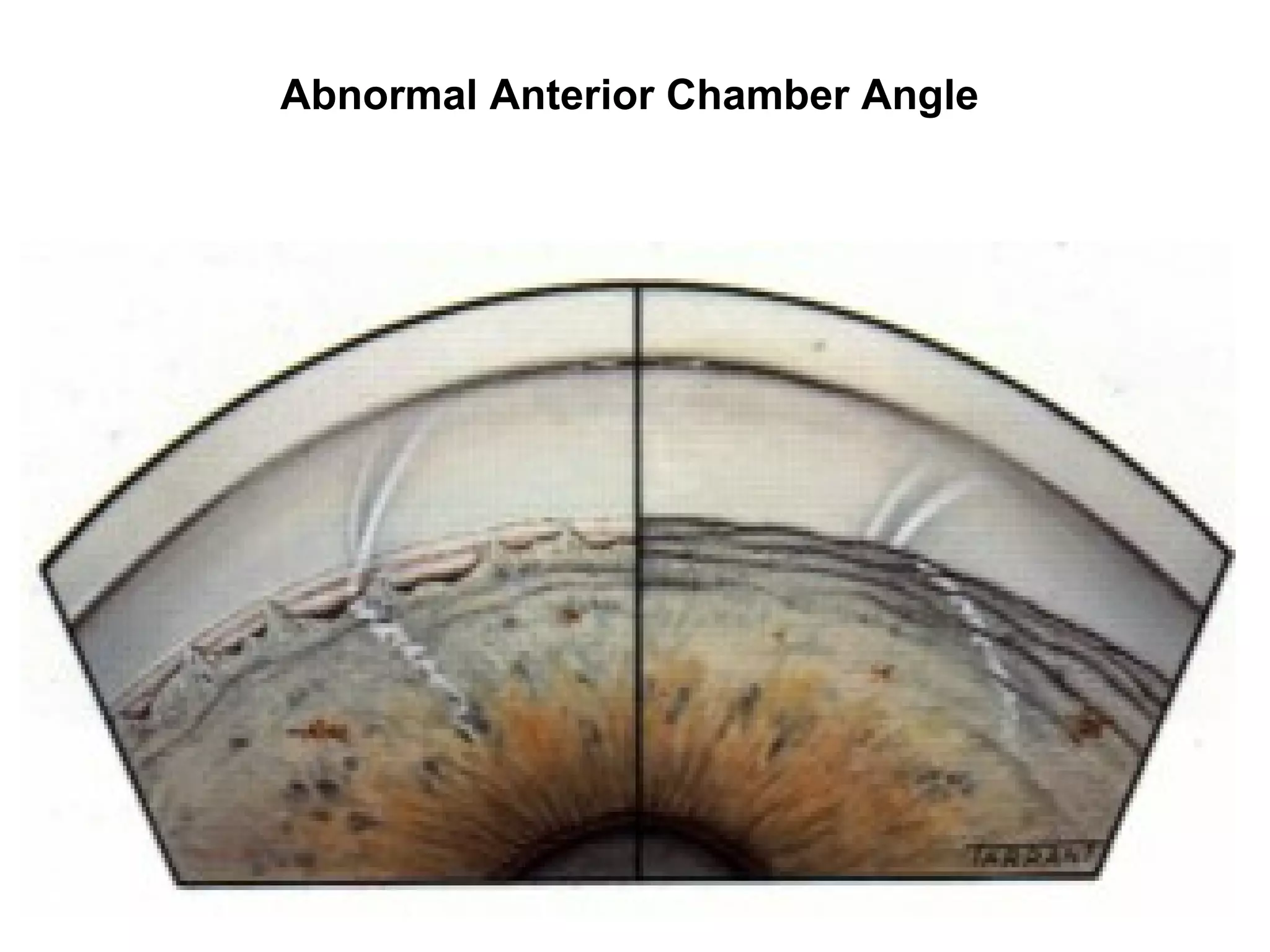

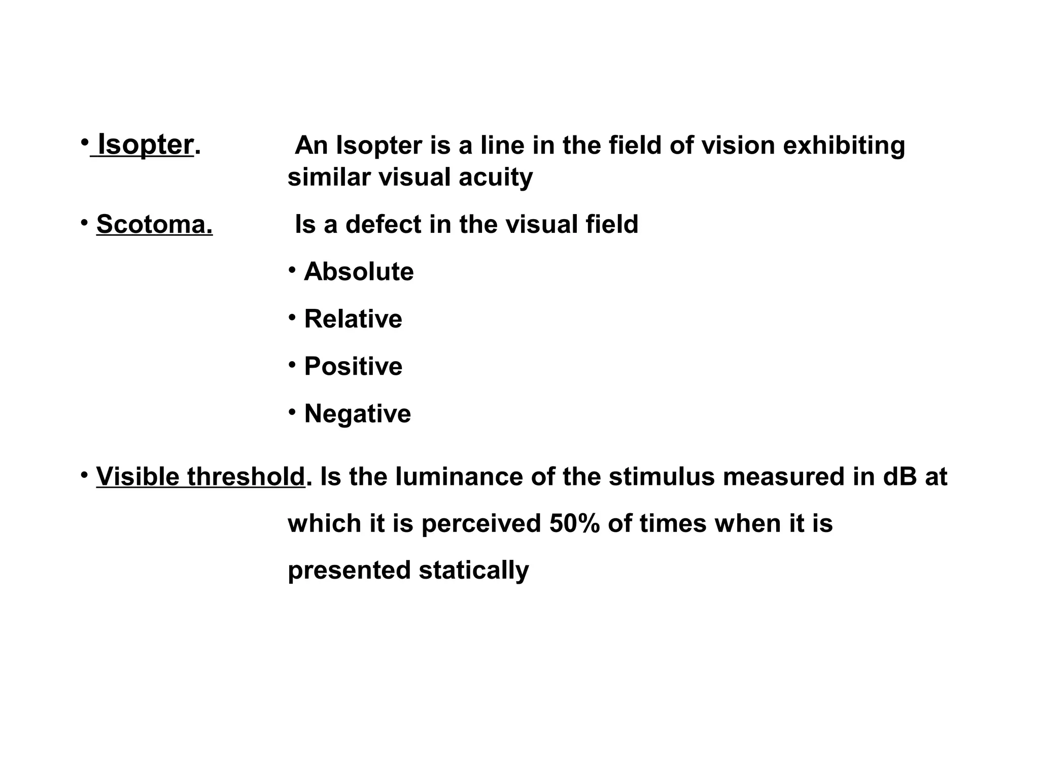


This document provides guidance on assessing patients for glaucoma through a comprehensive examination and testing. A thorough history, examination of visual functions and the eye, measurement of intraocular pressure, gonioscopy, and evaluation of the optic nerve and retinal nerve fiber layer are recommended. Additional tests such as visual field testing, HRT, OCT and GDx can further aid diagnosis. A multidisciplinary approach including evaluation of other organ systems may be warranted in some cases. The goal of assessment is to diagnose and classify glaucoma, identify risk factors, plan appropriate management, and monitor for disease progression.


