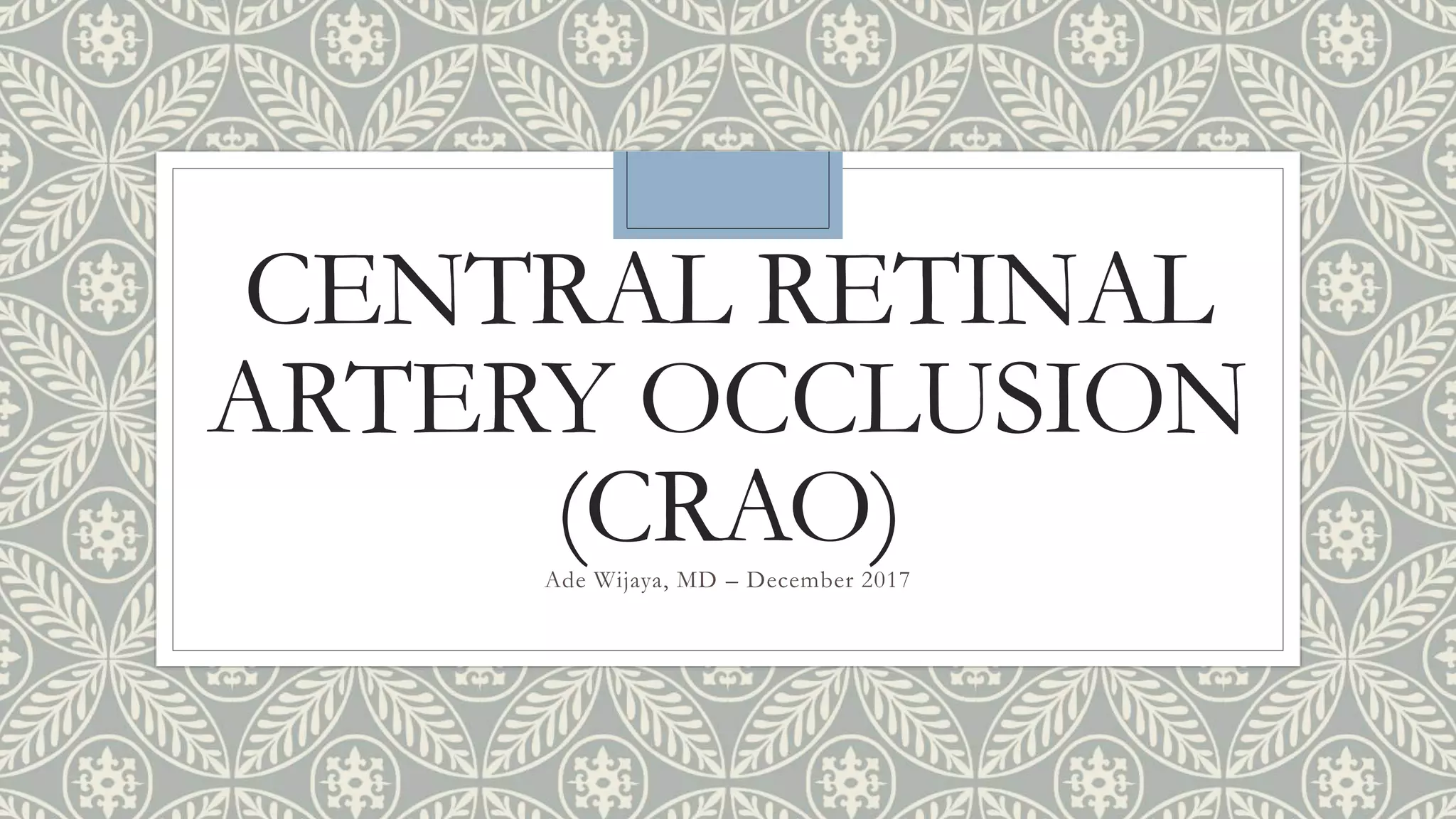
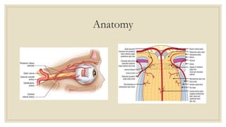
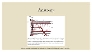
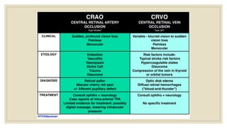
CENTRAL RETINAL
ARTERY OCCLUSION
(CRAO)Ade Wijaya, MD – December 2017
Outline:
◦ Introduction
◦ Epidemiology
◦ Anatomy
◦ Pathophysiology
◦ Type of CRAO
◦ Diagnosis
◦ Management
Introduction
◦ First described by von Graefes in 1859
◦ Acute stroke of the eye
◦ Ophtalmic emergency
◦ Increase fall risk  increase dependency
◦ Atherosclerotic / embolism
von Graefes A. Ueber Embolie der Arteria centralis retinae als Ursache plotzlicherErblindung. Arch Ophthalmol 1859; 5: 136–157.
Rumelt S, Dorenboim Y, Rehany U. Aggressive systematic treatment for central retinal artery occlusion. Am J Ophthalmol1999; 128: 733–738.
Vu H, Keeffe J, McCarty C, Taylor HR. Impact of unilateral and bilateral vision loss on quality of life. Br J Ophthalmol 2005; 89: 360–363.
Epidemiology
◦ 1 in 100 000 people
◦ 1 in 10 000 ophthalmological outpatient visits
◦ 80% of patients having a visual acuity (VA) of 20/400 or worse
◦ 1.9/100,000 in the United States
Leavitt JA, Larson TA, Hodge DO, Gullerud RE. The incidence of central retinal artery occlusion in Olmsted County, Minnesota. Am J Ophthalmol. 2011;152:820–3.
Hayreh SS, Zimmerman MB. Central retinal artery occlusion: visual outcome. Am J Ophthalmol 2005; 140(3): 376–391.
Rumelt S, Dorenboim Y, Rehany U. Aggressive systematic treatment for central retinal artery occlusion. Am J Ophthalmol1999; 128: 733–738.
Anatomy
Anatomy
Hayreh SS. Anatomy and physiology of the optic nerve head.Trans Am Acad Ophthalmol Otolaryngol 1974; 78: OP240–OP254.
Pathophysiology
Type of CRAO
Non-arteritic permanent
CRAO
Non-arteritic transient
CRAO
Non-arteritic CRAO
with cilioretinal sparing
Arteritic CRAO
CRAO
Non-arteritic Permanent CRAO
◦ 2/3 of all CRAO cases
◦ Caused by platelet fibrin thrombi and emboli
◦ Risk factors: arterial hypertension, diabetes mellitus, carotid artery disease, coronary artery disease, transient
ischaemic attacks (TIAs) or cerebral vascular accidents, and smoking tobacco.
◦ Other risk factors are a family history of any type of vascular disease.
◦ In younger patients (under 50 years), proatherogenic states, such as hyperhomocystenemia, factor V Leiden,
protein C and S and anti-thrombin deficiencies, anti-phospholipid antibodies or prothrombin gene mutations,
sickle cell disease, and migraine due to vasospasm and paraneoplastic syndromes may all contribute to non-
arteritic CRAO.
◦ Ocular risk factors can include raised intraocular pressure, optic nerve head drusen, and a preretinal arterial
loops. These result in reduced perfusion pressure across the optic nerve head
Varma, D. D., Cugati, S., Lee, A. W., & Chen, C. S. (2013). A review of central retinal artery occlusion: clinical presentation and management. Eye, 27(6), 688-697.
Non-arteritic transient CRAO
◦ Transient monocular blindness
◦ 15–17% of CRAOs
◦ Best prognosis
◦ Analogue to TIA
◦ Transient vasospasm due to serotonin release from platelets on atherosclerotic plaques
Hayreh SS, Zimmerman MB. Central retinal artery occlusion: visual outcome. Am J Ophthalmol 2005; 140(3): 376–391.
Kline L. The natural history of patients with amaurosis fugax.Ophthalmol Clin N Am 1996; 9: 351–358.
Hayreh SS, Piegors DJ, Heistad DD. Serotonin-induced constriction of ocular arteries in atherosclerotic monkeys: implications for ischemic disorders of the retina and optic nerve head. Arch Ophthalmol 1997; 115: 220–228.
Non-arteritic CRAO with cilioretinal sparing
◦ Preservation of the cilioretinal artery results in
preserved perfusion of the macula region
Arteritic CRAO
◦ < 5 %
◦ Related to giant cell arteritis
Hayreh SS, Zimmerman MB. Central retinal artery occlusion: visual outcome. Am J Ophthalmol 2005; 140(3): 376–391.
Diagnosis
◦ History of visual loss to confirm the diagnosis of CRAO.
◦ An evaluation of clinical risk factors that may need to be modified.
◦ General physical examination findings.
◦ Ocular examination/investigation findings.
◦ Ancillary investigation findings.
Varma, D. D., Cugati, S., Lee, A. W., & Chen, C. S. (2013). A review of central retinal artery occlusion: clinical presentation and management. Eye, 27(6), 688-697.
History
◦ Sudden,
◦ Painless monocular vision loss
◦ Snellen VA of counting fingers or worse is found in 74% of patients with a visual field defect
◦ Family history of cerebrovascular and cardiovascular disease, diabetes, hyperlipidaemia, a past history of
atherosclerotic cardiac or cerebrovascular disease, valvular heart disease, or transient ischaemic events, such
as transient monocular blindness, TIAs, or anginal symptoms
◦ The presence of vasculitis, sickle cell disease, myeloproliferative disorders, hypercoagualable states, and the
use of the oral contraceptive pills or intravenous drugs
Varma, D. D., Cugati, S., Lee, A. W., & Chen, C. S. (2013). A review of central retinal artery occlusion: clinical presentation and management. Eye, 27(6), 688-697.
Physical Examination
◦ Ocular findings to confirm the diagnosis and exclude other causes of monocular vision loss
◦ Assessment of vascular risk.
Varma, D. D., Cugati, S., Lee, A. W., & Chen, C. S. (2013). A review of central retinal artery occlusion: clinical presentation and management. Eye, 27(6), 688-697.
Ocular Evaluation:
Funduscopic at Early Stage of CRAO
◦ Retinal opacity in the posterior pole (58%),
◦ Cherry-red spot (90%),
◦ Cattle trucking (19%),
◦ Retinal arterial attenuation (32%),
◦ Optic disk oedema (22%)
◦ Pallor (39%).
Hayreh SS, Zimmerman MB. Fundus changes in central retinal artery occlusion. Retina 2007; 27: 276–289.
Ocular Evaluation:
Funduscopic at Late Stage of CRAO
◦ Optic atrophy (91%),
◦ Retinal arterial attenuation (58%),
◦ Cilioretinal collaterals (18%),
◦ Macular retinal pigment epithelial changes (11%)
Hayreh SS, Zimmerman MB. Fundus changes in central retinal artery occlusion. Retina 2007; 27: 276–289.
Ocular Evaluation:
Other Findings
◦ Intra-arterial (IA) emboli (20%)
◦ Small, yellow, and refractile plaques, the ‘Hollenhorst plaques’, suggest cholesterol emboli
◦ Single, white, non-scintillating plaques located in the proximal retinal vasculature are due to calcific emboli
◦ Fibrino-platelet emboli are seen as small pale bodies.
◦ It is important to look at the contralateral eye as there may be clues to possible underlying pathology, such as
hypertensive retinopathy, arteriole changes, or previous vaso-occlusive diseases.
◦ increased intraocular pressures, the presence of pre-retinal arterial loops, and drusen on the optic nerve head may
predispose to a CRAO as that reduces the mean arterial perfusion across the optic nerve head.
◦ If ophthalmoscopy reveals the presence of hypertensive or sickle cell retinopathy, it can suggest the presence of
small vessel disease.
◦ Cattle trucking
◦ OCT
Varma, D. D., Cugati, S., Lee, A. W., & Chen, C. S. (2013). A review of central retinal artery occlusion: clinical presentation and management. Eye, 27(6), 688-697.
Management
◦ Acute: Attempt to restore ocular perfusion to the CRA.
◦ Subacute: Preventing secondary neovascular complications to the eye.
◦ Long term: Preventing other vascular ischaemic events to the eye or other end organ.
Varma, D. D., Cugati, S., Lee, A. W., & Chen, C. S. (2013). A review of central retinal artery occlusion: clinical presentation and management. Eye, 27(6), 688-697.
Acute Stage Therapy
◦ Use of sublingual isosorbide dinitrate or systemic pentoxifylline or inhalation of a carbogen, hyperbaric
oxygen, to increase blood oxygen content and dilate retinal arteries.
◦ Ocular massage to attempt to dislodge emboli.
◦ Intravenous acetazolamide and mannitol, plus anterior chamber paracentesis, followed by withdrawal of a
small amount of aqueous fluid from the eye to increase retinal artery perfusion pressure by reducing
intraocular pressure.
◦ Multimodal stepwise conservative approaches involving combinations of: ocular massage, globe compression,
sublingual isosorbide dinitrate, intravenous acetazolamide, followed by intravenous mannitol,
methylprednisolone, streptokinase, retrobulbar tolazoline, and different anticoagulants.
◦ Thrombolytics
Varma, D. D., Cugati, S., Lee, A. W., & Chen, C. S. (2013). A review of central retinal artery occlusion: clinical presentation and management. Eye, 27(6), 688-697.
Cugati, S., Varma, D. D., Chen, C. S., & Lee, A. W. (2013). Treatment options for central retinal artery occlusion. Current treatment options in neurology, 15(1), 63-77.
Summary
◦ Opthalmic emergency
◦ Ocular analoque of cerebral stroke
◦ Atherosclerotic risk factors
◦ Acute treatment and secondary prevention
THANK YOU

Central Retinal Artery Occlusion

  • 1.
    CENTRAL RETINAL ARTERY OCCLUSION (CRAO)AdeWijaya, MD – December 2017
  • 2.
    Outline: ◦ Introduction ◦ Epidemiology ◦Anatomy ◦ Pathophysiology ◦ Type of CRAO ◦ Diagnosis ◦ Management
  • 3.
    Introduction ◦ First describedby von Graefes in 1859 ◦ Acute stroke of the eye ◦ Ophtalmic emergency ◦ Increase fall risk  increase dependency ◦ Atherosclerotic / embolism von Graefes A. Ueber Embolie der Arteria centralis retinae als Ursache plotzlicherErblindung. Arch Ophthalmol 1859; 5: 136–157. Rumelt S, Dorenboim Y, Rehany U. Aggressive systematic treatment for central retinal artery occlusion. Am J Ophthalmol1999; 128: 733–738. Vu H, Keeffe J, McCarty C, Taylor HR. Impact of unilateral and bilateral vision loss on quality of life. Br J Ophthalmol 2005; 89: 360–363.
  • 4.
    Epidemiology ◦ 1 in100 000 people ◦ 1 in 10 000 ophthalmological outpatient visits ◦ 80% of patients having a visual acuity (VA) of 20/400 or worse ◦ 1.9/100,000 in the United States Leavitt JA, Larson TA, Hodge DO, Gullerud RE. The incidence of central retinal artery occlusion in Olmsted County, Minnesota. Am J Ophthalmol. 2011;152:820–3. Hayreh SS, Zimmerman MB. Central retinal artery occlusion: visual outcome. Am J Ophthalmol 2005; 140(3): 376–391. Rumelt S, Dorenboim Y, Rehany U. Aggressive systematic treatment for central retinal artery occlusion. Am J Ophthalmol1999; 128: 733–738.
  • 5.
  • 6.
    Anatomy Hayreh SS. Anatomyand physiology of the optic nerve head.Trans Am Acad Ophthalmol Otolaryngol 1974; 78: OP240–OP254.
  • 7.
  • 8.
    Type of CRAO Non-arteriticpermanent CRAO Non-arteritic transient CRAO Non-arteritic CRAO with cilioretinal sparing Arteritic CRAO CRAO
  • 9.
    Non-arteritic Permanent CRAO ◦2/3 of all CRAO cases ◦ Caused by platelet fibrin thrombi and emboli ◦ Risk factors: arterial hypertension, diabetes mellitus, carotid artery disease, coronary artery disease, transient ischaemic attacks (TIAs) or cerebral vascular accidents, and smoking tobacco. ◦ Other risk factors are a family history of any type of vascular disease. ◦ In younger patients (under 50 years), proatherogenic states, such as hyperhomocystenemia, factor V Leiden, protein C and S and anti-thrombin deficiencies, anti-phospholipid antibodies or prothrombin gene mutations, sickle cell disease, and migraine due to vasospasm and paraneoplastic syndromes may all contribute to non- arteritic CRAO. ◦ Ocular risk factors can include raised intraocular pressure, optic nerve head drusen, and a preretinal arterial loops. These result in reduced perfusion pressure across the optic nerve head Varma, D. D., Cugati, S., Lee, A. W., & Chen, C. S. (2013). A review of central retinal artery occlusion: clinical presentation and management. Eye, 27(6), 688-697.
  • 10.
    Non-arteritic transient CRAO ◦Transient monocular blindness ◦ 15–17% of CRAOs ◦ Best prognosis ◦ Analogue to TIA ◦ Transient vasospasm due to serotonin release from platelets on atherosclerotic plaques Hayreh SS, Zimmerman MB. Central retinal artery occlusion: visual outcome. Am J Ophthalmol 2005; 140(3): 376–391. Kline L. The natural history of patients with amaurosis fugax.Ophthalmol Clin N Am 1996; 9: 351–358. Hayreh SS, Piegors DJ, Heistad DD. Serotonin-induced constriction of ocular arteries in atherosclerotic monkeys: implications for ischemic disorders of the retina and optic nerve head. Arch Ophthalmol 1997; 115: 220–228.
  • 11.
    Non-arteritic CRAO withcilioretinal sparing ◦ Preservation of the cilioretinal artery results in preserved perfusion of the macula region Arteritic CRAO ◦ < 5 % ◦ Related to giant cell arteritis Hayreh SS, Zimmerman MB. Central retinal artery occlusion: visual outcome. Am J Ophthalmol 2005; 140(3): 376–391.
  • 12.
    Diagnosis ◦ History ofvisual loss to confirm the diagnosis of CRAO. ◦ An evaluation of clinical risk factors that may need to be modified. ◦ General physical examination findings. ◦ Ocular examination/investigation findings. ◦ Ancillary investigation findings. Varma, D. D., Cugati, S., Lee, A. W., & Chen, C. S. (2013). A review of central retinal artery occlusion: clinical presentation and management. Eye, 27(6), 688-697.
  • 13.
    History ◦ Sudden, ◦ Painlessmonocular vision loss ◦ Snellen VA of counting fingers or worse is found in 74% of patients with a visual field defect ◦ Family history of cerebrovascular and cardiovascular disease, diabetes, hyperlipidaemia, a past history of atherosclerotic cardiac or cerebrovascular disease, valvular heart disease, or transient ischaemic events, such as transient monocular blindness, TIAs, or anginal symptoms ◦ The presence of vasculitis, sickle cell disease, myeloproliferative disorders, hypercoagualable states, and the use of the oral contraceptive pills or intravenous drugs Varma, D. D., Cugati, S., Lee, A. W., & Chen, C. S. (2013). A review of central retinal artery occlusion: clinical presentation and management. Eye, 27(6), 688-697.
  • 14.
    Physical Examination ◦ Ocularfindings to confirm the diagnosis and exclude other causes of monocular vision loss ◦ Assessment of vascular risk. Varma, D. D., Cugati, S., Lee, A. W., & Chen, C. S. (2013). A review of central retinal artery occlusion: clinical presentation and management. Eye, 27(6), 688-697.
  • 15.
    Ocular Evaluation: Funduscopic atEarly Stage of CRAO ◦ Retinal opacity in the posterior pole (58%), ◦ Cherry-red spot (90%), ◦ Cattle trucking (19%), ◦ Retinal arterial attenuation (32%), ◦ Optic disk oedema (22%) ◦ Pallor (39%). Hayreh SS, Zimmerman MB. Fundus changes in central retinal artery occlusion. Retina 2007; 27: 276–289.
  • 16.
    Ocular Evaluation: Funduscopic atLate Stage of CRAO ◦ Optic atrophy (91%), ◦ Retinal arterial attenuation (58%), ◦ Cilioretinal collaterals (18%), ◦ Macular retinal pigment epithelial changes (11%) Hayreh SS, Zimmerman MB. Fundus changes in central retinal artery occlusion. Retina 2007; 27: 276–289.
  • 17.
    Ocular Evaluation: Other Findings ◦Intra-arterial (IA) emboli (20%) ◦ Small, yellow, and refractile plaques, the ‘Hollenhorst plaques’, suggest cholesterol emboli ◦ Single, white, non-scintillating plaques located in the proximal retinal vasculature are due to calcific emboli ◦ Fibrino-platelet emboli are seen as small pale bodies. ◦ It is important to look at the contralateral eye as there may be clues to possible underlying pathology, such as hypertensive retinopathy, arteriole changes, or previous vaso-occlusive diseases. ◦ increased intraocular pressures, the presence of pre-retinal arterial loops, and drusen on the optic nerve head may predispose to a CRAO as that reduces the mean arterial perfusion across the optic nerve head. ◦ If ophthalmoscopy reveals the presence of hypertensive or sickle cell retinopathy, it can suggest the presence of small vessel disease. ◦ Cattle trucking ◦ OCT Varma, D. D., Cugati, S., Lee, A. W., & Chen, C. S. (2013). A review of central retinal artery occlusion: clinical presentation and management. Eye, 27(6), 688-697.
  • 20.
    Management ◦ Acute: Attemptto restore ocular perfusion to the CRA. ◦ Subacute: Preventing secondary neovascular complications to the eye. ◦ Long term: Preventing other vascular ischaemic events to the eye or other end organ. Varma, D. D., Cugati, S., Lee, A. W., & Chen, C. S. (2013). A review of central retinal artery occlusion: clinical presentation and management. Eye, 27(6), 688-697.
  • 21.
    Acute Stage Therapy ◦Use of sublingual isosorbide dinitrate or systemic pentoxifylline or inhalation of a carbogen, hyperbaric oxygen, to increase blood oxygen content and dilate retinal arteries. ◦ Ocular massage to attempt to dislodge emboli. ◦ Intravenous acetazolamide and mannitol, plus anterior chamber paracentesis, followed by withdrawal of a small amount of aqueous fluid from the eye to increase retinal artery perfusion pressure by reducing intraocular pressure. ◦ Multimodal stepwise conservative approaches involving combinations of: ocular massage, globe compression, sublingual isosorbide dinitrate, intravenous acetazolamide, followed by intravenous mannitol, methylprednisolone, streptokinase, retrobulbar tolazoline, and different anticoagulants. ◦ Thrombolytics Varma, D. D., Cugati, S., Lee, A. W., & Chen, C. S. (2013). A review of central retinal artery occlusion: clinical presentation and management. Eye, 27(6), 688-697.
  • 22.
    Cugati, S., Varma,D. D., Chen, C. S., & Lee, A. W. (2013). Treatment options for central retinal artery occlusion. Current treatment options in neurology, 15(1), 63-77.
  • 23.
    Summary ◦ Opthalmic emergency ◦Ocular analoque of cerebral stroke ◦ Atherosclerotic risk factors ◦ Acute treatment and secondary prevention
  • 24.