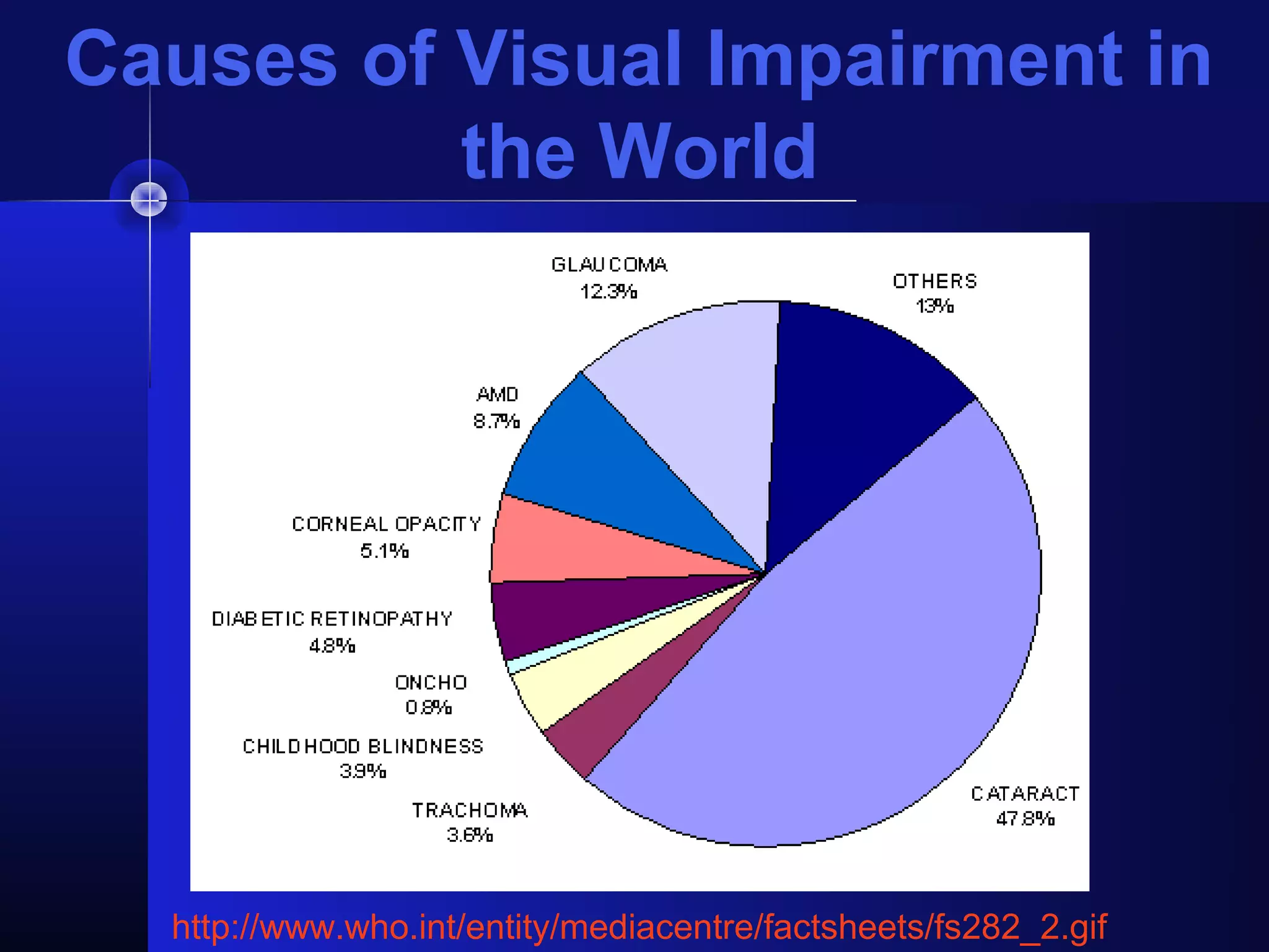
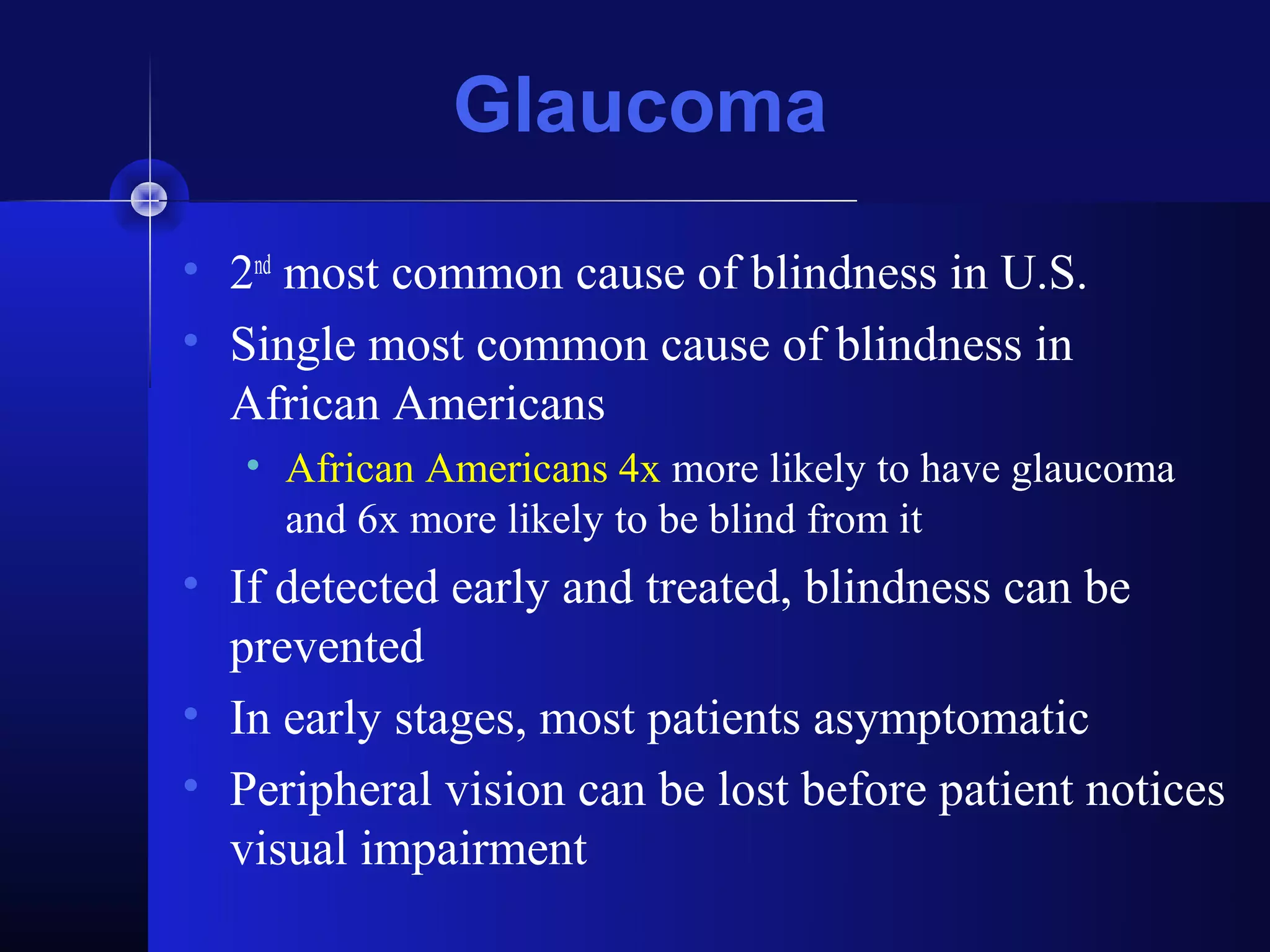
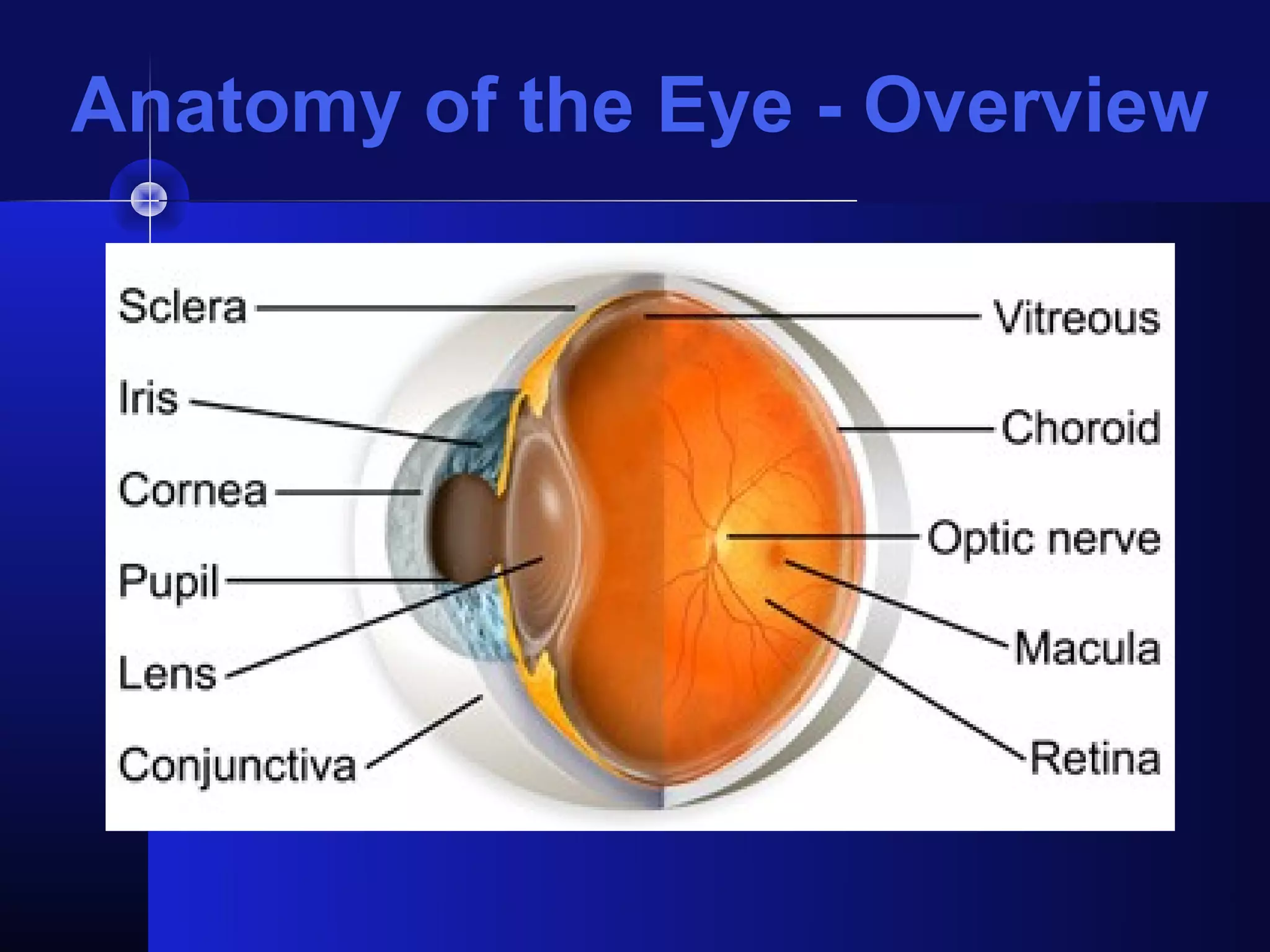
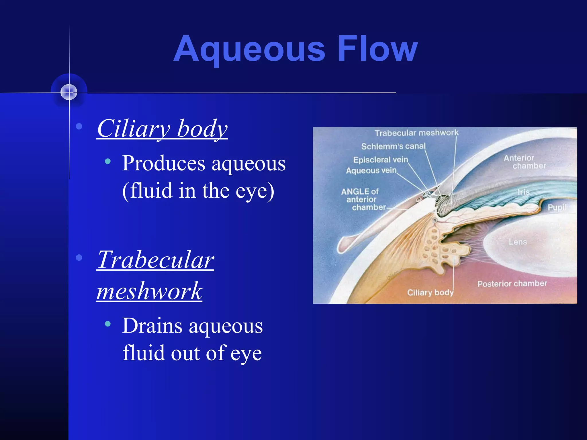
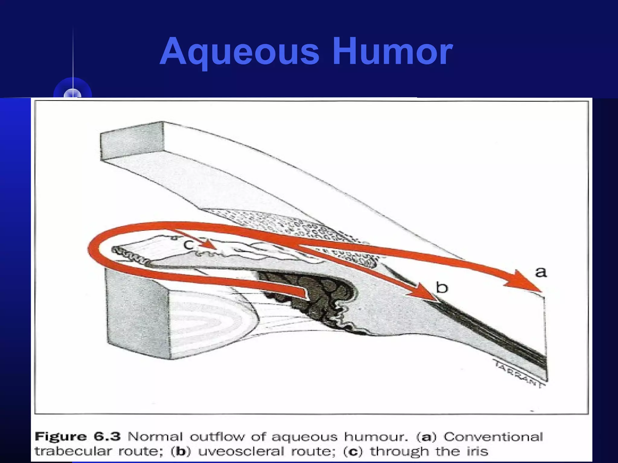
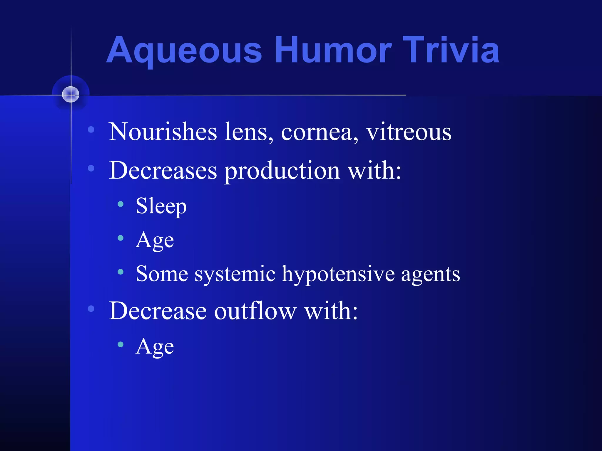
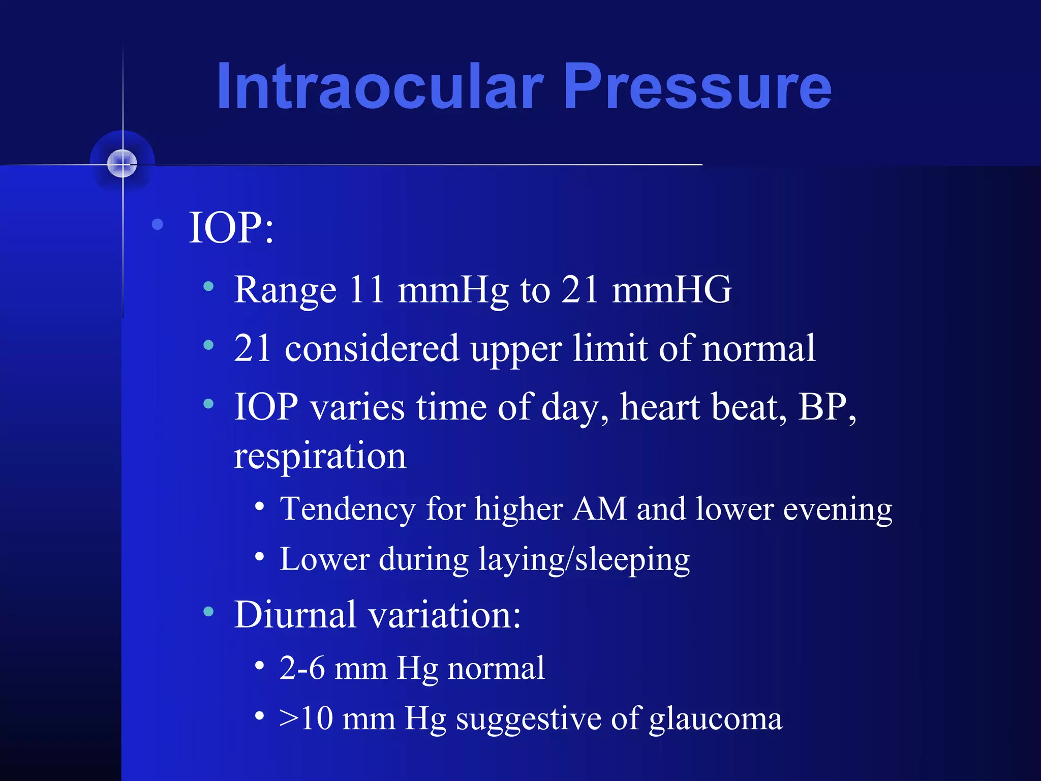
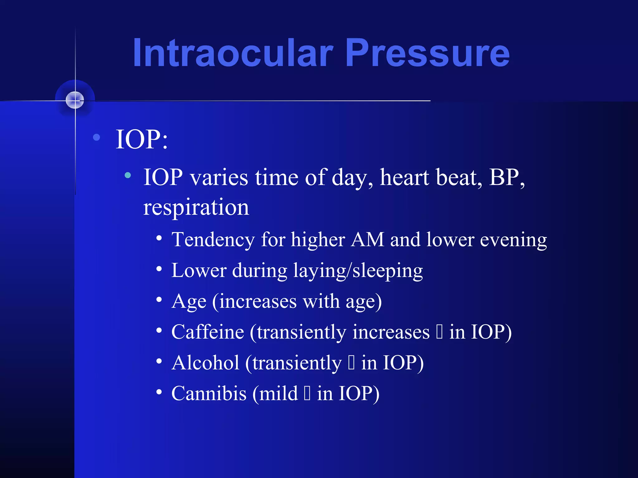
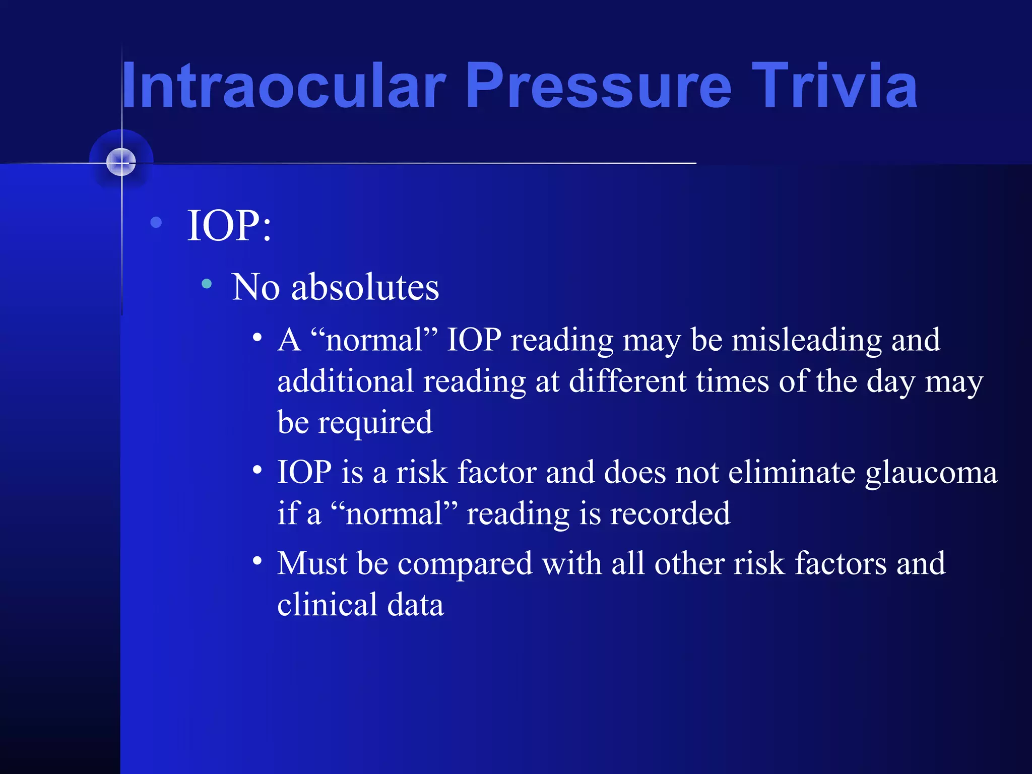
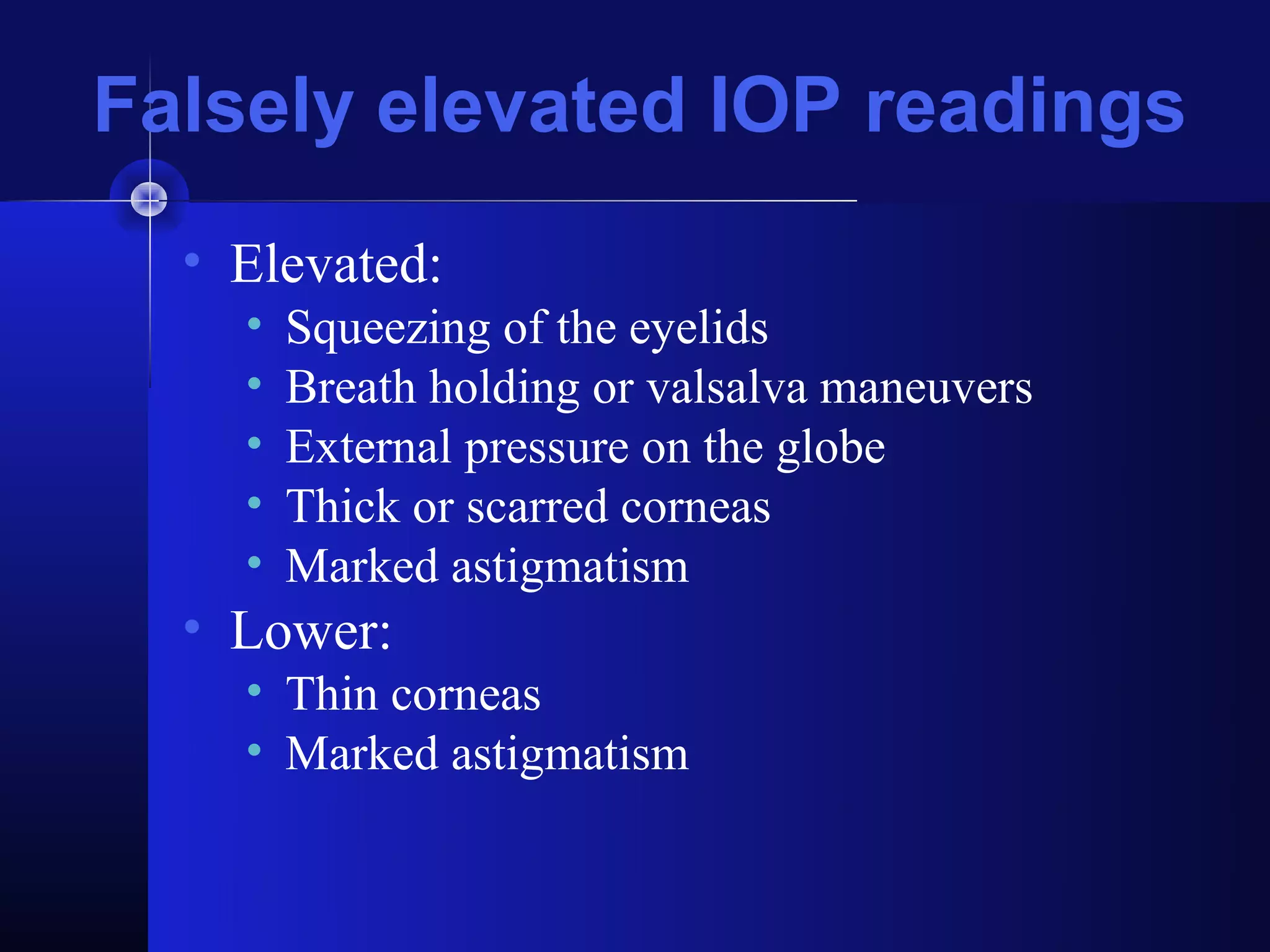
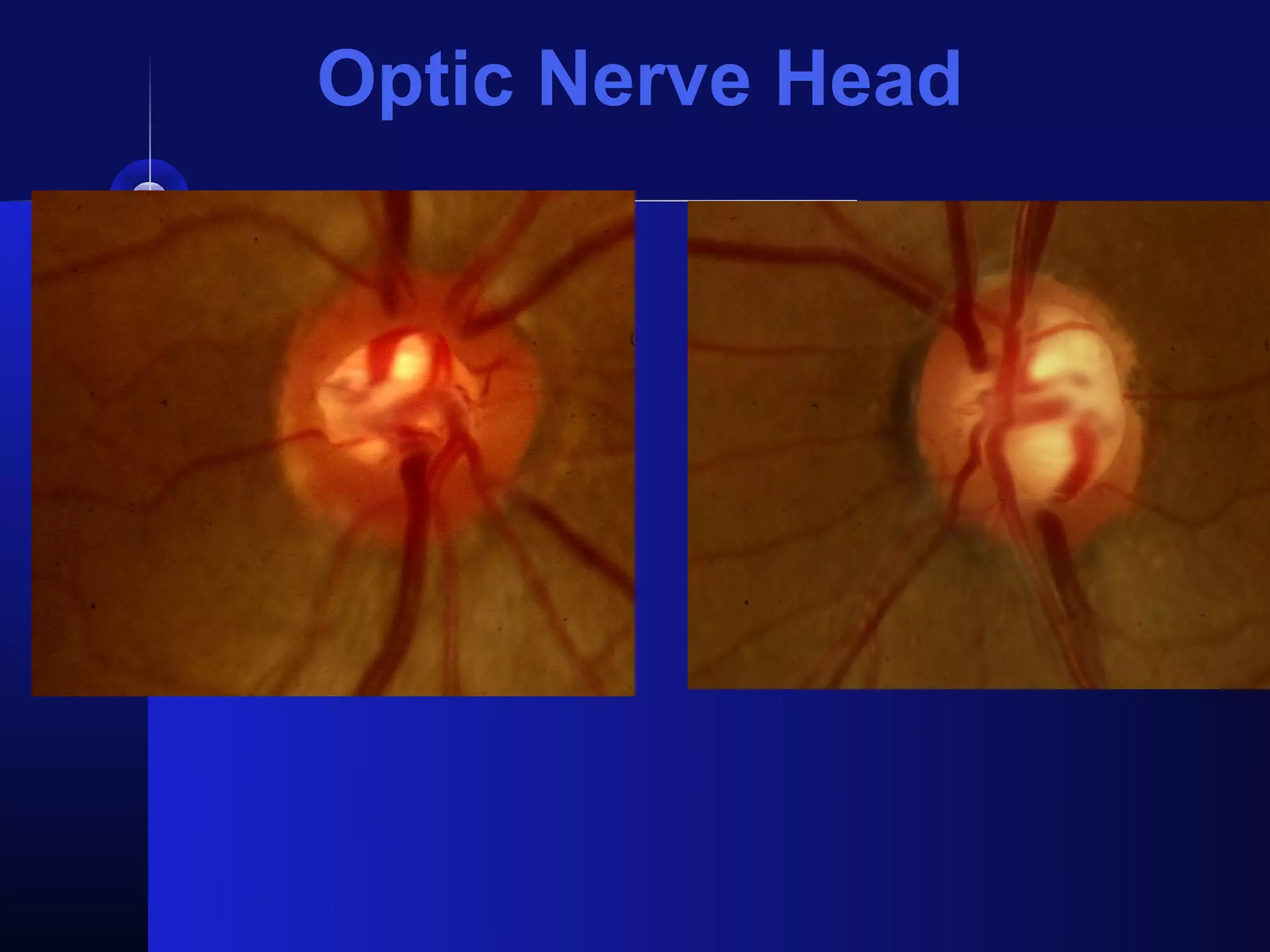
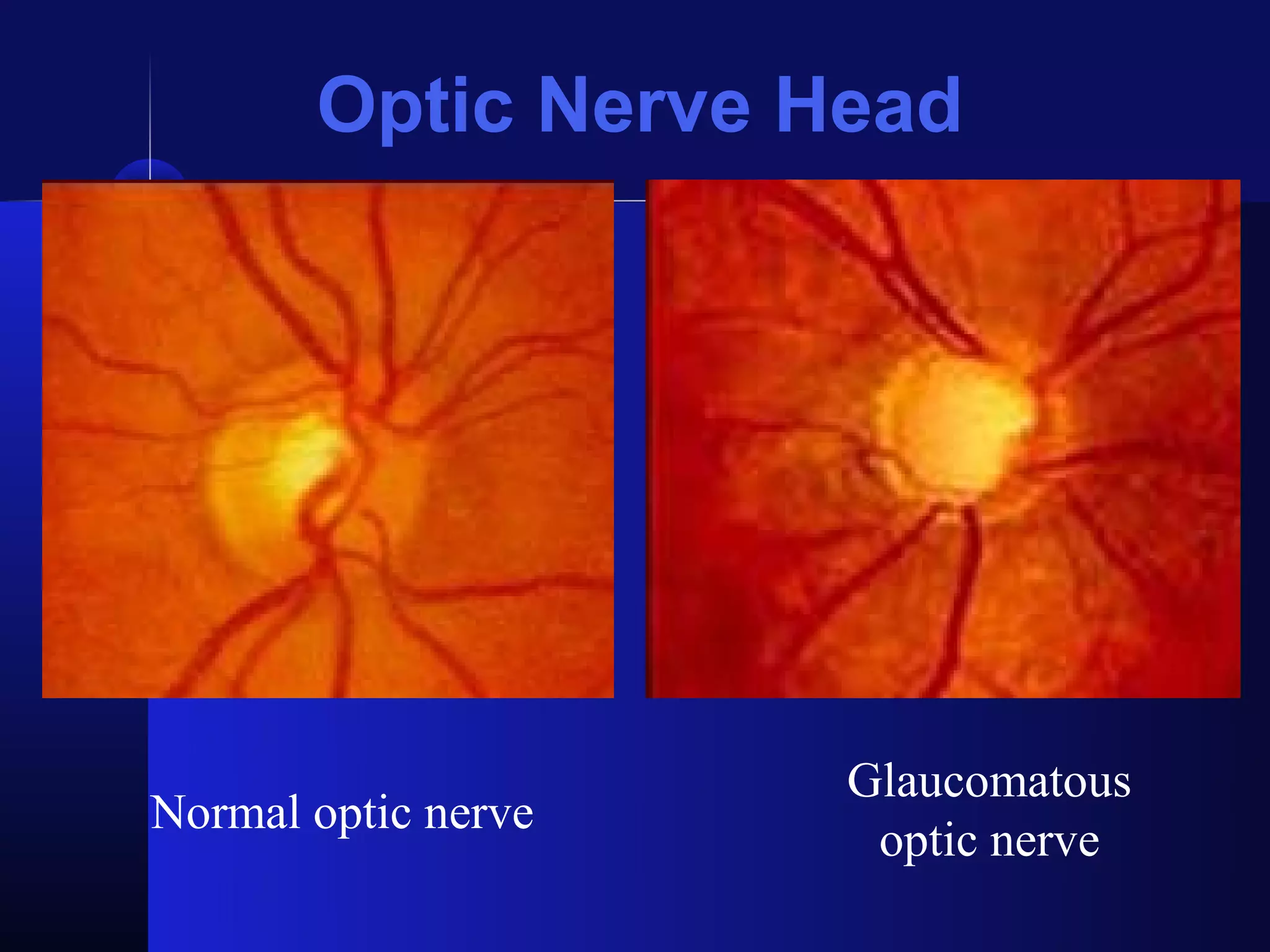
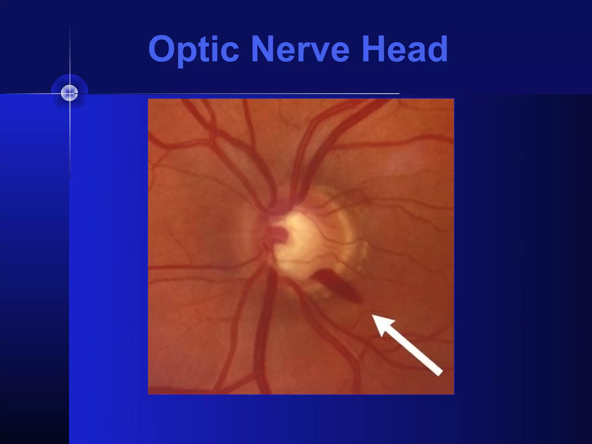
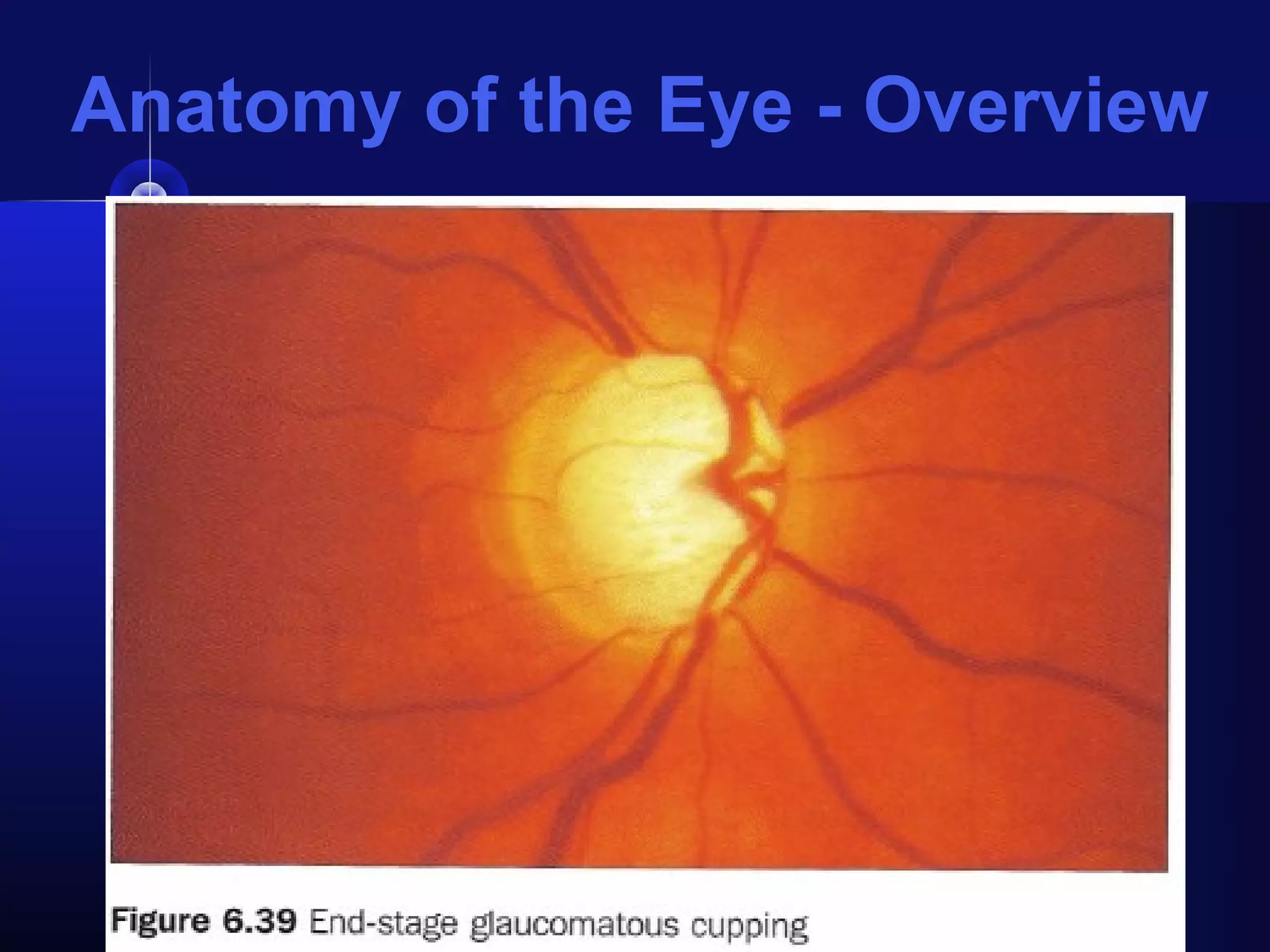
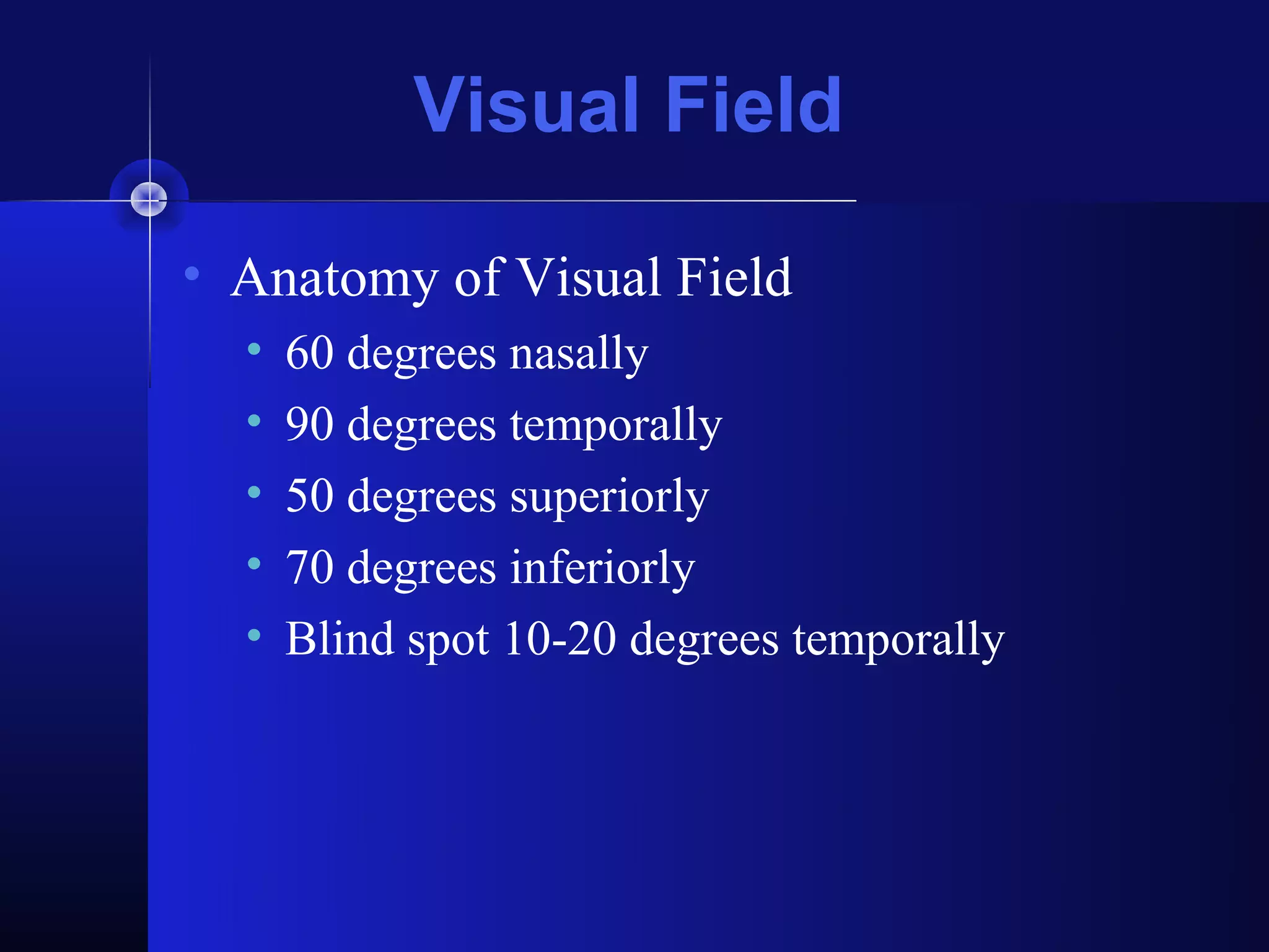
SlideShare a Scribd company logo
Glaucoma Screening


       Nicholas J. Silvestros, OD
               Clinical Instructor
Department of Ophthalmology and Vision Sciences
        Washington University St. Louis
               School of Medicine
Causes of Visual Impairment in
          the World


  http://www.who.int/entity/mediacentre/factsheets/fs282_2.gif
Glaucoma
• 2nd most common cause of blindness in U.S.
• Single most common cause of blindness in
  African Americans
   • African Americans 4x more likely to have glaucoma
     and 6x more likely to be blind from it
• If detected early and treated, blindness can be
  prevented
• In early stages, most patients asymptomatic
• Peripheral vision can be lost before patient notices
  visual impairment
Anatomy of the Eye - Overview
Aqueous Flow

• Ciliary body
  • Produces aqueous
    (fluid in the eye)


• Trabecular
  meshwork
  • Drains aqueous
    fluid out of eye
Aqueous Humor Formation

 • Involves the combination of 2 known
   processes:
   • Active transport (secretion)
      • 80% of Aqueous
   • Passive transport (ultrafiltration and diffusion)
      • 20% of Aqueous
      • Affected by topical glaucoma medications: beta-
        blockers, sympathomimetics and carbonic
        anhydrase inhibitors
Trabecular Outflow

• Conventional outflow
  • 80-90% outflow
  • Increased:
     • Drugs: Cholinergics (pilocarpine), Adrenergic
       agonists
     • Surgical: ALT/SLT, Trabeculotomy/goniotomy
• Unconventional outflow
  • 10-20% outflow
  • Increased:
     • Drugs: sympathomimetics and prostaglandins
Aqueous Humor
Aqueous Humor Trivia

• Nourishes lens, cornea, vitreous
• Decreases production with:
  • Sleep
  • Age
  • Some systemic hypotensive agents
• Decrease outflow with:
  • Age
Intraocular Pressure

• IOP:
  • Range 11 mmHg to 21 mmHG
  • 21 considered upper limit of normal
  • IOP varies time of day, heart beat, BP,
    respiration
     • Tendency for higher AM and lower evening
     • Lower during laying/sleeping
  • Diurnal variation:
     • 2-6 mm Hg normal
     • >10 mm Hg suggestive of glaucoma
Intraocular Pressure

• IOP:
  • IOP varies time of day, heart beat, BP,
    respiration
     •   Tendency for higher AM and lower evening
     •   Lower during laying/sleeping
     •   Age (increases with age)
     •   Caffeine (transiently increases  in IOP)
     •   Alcohol (transiently  in IOP)
     •   Cannibis (mild  in IOP)
Intraocular Pressure Trivia

• IOP:
  • No absolutes
     • A “normal” IOP reading may be misleading and
       additional reading at different times of the day may
       be required
     • IOP is a risk factor and does not eliminate glaucoma
       if a “normal” reading is recorded
     • Must be compared with all other risk factors and
       clinical data
Measurement of IOP
• Applanation Tonometry:
  • Measures the force necessary to flatten an area
    of cornea 3.06 mm diameter
  • Central part of cornea flattened while variable
    force records pressure
  • Central Corneal Thickness:
     • >540 micrometers produce falsely high IOP
       readings by TA
     • <540 micrometers produce falsely low IOP reading
       by TA
Measurement of IOP
Measurement of IOP
• Applanation Tonometry:
  • Goldmann tonometer
     • Most popular tonometer and accurate tonometer
  • Tono-Pen tonometer
     • Hand held portable tonometer
     • Over estimates low IOP and underestimates high
       IOP
Measurement of IOP
• Non-Contact Tonometry:
  • Air-Puff tonometer
     • Goldmann principles with air instead of prism
         time required to flatten cornea relates directly to level of
          IOP
     • Does not require topical anesthetic
     • Useful for screenings
     • Disadvantage – accurate low to mid IOP range
Anatomy of the Eye - Overview
Falsely elevated IOP readings
  • Elevated:
    •   Squeezing of the eyelids
    •   Breath holding or valsalva maneuvers
    •   External pressure on the globe
    •   Thick or scarred corneas
    •   Marked astigmatism
  • Lower:
    • Thin corneas
    • Marked astigmatism
Optic Nerve Head

• 1.2 million axons
  • Declines with age
  • Cell bodies are the ganglion cells
• Magnocellular (M) cells 10%
  • Large diameter (dim illumination)
• Parvocellular (P) cells 90%
  • Small diameter axons (color, fine detail)
Optic Nerve Head

•   Scleral Canal
•   Lamina Cribrosa
•   Optic Cup
•   Neuroretinal Rim
•   Size of ON:
    • AA>Asians>Hispanics>Whites
Optic Nerve Head
Optic Nerve Head

• Cup-Disc Ratio
  • Fraction of vertical and horizontal meridians
     • C/D=0.3/0.3
     • Normal is 0.3 or less
  • Ratio greater than 0.7 regarded suspicious
  • Asymmetry between two eyes of 0.2 or more
    regarded suspicious
  • Cup size is needed to evaluate progression not
    initial diagnosis
Optic nerve appearance in glaucoma

  • Glaucoma nerve damage ranges from
    localized to diffuse
    • Localized easier to recognize with notching
  • Description of nerve important
    • Neuralretinal rim tissue
       •   Thickness
       •   Symmetry
       •   Color
       •   Notching
       •   Hemorrhage disc margin
Optic Nerve Head
Optic Nerve Head
Optic Nerve Head
Optic Nerve Head
Optic Nerve Head


                     Glaucomatous
Normal optic nerve
                      optic nerve
Optic Nerve Head
Anatomy of the Eye - Overview
Visual Field

• Anatomy of Visual Field
  •   60 degrees nasally
  •   90 degrees temporally
  •   50 degrees superiorly
  •   70 degrees inferiorly
  •   Blind spot 10-20 degrees temporally
Anatomy of the Nerve Fibers

More Related Content

What's hot

Esotropia
EsotropiaEsotropia
Esotropia
PRAKRITIYAGNAM
 
Diplopia charting
Diplopia chartingDiplopia charting
Diplopia charting
SSSIHMS-PG
 
Indirect ophthalmoscopy
Indirect ophthalmoscopy Indirect ophthalmoscopy
Indirect ophthalmoscopy
Shruti Laddha
 
Clinical examination of squint
Clinical examination of squintClinical examination of squint
Clinical examination of squint
Reshma Peter
 
Corneal physiology in relation to contact lens wear
Corneal physiology in relation to contact lens wearCorneal physiology in relation to contact lens wear
Corneal physiology in relation to contact lens wear
Hira Dahal
 
History taking of low vision
History taking of low visionHistory taking of low vision
History taking of low vision
sagarkalamkar05
 
GDx
GDxGDx
Soft Contact Lens Fitting
Soft Contact Lens FittingSoft Contact Lens Fitting
Soft Contact Lens FittingVishakh Nair
 
Rgp lens
Rgp lensRgp lens
Synoptophore
SynoptophoreSynoptophore
Synoptophore
Nikhil Oza
 
Test for stereopsis
Test for stereopsisTest for stereopsis
Test for stereopsis
Aliasger Fakhruddin
 
Ac/a ratio
Ac/a ratio Ac/a ratio
Ac/a ratio
anupama manoharan
 
Optical aberrations
Optical aberrationsOptical aberrations
Optical aberrations
Jagdish Dukre
 
Hess chart
Hess chartHess chart
Active Vision Therapy in Management of Amblyopia (healthkura.com)
Active Vision Therapy in Management of Amblyopia (healthkura.com)Active Vision Therapy in Management of Amblyopia (healthkura.com)
Active Vision Therapy in Management of Amblyopia (healthkura.com)
Bikash Sapkota
 
Dynamic retinoscopy
Dynamic retinoscopyDynamic retinoscopy
Dynamic retinoscopy
Jinal chauhan
 
Techniques of tear film evaluation by Raju Kaiti
Techniques of tear film evaluation  by Raju KaitiTechniques of tear film evaluation  by Raju Kaiti
Techniques of tear film evaluation by Raju Kaiti
Raju Kaiti
 
Ophthalmic Viscoelastic devices
Ophthalmic Viscoelastic devicesOphthalmic Viscoelastic devices
Ophthalmic Viscoelastic devices
Binny Tyagi
 
Slit Lamp Illumination Techniques
Slit Lamp Illumination TechniquesSlit Lamp Illumination Techniques
Slit Lamp Illumination Techniques
Irina Kezik
 

What's hot (20)

Esotropia
EsotropiaEsotropia
Esotropia
 
Diplopia charting
Diplopia chartingDiplopia charting
Diplopia charting
 
Indirect ophthalmoscopy
Indirect ophthalmoscopy Indirect ophthalmoscopy
Indirect ophthalmoscopy
 
Clinical examination of squint
Clinical examination of squintClinical examination of squint
Clinical examination of squint
 
Corneal physiology in relation to contact lens wear
Corneal physiology in relation to contact lens wearCorneal physiology in relation to contact lens wear
Corneal physiology in relation to contact lens wear
 
History taking of low vision
History taking of low visionHistory taking of low vision
History taking of low vision
 
GDx
GDxGDx
GDx
 
fitting RGP lenses
fitting RGP lensesfitting RGP lenses
fitting RGP lenses
 
Soft Contact Lens Fitting
Soft Contact Lens FittingSoft Contact Lens Fitting
Soft Contact Lens Fitting
 
Rgp lens
Rgp lensRgp lens
Rgp lens
 
Synoptophore
SynoptophoreSynoptophore
Synoptophore
 
Test for stereopsis
Test for stereopsisTest for stereopsis
Test for stereopsis
 
Ac/a ratio
Ac/a ratio Ac/a ratio
Ac/a ratio
 
Optical aberrations
Optical aberrationsOptical aberrations
Optical aberrations
 
Hess chart
Hess chartHess chart
Hess chart
 
Active Vision Therapy in Management of Amblyopia (healthkura.com)
Active Vision Therapy in Management of Amblyopia (healthkura.com)Active Vision Therapy in Management of Amblyopia (healthkura.com)
Active Vision Therapy in Management of Amblyopia (healthkura.com)
 
Dynamic retinoscopy
Dynamic retinoscopyDynamic retinoscopy
Dynamic retinoscopy
 
Techniques of tear film evaluation by Raju Kaiti
Techniques of tear film evaluation  by Raju KaitiTechniques of tear film evaluation  by Raju Kaiti
Techniques of tear film evaluation by Raju Kaiti
 
Ophthalmic Viscoelastic devices
Ophthalmic Viscoelastic devicesOphthalmic Viscoelastic devices
Ophthalmic Viscoelastic devices
 
Slit Lamp Illumination Techniques
Slit Lamp Illumination TechniquesSlit Lamp Illumination Techniques
Slit Lamp Illumination Techniques
 

Viewers also liked

opthalmology.Cornea&sclera.(dr.tara)
opthalmology.Cornea&sclera.(dr.tara)opthalmology.Cornea&sclera.(dr.tara)
opthalmology.Cornea&sclera.(dr.tara)student
 
Cornea and ocular surface anatomy and physiology
Cornea and ocular surface anatomy and physiologyCornea and ocular surface anatomy and physiology
Cornea and ocular surface anatomy and physiologyLaurence Sullivan
 
Ophthalmology 5th year, 3rd lecture (Dr. Bakhtyar)
Ophthalmology 5th year, 3rd lecture (Dr. Bakhtyar)Ophthalmology 5th year, 3rd lecture (Dr. Bakhtyar)
Ophthalmology 5th year, 3rd lecture (Dr. Bakhtyar)
College of Medicine, Sulaymaniyah
 
Anatomy of cornea & corneal tranparency dr.ysr
Anatomy of cornea &  corneal tranparency dr.ysrAnatomy of cornea &  corneal tranparency dr.ysr
Anatomy of cornea & corneal tranparency dr.ysrDrYajuvendra Rathore
 
Evaluation of anti inflammatory drugs
Evaluation of anti inflammatory drugsEvaluation of anti inflammatory drugs
Evaluation of anti inflammatory drugs
Dr. Jill Mathew
 
Screening Methods of Antihypertensive Agents
Screening Methods of Antihypertensive AgentsScreening Methods of Antihypertensive Agents
Screening Methods of Antihypertensive Agents
Dr. Advaitha MV
 
pachymetry confocal microscopy cornea ophthalmology diagnostics
pachymetry confocal microscopy cornea ophthalmology diagnosticspachymetry confocal microscopy cornea ophthalmology diagnostics
pachymetry confocal microscopy cornea ophthalmology diagnosticsPaavan Kalra
 
Visual Field Examination
Visual Field ExaminationVisual Field Examination
Visual Field ExaminationPaavan Kalra
 

Viewers also liked (8)

opthalmology.Cornea&sclera.(dr.tara)
opthalmology.Cornea&sclera.(dr.tara)opthalmology.Cornea&sclera.(dr.tara)
opthalmology.Cornea&sclera.(dr.tara)
 
Cornea and ocular surface anatomy and physiology
Cornea and ocular surface anatomy and physiologyCornea and ocular surface anatomy and physiology
Cornea and ocular surface anatomy and physiology
 
Ophthalmology 5th year, 3rd lecture (Dr. Bakhtyar)
Ophthalmology 5th year, 3rd lecture (Dr. Bakhtyar)Ophthalmology 5th year, 3rd lecture (Dr. Bakhtyar)
Ophthalmology 5th year, 3rd lecture (Dr. Bakhtyar)
 
Anatomy of cornea & corneal tranparency dr.ysr
Anatomy of cornea &  corneal tranparency dr.ysrAnatomy of cornea &  corneal tranparency dr.ysr
Anatomy of cornea & corneal tranparency dr.ysr
 
Evaluation of anti inflammatory drugs
Evaluation of anti inflammatory drugsEvaluation of anti inflammatory drugs
Evaluation of anti inflammatory drugs
 
Screening Methods of Antihypertensive Agents
Screening Methods of Antihypertensive AgentsScreening Methods of Antihypertensive Agents
Screening Methods of Antihypertensive Agents
 
pachymetry confocal microscopy cornea ophthalmology diagnostics
pachymetry confocal microscopy cornea ophthalmology diagnosticspachymetry confocal microscopy cornea ophthalmology diagnostics
pachymetry confocal microscopy cornea ophthalmology diagnostics
 
Visual Field Examination
Visual Field ExaminationVisual Field Examination
Visual Field Examination
 

Similar to Glaucoma Screening

Glaucoma ppt kiran oli
Glaucoma ppt kiran oliGlaucoma ppt kiran oli
Glaucoma ppt kiran oli
optoli
 
POAG AND PACG
POAG AND PACGPOAG AND PACG
POAG AND PACG
Jayendra Jha
 
Anatomy of the angle structure (glaucoma)
Anatomy of the angle structure (glaucoma) Anatomy of the angle structure (glaucoma)
Anatomy of the angle structure (glaucoma)
Suleman Muhammad
 
Anatomy of the angle structure (glaucoma)
Anatomy of the angle structure (glaucoma)Anatomy of the angle structure (glaucoma)
Anatomy of the angle structure (glaucoma)
Suleman Muhammad
 
Glaucoma-Presentation new.pptx
Glaucoma-Presentation new.pptxGlaucoma-Presentation new.pptx
Glaucoma-Presentation new.pptx
yashabandil155
 
Poag 28.04.16 - dr.a.r.rajalakshmi
Poag 28.04.16 - dr.a.r.rajalakshmiPoag 28.04.16 - dr.a.r.rajalakshmi
Poag 28.04.16 - dr.a.r.rajalakshmi
ophthalmgmcri
 
Primary open angle glaucoma
Primary open angle glaucomaPrimary open angle glaucoma
Primary open angle glaucoma
faculty of medicine -benha university
 
Ophthalmoscope workshop
Ophthalmoscope workshopOphthalmoscope workshop
Ophthalmoscope workshop
azza mokhtar
 
Glaucoma
GlaucomaGlaucoma
Glaucoma
nooralsoub1
 
HISTORY TAKING SKILLS ---1.pptx
HISTORY TAKING SKILLS ---1.pptxHISTORY TAKING SKILLS ---1.pptx
HISTORY TAKING SKILLS ---1.pptx
LavanyaMadabushi
 
Introduction to Glaucoma by Dr. Iddi.pptx
Introduction to Glaucoma by Dr. Iddi.pptxIntroduction to Glaucoma by Dr. Iddi.pptx
Introduction to Glaucoma by Dr. Iddi.pptx
Iddi Ndyabawe
 
THE PATIENT WITH DECREASED VISION Classification and Management by Iddi.pptx
THE PATIENT WITH DECREASED VISION Classification and Management by Iddi.pptxTHE PATIENT WITH DECREASED VISION Classification and Management by Iddi.pptx
THE PATIENT WITH DECREASED VISION Classification and Management by Iddi.pptx
Iddi Ndyabawe
 
primary closed angle glaucoma (Acute congestive glaucoma)
primary closed angle glaucoma (Acute congestive glaucoma)primary closed angle glaucoma (Acute congestive glaucoma)
primary closed angle glaucoma (Acute congestive glaucoma)
BlueO_O
 
20-eye-diseases.ppt diseae\ses of the eye
20-eye-diseases.ppt diseae\ses of the eye20-eye-diseases.ppt diseae\ses of the eye
20-eye-diseases.ppt diseae\ses of the eye
MamataStephen
 
INTRAOCULAR PRESSURE &TONOMETRY Dr. ZAW MIN HTET (OPHTHALMOLOGIST)
INTRAOCULAR PRESSURE &TONOMETRY Dr. ZAW MIN HTET (OPHTHALMOLOGIST)INTRAOCULAR PRESSURE &TONOMETRY Dr. ZAW MIN HTET (OPHTHALMOLOGIST)
INTRAOCULAR PRESSURE &TONOMETRY Dr. ZAW MIN HTET (OPHTHALMOLOGIST)
EricHtet
 
Some problem solving in neuro imaging updated Oct 2021
Some problem solving in neuro imaging updated Oct 2021 Some problem solving in neuro imaging updated Oct 2021
Some problem solving in neuro imaging updated Oct 2021
VincentBatista2
 
GLAUCOMA.pdf
GLAUCOMA.pdfGLAUCOMA.pdf
GLAUCOMA.pdf
Meghna Verma
 
Occular emergencies
Occular emergenciesOccular emergencies
Occular emergencies
ASHMAL
 
Fundus examination
Fundus examinationFundus examination
Fundus examination
akhil deshmukh
 

Similar to Glaucoma Screening (20)

Glaucoma ppt kiran oli
Glaucoma ppt kiran oliGlaucoma ppt kiran oli
Glaucoma ppt kiran oli
 
POAG AND PACG
POAG AND PACGPOAG AND PACG
POAG AND PACG
 
Anatomy of the angle structure (glaucoma)
Anatomy of the angle structure (glaucoma) Anatomy of the angle structure (glaucoma)
Anatomy of the angle structure (glaucoma)
 
Anatomy of the angle structure (glaucoma)
Anatomy of the angle structure (glaucoma)Anatomy of the angle structure (glaucoma)
Anatomy of the angle structure (glaucoma)
 
Glaucoma-Presentation new.pptx
Glaucoma-Presentation new.pptxGlaucoma-Presentation new.pptx
Glaucoma-Presentation new.pptx
 
Poag 28.04.16 - dr.a.r.rajalakshmi
Poag 28.04.16 - dr.a.r.rajalakshmiPoag 28.04.16 - dr.a.r.rajalakshmi
Poag 28.04.16 - dr.a.r.rajalakshmi
 
Primary open angle glaucoma
Primary open angle glaucomaPrimary open angle glaucoma
Primary open angle glaucoma
 
Ophthalmoscope workshop
Ophthalmoscope workshopOphthalmoscope workshop
Ophthalmoscope workshop
 
Glaucoma
GlaucomaGlaucoma
Glaucoma
 
HISTORY TAKING SKILLS ---1.pptx
HISTORY TAKING SKILLS ---1.pptxHISTORY TAKING SKILLS ---1.pptx
HISTORY TAKING SKILLS ---1.pptx
 
Glaucoma diska
Glaucoma diskaGlaucoma diska
Glaucoma diska
 
Introduction to Glaucoma by Dr. Iddi.pptx
Introduction to Glaucoma by Dr. Iddi.pptxIntroduction to Glaucoma by Dr. Iddi.pptx
Introduction to Glaucoma by Dr. Iddi.pptx
 
THE PATIENT WITH DECREASED VISION Classification and Management by Iddi.pptx
THE PATIENT WITH DECREASED VISION Classification and Management by Iddi.pptxTHE PATIENT WITH DECREASED VISION Classification and Management by Iddi.pptx
THE PATIENT WITH DECREASED VISION Classification and Management by Iddi.pptx
 
primary closed angle glaucoma (Acute congestive glaucoma)
primary closed angle glaucoma (Acute congestive glaucoma)primary closed angle glaucoma (Acute congestive glaucoma)
primary closed angle glaucoma (Acute congestive glaucoma)
 
20-eye-diseases.ppt diseae\ses of the eye
20-eye-diseases.ppt diseae\ses of the eye20-eye-diseases.ppt diseae\ses of the eye
20-eye-diseases.ppt diseae\ses of the eye
 
INTRAOCULAR PRESSURE &TONOMETRY Dr. ZAW MIN HTET (OPHTHALMOLOGIST)
INTRAOCULAR PRESSURE &TONOMETRY Dr. ZAW MIN HTET (OPHTHALMOLOGIST)INTRAOCULAR PRESSURE &TONOMETRY Dr. ZAW MIN HTET (OPHTHALMOLOGIST)
INTRAOCULAR PRESSURE &TONOMETRY Dr. ZAW MIN HTET (OPHTHALMOLOGIST)
 
Some problem solving in neuro imaging updated Oct 2021
Some problem solving in neuro imaging updated Oct 2021 Some problem solving in neuro imaging updated Oct 2021
Some problem solving in neuro imaging updated Oct 2021
 
GLAUCOMA.pdf
GLAUCOMA.pdfGLAUCOMA.pdf
GLAUCOMA.pdf
 
Occular emergencies
Occular emergenciesOccular emergencies
Occular emergencies
 
Fundus examination
Fundus examinationFundus examination
Fundus examination
 

Recently uploaded

Triangles of Neck and Clinical Correlation by Dr. RIG.pptx
Triangles of Neck and Clinical Correlation by Dr. RIG.pptxTriangles of Neck and Clinical Correlation by Dr. RIG.pptx
Triangles of Neck and Clinical Correlation by Dr. RIG.pptx
Dr. Rabia Inam Gandapore
 
Ozempic: Preoperative Management of Patients on GLP-1 Receptor Agonists
Ozempic: Preoperative Management of Patients on GLP-1 Receptor Agonists  Ozempic: Preoperative Management of Patients on GLP-1 Receptor Agonists
Ozempic: Preoperative Management of Patients on GLP-1 Receptor Agonists
Saeid Safari
 
Pharma Pcd Franchise in Jharkhand - Yodley Lifesciences
Pharma Pcd Franchise in Jharkhand - Yodley LifesciencesPharma Pcd Franchise in Jharkhand - Yodley Lifesciences
Pharma Pcd Franchise in Jharkhand - Yodley Lifesciences
Yodley Lifesciences
 
Physiology of Special Chemical Sensation of Taste
Physiology of Special Chemical Sensation of TastePhysiology of Special Chemical Sensation of Taste
Physiology of Special Chemical Sensation of Taste
MedicoseAcademics
 
Top 10 Best Ayurvedic Kidney Stone Syrups in India
Top 10 Best Ayurvedic Kidney Stone Syrups in IndiaTop 10 Best Ayurvedic Kidney Stone Syrups in India
Top 10 Best Ayurvedic Kidney Stone Syrups in India
SwastikAyurveda
 
Superficial & Deep Fascia of the NECK.pptx
Superficial & Deep Fascia of the NECK.pptxSuperficial & Deep Fascia of the NECK.pptx
Superficial & Deep Fascia of the NECK.pptx
Dr. Rabia Inam Gandapore
 
KDIGO 2024 guidelines for diabetologists
KDIGO 2024 guidelines for diabetologistsKDIGO 2024 guidelines for diabetologists
KDIGO 2024 guidelines for diabetologists
د.محمود نجيب
 
NVBDCP.pptx Nation vector borne disease control program
NVBDCP.pptx Nation vector borne disease control programNVBDCP.pptx Nation vector borne disease control program
NVBDCP.pptx Nation vector borne disease control program
Sapna Thakur
 
Pictures of Superficial & Deep Fascia.ppt.pdf
Pictures of Superficial & Deep Fascia.ppt.pdfPictures of Superficial & Deep Fascia.ppt.pdf
Pictures of Superficial & Deep Fascia.ppt.pdf
Dr. Rabia Inam Gandapore
 
Dehradun #ℂall #gIRLS Oyo Hotel 9719300533 #ℂall #gIRL in Dehradun
Dehradun #ℂall #gIRLS Oyo Hotel 9719300533 #ℂall #gIRL in DehradunDehradun #ℂall #gIRLS Oyo Hotel 9719300533 #ℂall #gIRL in Dehradun
Dehradun #ℂall #gIRLS Oyo Hotel 9719300533 #ℂall #gIRL in Dehradun
chandankumarsmartiso
 
Pharynx and Clinical Correlations BY Dr.Rabia Inam Gandapore.pptx
Pharynx and Clinical Correlations BY Dr.Rabia Inam Gandapore.pptxPharynx and Clinical Correlations BY Dr.Rabia Inam Gandapore.pptx
Pharynx and Clinical Correlations BY Dr.Rabia Inam Gandapore.pptx
Dr. Rabia Inam Gandapore
 
Ocular injury ppt Upendra pal optometrist upums saifai etawah
Ocular injury  ppt  Upendra pal  optometrist upums saifai etawahOcular injury  ppt  Upendra pal  optometrist upums saifai etawah
Ocular injury ppt Upendra pal optometrist upums saifai etawah
pal078100
 
ARTHROLOGY PPT NCISM SYLLABUS AYURVEDA STUDENTS
ARTHROLOGY PPT NCISM SYLLABUS AYURVEDA STUDENTSARTHROLOGY PPT NCISM SYLLABUS AYURVEDA STUDENTS
ARTHROLOGY PPT NCISM SYLLABUS AYURVEDA STUDENTS
Dr. Vinay Pareek
 
How STIs Influence the Development of Pelvic Inflammatory Disease.pptx
How STIs Influence the Development of Pelvic Inflammatory Disease.pptxHow STIs Influence the Development of Pelvic Inflammatory Disease.pptx
How STIs Influence the Development of Pelvic Inflammatory Disease.pptx
FFragrant
 
basicmodesofventilation2022-220313203758.pdf
basicmodesofventilation2022-220313203758.pdfbasicmodesofventilation2022-220313203758.pdf
basicmodesofventilation2022-220313203758.pdf
aljamhori teaching hospital
 
ANATOMY AND PHYSIOLOGY OF URINARY SYSTEM.pptx
ANATOMY AND PHYSIOLOGY OF URINARY SYSTEM.pptxANATOMY AND PHYSIOLOGY OF URINARY SYSTEM.pptx
ANATOMY AND PHYSIOLOGY OF URINARY SYSTEM.pptx
Swetaba Besh
 
micro teaching on communication m.sc nursing.pdf
micro teaching on communication m.sc nursing.pdfmicro teaching on communication m.sc nursing.pdf
micro teaching on communication m.sc nursing.pdf
Anurag Sharma
 
Novas diretrizes da OMS para os cuidados perinatais de mais qualidade
Novas diretrizes da OMS para os cuidados perinatais de mais qualidadeNovas diretrizes da OMS para os cuidados perinatais de mais qualidade
Novas diretrizes da OMS para os cuidados perinatais de mais qualidade
Prof. Marcus Renato de Carvalho
 
Basavarajeeyam - Ayurvedic heritage book of Andhra pradesh
Basavarajeeyam - Ayurvedic heritage book of Andhra pradeshBasavarajeeyam - Ayurvedic heritage book of Andhra pradesh
Basavarajeeyam - Ayurvedic heritage book of Andhra pradesh
Dr. Madduru Muni Haritha
 
Non-respiratory Functions of the Lungs.pdf
Non-respiratory Functions of the Lungs.pdfNon-respiratory Functions of the Lungs.pdf
Non-respiratory Functions of the Lungs.pdf
MedicoseAcademics
 

Recently uploaded (20)

Triangles of Neck and Clinical Correlation by Dr. RIG.pptx
Triangles of Neck and Clinical Correlation by Dr. RIG.pptxTriangles of Neck and Clinical Correlation by Dr. RIG.pptx
Triangles of Neck and Clinical Correlation by Dr. RIG.pptx
 
Ozempic: Preoperative Management of Patients on GLP-1 Receptor Agonists
Ozempic: Preoperative Management of Patients on GLP-1 Receptor Agonists  Ozempic: Preoperative Management of Patients on GLP-1 Receptor Agonists
Ozempic: Preoperative Management of Patients on GLP-1 Receptor Agonists
 
Pharma Pcd Franchise in Jharkhand - Yodley Lifesciences
Pharma Pcd Franchise in Jharkhand - Yodley LifesciencesPharma Pcd Franchise in Jharkhand - Yodley Lifesciences
Pharma Pcd Franchise in Jharkhand - Yodley Lifesciences
 
Physiology of Special Chemical Sensation of Taste
Physiology of Special Chemical Sensation of TastePhysiology of Special Chemical Sensation of Taste
Physiology of Special Chemical Sensation of Taste
 
Top 10 Best Ayurvedic Kidney Stone Syrups in India
Top 10 Best Ayurvedic Kidney Stone Syrups in IndiaTop 10 Best Ayurvedic Kidney Stone Syrups in India
Top 10 Best Ayurvedic Kidney Stone Syrups in India
 
Superficial & Deep Fascia of the NECK.pptx
Superficial & Deep Fascia of the NECK.pptxSuperficial & Deep Fascia of the NECK.pptx
Superficial & Deep Fascia of the NECK.pptx
 
KDIGO 2024 guidelines for diabetologists
KDIGO 2024 guidelines for diabetologistsKDIGO 2024 guidelines for diabetologists
KDIGO 2024 guidelines for diabetologists
 
NVBDCP.pptx Nation vector borne disease control program
NVBDCP.pptx Nation vector borne disease control programNVBDCP.pptx Nation vector borne disease control program
NVBDCP.pptx Nation vector borne disease control program
 
Pictures of Superficial & Deep Fascia.ppt.pdf
Pictures of Superficial & Deep Fascia.ppt.pdfPictures of Superficial & Deep Fascia.ppt.pdf
Pictures of Superficial & Deep Fascia.ppt.pdf
 
Dehradun #ℂall #gIRLS Oyo Hotel 9719300533 #ℂall #gIRL in Dehradun
Dehradun #ℂall #gIRLS Oyo Hotel 9719300533 #ℂall #gIRL in DehradunDehradun #ℂall #gIRLS Oyo Hotel 9719300533 #ℂall #gIRL in Dehradun
Dehradun #ℂall #gIRLS Oyo Hotel 9719300533 #ℂall #gIRL in Dehradun
 
Pharynx and Clinical Correlations BY Dr.Rabia Inam Gandapore.pptx
Pharynx and Clinical Correlations BY Dr.Rabia Inam Gandapore.pptxPharynx and Clinical Correlations BY Dr.Rabia Inam Gandapore.pptx
Pharynx and Clinical Correlations BY Dr.Rabia Inam Gandapore.pptx
 
Ocular injury ppt Upendra pal optometrist upums saifai etawah
Ocular injury  ppt  Upendra pal  optometrist upums saifai etawahOcular injury  ppt  Upendra pal  optometrist upums saifai etawah
Ocular injury ppt Upendra pal optometrist upums saifai etawah
 
ARTHROLOGY PPT NCISM SYLLABUS AYURVEDA STUDENTS
ARTHROLOGY PPT NCISM SYLLABUS AYURVEDA STUDENTSARTHROLOGY PPT NCISM SYLLABUS AYURVEDA STUDENTS
ARTHROLOGY PPT NCISM SYLLABUS AYURVEDA STUDENTS
 
How STIs Influence the Development of Pelvic Inflammatory Disease.pptx
How STIs Influence the Development of Pelvic Inflammatory Disease.pptxHow STIs Influence the Development of Pelvic Inflammatory Disease.pptx
How STIs Influence the Development of Pelvic Inflammatory Disease.pptx
 
basicmodesofventilation2022-220313203758.pdf
basicmodesofventilation2022-220313203758.pdfbasicmodesofventilation2022-220313203758.pdf
basicmodesofventilation2022-220313203758.pdf
 
ANATOMY AND PHYSIOLOGY OF URINARY SYSTEM.pptx
ANATOMY AND PHYSIOLOGY OF URINARY SYSTEM.pptxANATOMY AND PHYSIOLOGY OF URINARY SYSTEM.pptx
ANATOMY AND PHYSIOLOGY OF URINARY SYSTEM.pptx
 
micro teaching on communication m.sc nursing.pdf
micro teaching on communication m.sc nursing.pdfmicro teaching on communication m.sc nursing.pdf
micro teaching on communication m.sc nursing.pdf
 
Novas diretrizes da OMS para os cuidados perinatais de mais qualidade
Novas diretrizes da OMS para os cuidados perinatais de mais qualidadeNovas diretrizes da OMS para os cuidados perinatais de mais qualidade
Novas diretrizes da OMS para os cuidados perinatais de mais qualidade
 
Basavarajeeyam - Ayurvedic heritage book of Andhra pradesh
Basavarajeeyam - Ayurvedic heritage book of Andhra pradeshBasavarajeeyam - Ayurvedic heritage book of Andhra pradesh
Basavarajeeyam - Ayurvedic heritage book of Andhra pradesh
 
Non-respiratory Functions of the Lungs.pdf
Non-respiratory Functions of the Lungs.pdfNon-respiratory Functions of the Lungs.pdf
Non-respiratory Functions of the Lungs.pdf
 

Glaucoma Screening

  • 1. Glaucoma Screening Nicholas J. Silvestros, OD Clinical Instructor Department of Ophthalmology and Vision Sciences Washington University St. Louis School of Medicine
  • 2. Causes of Visual Impairment in the World http://www.who.int/entity/mediacentre/factsheets/fs282_2.gif
  • 3. Glaucoma • 2nd most common cause of blindness in U.S. • Single most common cause of blindness in African Americans • African Americans 4x more likely to have glaucoma and 6x more likely to be blind from it • If detected early and treated, blindness can be prevented • In early stages, most patients asymptomatic • Peripheral vision can be lost before patient notices visual impairment
  • 4. Anatomy of the Eye - Overview
  • 5. Aqueous Flow • Ciliary body • Produces aqueous (fluid in the eye) • Trabecular meshwork • Drains aqueous fluid out of eye
  • 6. Aqueous Humor Formation • Involves the combination of 2 known processes: • Active transport (secretion) • 80% of Aqueous • Passive transport (ultrafiltration and diffusion) • 20% of Aqueous • Affected by topical glaucoma medications: beta- blockers, sympathomimetics and carbonic anhydrase inhibitors
  • 7. Trabecular Outflow • Conventional outflow • 80-90% outflow • Increased: • Drugs: Cholinergics (pilocarpine), Adrenergic agonists • Surgical: ALT/SLT, Trabeculotomy/goniotomy • Unconventional outflow • 10-20% outflow • Increased: • Drugs: sympathomimetics and prostaglandins
  • 9. Aqueous Humor Trivia • Nourishes lens, cornea, vitreous • Decreases production with: • Sleep • Age • Some systemic hypotensive agents • Decrease outflow with: • Age
  • 10. Intraocular Pressure • IOP: • Range 11 mmHg to 21 mmHG • 21 considered upper limit of normal • IOP varies time of day, heart beat, BP, respiration • Tendency for higher AM and lower evening • Lower during laying/sleeping • Diurnal variation: • 2-6 mm Hg normal • >10 mm Hg suggestive of glaucoma
  • 11. Intraocular Pressure • IOP: • IOP varies time of day, heart beat, BP, respiration • Tendency for higher AM and lower evening • Lower during laying/sleeping • Age (increases with age) • Caffeine (transiently increases  in IOP) • Alcohol (transiently  in IOP) • Cannibis (mild  in IOP)
  • 12. Intraocular Pressure Trivia • IOP: • No absolutes • A “normal” IOP reading may be misleading and additional reading at different times of the day may be required • IOP is a risk factor and does not eliminate glaucoma if a “normal” reading is recorded • Must be compared with all other risk factors and clinical data
  • 13. Measurement of IOP • Applanation Tonometry: • Measures the force necessary to flatten an area of cornea 3.06 mm diameter • Central part of cornea flattened while variable force records pressure • Central Corneal Thickness: • >540 micrometers produce falsely high IOP readings by TA • <540 micrometers produce falsely low IOP reading by TA
  • 15. Measurement of IOP • Applanation Tonometry: • Goldmann tonometer • Most popular tonometer and accurate tonometer • Tono-Pen tonometer • Hand held portable tonometer • Over estimates low IOP and underestimates high IOP
  • 16. Measurement of IOP • Non-Contact Tonometry: • Air-Puff tonometer • Goldmann principles with air instead of prism  time required to flatten cornea relates directly to level of IOP • Does not require topical anesthetic • Useful for screenings • Disadvantage – accurate low to mid IOP range
  • 17. Anatomy of the Eye - Overview
  • 18. Anatomy of the Eye - Overview
  • 19. Anatomy of the Eye - Overview
  • 20. Falsely elevated IOP readings • Elevated: • Squeezing of the eyelids • Breath holding or valsalva maneuvers • External pressure on the globe • Thick or scarred corneas • Marked astigmatism • Lower: • Thin corneas • Marked astigmatism
  • 21. Optic Nerve Head • 1.2 million axons • Declines with age • Cell bodies are the ganglion cells • Magnocellular (M) cells 10% • Large diameter (dim illumination) • Parvocellular (P) cells 90% • Small diameter axons (color, fine detail)
  • 22. Optic Nerve Head • Scleral Canal • Lamina Cribrosa • Optic Cup • Neuroretinal Rim • Size of ON: • AA>Asians>Hispanics>Whites
  • 24. Optic Nerve Head • Cup-Disc Ratio • Fraction of vertical and horizontal meridians • C/D=0.3/0.3 • Normal is 0.3 or less • Ratio greater than 0.7 regarded suspicious • Asymmetry between two eyes of 0.2 or more regarded suspicious • Cup size is needed to evaluate progression not initial diagnosis
  • 25. Optic nerve appearance in glaucoma • Glaucoma nerve damage ranges from localized to diffuse • Localized easier to recognize with notching • Description of nerve important • Neuralretinal rim tissue • Thickness • Symmetry • Color • Notching • Hemorrhage disc margin
  • 30. Optic Nerve Head Glaucomatous Normal optic nerve optic nerve
  • 32. Anatomy of the Eye - Overview
  • 33. Visual Field • Anatomy of Visual Field • 60 degrees nasally • 90 degrees temporally • 50 degrees superiorly • 70 degrees inferiorly • Blind spot 10-20 degrees temporally
  • 34. Anatomy of the Nerve Fibers

Editor's Notes

  1. Cataract leading cause of visual impairment in the world. ARMD leading cause of irreversible blindness in Caucasians and glaucoma is the leading cause of blindness in AA and Hispanics in the U.S.
  2. Let’s now look at the anatomy of the eye to better understand the diseases and age-related changes which occur in the eye.
  3. Glaucoma is a problem with drainage of aqueous through the TM resulting in increased IOP and….damage to the optic nerve.
  4. AH formed by the inner nonpigmented
  5. TM (uveoscleral, corneosclera, juxtacanalicular), schlemm’s canal, collector channels,aquous veins, episclearal or conj veins, santerior ciliary and superior ophthalmic veins and cavernous sinus
  6. Let’s now look at the anatomy of the eye to better understand the diseases and age-related changes which occur in the eye.
  7. Rate of formation and outflow facility decline with age!
  8. Rate of formation and outflow facility decline with age!
  9. Rate of formation and outflow facility decline with age!
  10. Rate of formation and outflow facility decline with age!
  11. Perkins- portable Golmann Non-contact – air puff
  12. Perkins- portable Golmann Non-contact – air puff
  13. Perkins- portable Golmann Non-contact – air puff
  14. Perkins- portable Golmann Non-contact – air puff
  15. Let’s now look at the anatomy of the eye to better understand the diseases and age-related changes which occur in the eye.
  16. Let’s now look at the anatomy of the eye to better understand the diseases and age-related changes which occur in the eye.
  17. Let’s now look at the anatomy of the eye to better understand the diseases and age-related changes which occur in the eye.
  18. Digital external pressure by examiner Restricted myopathy (thyroid) – &gt;6 mm Hg elevation in IOP in upgaze
  19. So normal on is slightly vertically oval
  20. Hopewell – c/d OS
  21. So normal on is slightly vertically oval
  22. Let’s now look at the anatomy of the eye to better understand the diseases and age-related changes which occur in the eye.
  23. Let’s now look at the anatomy of the eye to better understand the diseases and age-related changes which occur in the eye.
  24. Let’s now look at the anatomy of the eye to better understand the diseases and age-related changes which occur in the eye.
  25. Nerve fiber layers affecting peripheral vision damaged early
  26. Let’s now look at the anatomy of the eye to better understand the diseases and age-related changes which occur in the eye.
  27. Let’s now look at the anatomy of the eye to better understand the diseases and age-related changes which occur in the eye.