Anatomy of the tracheo broncheal tree
•Download as PPT, PDF•
5 likes•1,219 views

This presentation is orginaly uploaded to http://kpkmedicalcolleges.tk by Dr.Suleman

Recommended
More Related Content
What's hot
What's hot (20)
Viewers also liked
Similar to Anatomy of the tracheo broncheal tree
Similar to Anatomy of the tracheo broncheal tree (20)
More from Suleman Muhammad
More from Suleman Muhammad (16)
Recently uploaded
Recently uploaded (20)
Anatomy of the tracheo broncheal tree
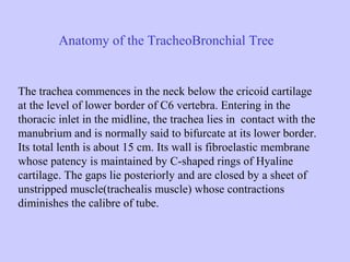
- 1. Anatomy of the TracheoBronchial Tree The trachea commences in the neck below the cricoid cartilage at the level of lower border of C6 vertebra. Entering in the thoracic inlet in the midline, the trachea lies in contact with the manubrium and is normally said to bifurcate at its lower border. Its total lenth is about 15 cm. Its wall is fibroelastic membrane whose patency is maintained by C-shaped rings of Hyaline cartilage. The gaps lie posteriorly and are closed by a sheet of unstripped muscle(trachealis muscle) whose contractions diminishes the calibre of tube.
- 2. The Bronchi are symmetrical, the right being one third wider and a little shorter then left. At the bifurcation an anteroposterior ridge the carina, lies to the left of the midline. The right bronchus slopes more steeply(25 degrees off vertical) then the left(45 degrees off the vertical), so that the foreign bodies of whatever shape are statistically more likely to enter the right bronchus. In the structure the bronchi are identical with the trachea.
- 4. Foreign bodies in the larynx and trachea The maximum incidence in the inhalation of the foreign bodies occurs between the age of one and three years. The most common cause of accidental death in the home in children under 06 years of age , is the inhalation of foreign bodies.
- 6. The peak incidence of inhalation of foreign bodies in early childhood is of course related to the fact that children have the habbit of putting objects into their mouths to determine their texture and taste and to chew on when teething. It is extremely important, therefore when possible to keep objects which might be inhaled out of the reach of small children.
- 7. Types of foreign bodies inhaled 1) 2) 3) 4) 5) 6) 7) 8) 9) 10) 11) 12) 13) 14) 15) 16) Portion of Nut. Whole Nut(peanut, bean, channa, cashio, pistachio). Food. Carrots. Popcorn. Fruit(stem,seed,peel). Bone. Plastic(whistle). Metal. Tooth. Stone. Bead. Balloon. Crayon. Wood. Paper.
- 8. History In most cases of inhaled foreign body, there is a definite history of chocking followed by paroxysmal coughing which then subsides. After the initial paroxysm of coughing the tracheobroncheal mucosa becomes tolerant of the foreign body and coughing ceases. This feature is often responsible for delays in diagnosis.other symptomps are -history of wheeze. -Unexplained persistent fever. - unsuspected foreign body at routine Endoscopy for other reasons. -Acute respiratory distress. -Pain at the root of neck. -Sharp and long standing oesophageal foreign bodies may produce a fistula between the oesophagus and the trachea and cause
- 9. Clinical examination of foreign bodies of Larynx and Trachea. -General examination of child is essential, Respiratory distress or cyanosis demands immediate action. -If there is a change in child’s cry or if the cry becomes hoarse or stridulous, a laryngeal foreign body should be suspected. -Excessive salivation may also occur. -An audible click may be heard due to movement of foreign body up and down the trachea. -A fluttering noise may also be heard. -A unilateral expiratory wheeze and reduced air entry may indicate foreign body in the bronchus. -If foreign body is not removed within 24 hours, pneumonic signs occurs. -Dry vegetable foreign bodies e.g: bean cause very rapid obstructive changes. -Atelectasis of occluded segment of the lung occurs. -The presence of florid granulation tissue around the inhaled foreign body may also cause haemoptysis.
- 10. Radiological findings. X-ray neck should be taken with the neck extended with anteroposterior and lateral views. Anteroposterior view of the chest in inspiration and expiration should be obtained. Obstructive emphysema is produced by a valvular obstruction to the expiratory air stream due the presence of the foreign body in the lumen of air passage.
- 15. Management of foreign bodies of Larynx and Trachea. If foreign body in the respiratory tract is suspected or diagnosed radiologically, endoscopic examination and removal under general anesthesia is the method of choice. In case of Laryngeal or large tracheal foreign bodies, this should be performed as an emergency procedure. If the airway is compromised, the endoscopy must be performed immediately with the facilities for performing an emergency tracheostomy. Large tracheal foreign bodies may have to be delivered through a tracheostomy.
- 16. Laryngeal foreign bodies are removed by direct laryngoscopy. Tracheal and Bronchial foreign bodies are best removed using a rigid bronchoscope. In the absence of respiratory distress the operation should be performed as an elective procedure by the Surgical team that are used to working together in their accustomed operating theatre.
- 17. In the rare event if surgeon is unable to remove a foreign body endoscopically, inspite of satisfactory operating conditions, it must be removed by thoracotomy and bronchotomy.