Downloaded 1,217 times
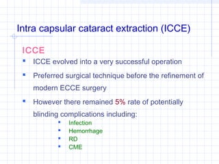


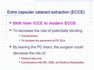


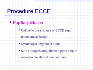


Intra capsular cataract extraction (ICCE) was an early surgical technique for removing cataracts but had high complication rates. Extra capsular cataract extraction (ECCE) was developed to address these issues by leaving the posterior capsule intact. ECCE became the standard technique with improvements in microscopes, irrigation/aspiration systems, and intraocular lenses. Phacoemulsification, an ECCE variant using ultrasonic fragmentation, further reduced complications through smaller incisions allowing faster recovery.


