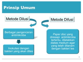
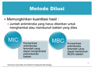
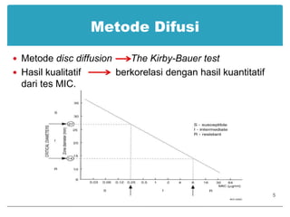
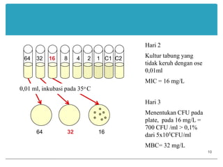
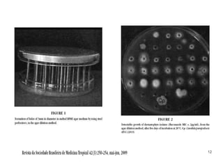
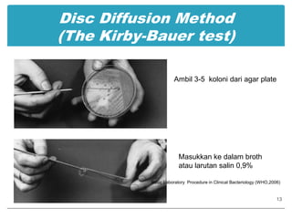
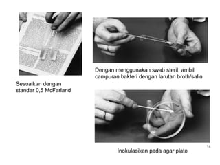
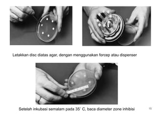
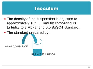
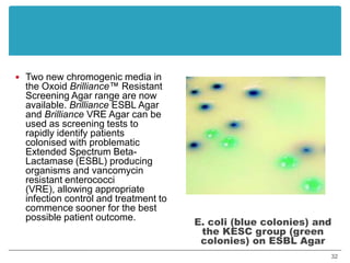
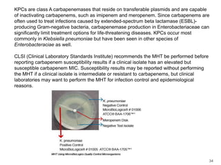
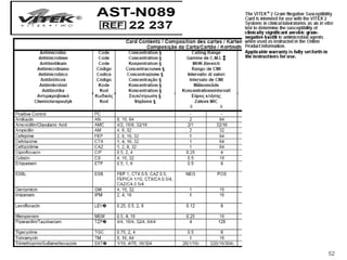
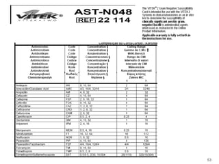
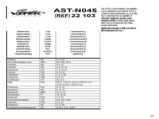
Tutor MikrobiologiTES KEPEKAAN ANTIMIKROBASri Kartika Sari / Jusak NugrahaKamis, 11 Maret 20101
PENDAHULUANTujuan tes kepekaan antimikroba2
Prinsip UmumMetode DilusiMetode Difusi3
Metode DilusiMemungkinkan kuantitasi hasil :Jumlah antimikroba yang harus diberikan untuk menghambat atau membunuh bakteri yang dites.4Koneman’s Color Atlas and Textbook of Diagnostic Microbiology
Metode DifusiMetode disc diffusionThe Kirby-Bauer testHasil kualitatif             berkorelasi dengan hasil kuantitatif dari tes MIC.5
Persiapan inokulumBakteri diambil dari Pure CultureJumlah bakteri yang akan diinokulasi          standar 0,5 McFarland.6
Pemilihan antimikrobaJenis obat yang rutin digunakan untuk tes kepekaan :7
8Basic Laboratory Procedure in Clinical Bacteriology (WHO,2006)
Broth Dilution methode 128   64    32    16     8     4     2   C1  C2 64    32    16      8      4     2    1   C1  C2 Hari 11 ml organisme (1x10⁶ CFU/ml) ditambah 1ml  konsentrasi broth dan antimikroba (mg/L)C1 : tanpa antibiotikC2: tanpa organismeKonsentrasi organisme 5x10⁵CFU/mlInkubasi pada 35oC9
64    32    16      8      4     2    1   C1  C2 64                32                16Hari 2Kultur tabung yang tidak keruh dengan ose 0,01mlMIC = 16 mg/LHari 3Menentukan CFU pada plate, pada 16 mg/L = 700 CFU /ml > 0,1% dari 5x10⁵CFU/mlMBC= 32 mg/L0,01 ml, inkubasi pada 35o C10
Agar Dilution MethodeProsedur :64 μg/ml16 μg/ml32 μg/ml1132μg/ml
12
Disc Diffusion Method(The Kirby-Bauer test)Ambil 3-5  koloni dari agar plateMasukkan ke dalam broth atau larutan salin 0,9%Basic Laboratory  Procedure in Clinical Bacteriology (WHO,2006)13
Dengan menggunakan swab steril, ambil campuran bakteri dengan larutan broth/salin Sesuaikan dengan standar 0,5 McFarland14Inokulasikan pada agar plate
Letakkan disc diatas agar, dengan menggunakan forcep atau dispenserSetelah inkubasi semalam pada 35˚ C, baca diameter zone inhibisi15
Pembacaan hasilCatatan :Pada tes oxacillin dengan Staphylococci dan vancomycin dengan Enterococci         bila dilihat dengan penyinaran/alat pembesar, koloni kecil diabaikan.Swarming Proteus spp diabaikanAdanya pertumbuhan tipis pada zone sulfonamide, trimetoprim-sulfametokzasol diabaikan,          ukur tepi pertumbuhan tebal.Adanya pertumbuhan koloni yang besar dalam zone inhibisi menunjukkan mixed culture        ulang16
17Basic Laboratory  Procedure in Clinical Bacteriology (WHO,2006)
Faktor yang mempengaruhi zone inhibisi :Media pertumbuhanpHTemperaturCation consentrationAtmosphereInokulumAntibiotic disc18
EtestEtest        strip berisi gradien konsentrasi antimikrobaPrinsip :Standar inokulum bakteri diinokulasikan pada Muller Hinton Agar. Strip Etest diletakkan diatas permukaan agar, setelah inkubasi akan terbentuk zone inhibisi.MIC dibaca pada pertumbuhan yang menyilang strip Etest.19
Prosedur :20
21
PenutupTes kepekaan antimikroba merupakan hal yang sangat penting untuk terapi dan menurunkan prevalensi resistensi kuman.22
TERIMAKASIH23
24
25
InoculumThe density of the suspension is adjusted to approximately 108 CFU/ml by comparing its turbidity to a McFarland 0,5 BaSO4 standard.The standard prepared by :0,5 ml  0,048 M BaCl299,5 ml 0,36 N H2SO426
http: //en.wikipedia.org./wiki/file: antiboitic _action.png27
28
29
30
31
Two new chromogenic media in the Oxoid Brilliance™ Resistant Screening Agar range are now available. Brilliance ESBL Agar and Brilliance VRE Agar can be used as screening tests to rapidly identify patients colonised with problematic Extended Spectrum Beta-Lactamase (ESBL) producing organisms and vancomycin resistant enterococci (VRE), allowing appropriate infection control and treatment to commence sooner for the best possible patient outcome.E. coli (blue colonies) and the KESC group (green colonies) on ESBL Agar32
Brilliance VRE Agar is a chromogenic screening plate for the detection of VRE. Following direct inoculation from faecal sample, swab, isolate or suspension, the medium provides presumptive identification of Enterococcusfaecium and Enterococcusfaecaliswithin 24 hours. Differentiation of vancomycin resistant E. faecium from E. faecalis is achieved through the inclusion of two chromogens that are targeted by specific enzymes. The action of these enzymes on the chromogens results in a build-up of colour within the colony. The colour produced depends on which enzymes the organisms possess. Additional antibiotics are present in combination with vancomycin, to suppress the growth of competing flora including E. gallinarum and E. casseliflavus, both of which are intrinsically resistant to vancomycin.Rapidmicrobiology.com33
KPCs are class A carbapenemases that reside on transferable plasmids and are capable of inactivating carbapenems, such as imipenem and meropenem. Since carbapenems are often used to treat infections caused by extended-spectrum beta lactamase (ESBL)-producing Gram-negative bacteria, carbapenemase production in Enterobacteriaceae can significantly limit treatment options for life-threatening diseases. KPCs occur most commonly in Klebsiellapneumoniae but have been seen in other species of Enterobacteraciae as well.CLSI (Clinical Laboratory Standards Institute) recommends the MHT be performed before reporting carbapenem susceptibility results if a clinical isolate has an elevated but susceptible carbapenem MIC. Susceptibility results may be reported without performing the MHT if a clinical isolate is intermediate or resistant to carbapenems, but clinical laboratories may want to perform the MHT for infection control and epidemiological reasons.34
35
36
37
38
39
40
41
In the Stokes controlled sensitivity test, a control organism is inoculated on part of a plate and the test organism is plated on the remainder. Disks are placed at the interface and the zones of inhibition are compared. The use of a sensitive control shows that the antibiotic is active, so that if the test organism grows up to the disk it may safely be assumed that the test organism is resistant to that drug.  Stokes' sensitivity testThe test bacterium in the diagram is susceptible to drug "x" but resistant to drug "y". The disk containing drug "y" contains active antibiotic as shown by the zone of inhibition it causes in the control bacteriumhttp://.www.bmb.leed.ac.uk/mbiol...sts.html42
  The VITEK® 2 compact is an automated bacterial identification and antibiotic susceptibility testing (ID/AST) system. Designed for small to midsize microbiology laboratories, the VITEK 2 compact offers an extensive ID and AST database to improve laboratory efficiency.      Using multi-spectrum colorimetric and transmittance optic systems, the VITEK 2 compact detects changes in organism growth under various media and antibiotic concentrations. The instrument’s advanced expert system interprets the antibiotic-resistance patterns, validates the results, and reports the resistance phenotype useful to patient treatment and infection control. The system offers rapid results, providing increased productivity and improved workflow. bioMerieux43
Antimicrobial Susceptibility Test (Broth Dilution) This test used to test all the ABC isolates. This picture shows the most accurate method of determining the antimicrobial susceptibility of GBS. This test is called the broth dilution antimicrobial susceptibility test. It is standardized by the the National Committee on Clinical Laboratory Standards (NCCLS). The picture above shows a microtiter plate with 96 wells. There are 8 rows of wells, labeled A through G. Each row of wells contains one antibiotic. In each row there are 12 wells. Each well contains a different concentration of the specific antibiotic, usually doubling dilutions. In this case, row A contains penicillin starting at the right at 16 ?g/ml and reducing to 0.008 well 12. Row B contains ampicillin starting in well 1 with 16 ?g/ml and reducing to 0.008 ?g/ml to well 12 in the same row. The other rows contain different antibiotics. All 96 wells are inoculated at the same time and the panel is incubated overnight (20-24h). The next day the wells are examined for growth. If there is growth in the well, the antibiotic did not inhibit the bacteria. If you take a close look in row A you will see growth (a small white button at the bottom of the well) only in well 12. In row B you see growth in both well 11 and 12. This means the minimum inhibitory concentration (MIC) of penicillin is 0.016 ?g/ml and the MIC for ampicillin is 0.032 ?g/ml. The MIC’s of these antimicrobials are considered susceptible for,GBS. Erythromycin is shown is row F. The concentration of erythromycin starts at 32 ?g/ml in well 1 and goes to 0.016 ?g/ml in well 12. You can see that there is growth in wells 12 to 4, meaning the MIC of erythromycin to this strain is 8 ?g/ml. This MIC is considered resistant for GBS. Since this is a standardized test results can be compared world-wide if the standard procedures are used. Consult the NCCLS manual for reporting interpretive standards (sensitive, intermediate, and resistance).NCCLS Performance Standards for Antimicrobial Susceptibility Testing; Twelfth Informational Supplement: M100-12 (2002) M2-A7 and M7-A5. 44
Testing of streptococcal isolates with erythromycin and clindamycin disks applied close together can often yield phenotypic information, although it is not always possible to differentiate between phenotypes using this method. See J. Clin. Microbiol 2001;39:1311-1315 for details of this method for phenotypic analysis of S. pneumoniae. The figures below are taken from that paper, with some major modifications made to simplify this discussion; see the paper for the real and more complicated story.                         45
DensitometerDescriptionA compact and efficient benchtop densitometer, the DEN-1 is designed to measure turbidity in the range of 0.3 to 5.0 McFarland units with a small standard deviation. If required, it can deliver a wider measurement range (up to 15.0 McFarland units) but with a greater standard deviation. Typical applications of the DEN-1 Densitometer includes: Determining concentration of cells (bactorial and yeast) in the fermentation process Detecting the susceptibility of micro-organisms to antibiotics Identifying micro-organisms with various test systems Measuring optical density at fixed wavelength46
47
48
49
50
51
52
53
54

Tpibaru8

  • 1.
    Tutor MikrobiologiTES KEPEKAANANTIMIKROBASri Kartika Sari / Jusak NugrahaKamis, 11 Maret 20101
  • 2.
  • 3.
  • 4.
    Metode DilusiMemungkinkan kuantitasihasil :Jumlah antimikroba yang harus diberikan untuk menghambat atau membunuh bakteri yang dites.4Koneman’s Color Atlas and Textbook of Diagnostic Microbiology
  • 5.
    Metode DifusiMetode discdiffusionThe Kirby-Bauer testHasil kualitatif berkorelasi dengan hasil kuantitatif dari tes MIC.5
  • 6.
    Persiapan inokulumBakteri diambildari Pure CultureJumlah bakteri yang akan diinokulasi standar 0,5 McFarland.6
  • 7.
    Pemilihan antimikrobaJenis obatyang rutin digunakan untuk tes kepekaan :7
  • 8.
    8Basic Laboratory Procedurein Clinical Bacteriology (WHO,2006)
  • 9.
    Broth Dilution methode128 64 32 16 8 4 2 C1 C2 64 32 16 8 4 2 1 C1 C2 Hari 11 ml organisme (1x10⁶ CFU/ml) ditambah 1ml konsentrasi broth dan antimikroba (mg/L)C1 : tanpa antibiotikC2: tanpa organismeKonsentrasi organisme 5x10⁵CFU/mlInkubasi pada 35oC9
  • 10.
    64 32 16 8 4 2 1 C1 C2 64 32 16Hari 2Kultur tabung yang tidak keruh dengan ose 0,01mlMIC = 16 mg/LHari 3Menentukan CFU pada plate, pada 16 mg/L = 700 CFU /ml > 0,1% dari 5x10⁵CFU/mlMBC= 32 mg/L0,01 ml, inkubasi pada 35o C10
  • 11.
    Agar Dilution MethodeProsedur:64 μg/ml16 μg/ml32 μg/ml1132μg/ml
  • 12.
  • 13.
    Disc Diffusion Method(TheKirby-Bauer test)Ambil 3-5 koloni dari agar plateMasukkan ke dalam broth atau larutan salin 0,9%Basic Laboratory Procedure in Clinical Bacteriology (WHO,2006)13
  • 14.
    Dengan menggunakan swabsteril, ambil campuran bakteri dengan larutan broth/salin Sesuaikan dengan standar 0,5 McFarland14Inokulasikan pada agar plate
  • 15.
    Letakkan disc diatasagar, dengan menggunakan forcep atau dispenserSetelah inkubasi semalam pada 35˚ C, baca diameter zone inhibisi15
  • 16.
    Pembacaan hasilCatatan :Padates oxacillin dengan Staphylococci dan vancomycin dengan Enterococci bila dilihat dengan penyinaran/alat pembesar, koloni kecil diabaikan.Swarming Proteus spp diabaikanAdanya pertumbuhan tipis pada zone sulfonamide, trimetoprim-sulfametokzasol diabaikan, ukur tepi pertumbuhan tebal.Adanya pertumbuhan koloni yang besar dalam zone inhibisi menunjukkan mixed culture ulang16
  • 17.
    17Basic Laboratory Procedure in Clinical Bacteriology (WHO,2006)
  • 18.
    Faktor yang mempengaruhizone inhibisi :Media pertumbuhanpHTemperaturCation consentrationAtmosphereInokulumAntibiotic disc18
  • 19.
    EtestEtest strip berisi gradien konsentrasi antimikrobaPrinsip :Standar inokulum bakteri diinokulasikan pada Muller Hinton Agar. Strip Etest diletakkan diatas permukaan agar, setelah inkubasi akan terbentuk zone inhibisi.MIC dibaca pada pertumbuhan yang menyilang strip Etest.19
  • 20.
  • 21.
  • 22.
    PenutupTes kepekaan antimikrobamerupakan hal yang sangat penting untuk terapi dan menurunkan prevalensi resistensi kuman.22
  • 23.
  • 24.
  • 25.
  • 26.
    InoculumThe density ofthe suspension is adjusted to approximately 108 CFU/ml by comparing its turbidity to a McFarland 0,5 BaSO4 standard.The standard prepared by :0,5 ml 0,048 M BaCl299,5 ml 0,36 N H2SO426
  • 27.
  • 28.
  • 29.
  • 30.
  • 31.
  • 32.
    Two new chromogenicmedia in the Oxoid Brilliance™ Resistant Screening Agar range are now available. Brilliance ESBL Agar and Brilliance VRE Agar can be used as screening tests to rapidly identify patients colonised with problematic Extended Spectrum Beta-Lactamase (ESBL) producing organisms and vancomycin resistant enterococci (VRE), allowing appropriate infection control and treatment to commence sooner for the best possible patient outcome.E. coli (blue colonies) and the KESC group (green colonies) on ESBL Agar32
  • 33.
    Brilliance VRE Agaris a chromogenic screening plate for the detection of VRE. Following direct inoculation from faecal sample, swab, isolate or suspension, the medium provides presumptive identification of Enterococcusfaecium and Enterococcusfaecaliswithin 24 hours. Differentiation of vancomycin resistant E. faecium from E. faecalis is achieved through the inclusion of two chromogens that are targeted by specific enzymes. The action of these enzymes on the chromogens results in a build-up of colour within the colony. The colour produced depends on which enzymes the organisms possess. Additional antibiotics are present in combination with vancomycin, to suppress the growth of competing flora including E. gallinarum and E. casseliflavus, both of which are intrinsically resistant to vancomycin.Rapidmicrobiology.com33
  • 34.
    KPCs are classA carbapenemases that reside on transferable plasmids and are capable of inactivating carbapenems, such as imipenem and meropenem. Since carbapenems are often used to treat infections caused by extended-spectrum beta lactamase (ESBL)-producing Gram-negative bacteria, carbapenemase production in Enterobacteriaceae can significantly limit treatment options for life-threatening diseases. KPCs occur most commonly in Klebsiellapneumoniae but have been seen in other species of Enterobacteraciae as well.CLSI (Clinical Laboratory Standards Institute) recommends the MHT be performed before reporting carbapenem susceptibility results if a clinical isolate has an elevated but susceptible carbapenem MIC. Susceptibility results may be reported without performing the MHT if a clinical isolate is intermediate or resistant to carbapenems, but clinical laboratories may want to perform the MHT for infection control and epidemiological reasons.34
  • 35.
  • 36.
  • 37.
  • 38.
  • 39.
  • 40.
  • 41.
  • 42.
    In the Stokescontrolled sensitivity test, a control organism is inoculated on part of a plate and the test organism is plated on the remainder. Disks are placed at the interface and the zones of inhibition are compared. The use of a sensitive control shows that the antibiotic is active, so that if the test organism grows up to the disk it may safely be assumed that the test organism is resistant to that drug.  Stokes' sensitivity testThe test bacterium in the diagram is susceptible to drug "x" but resistant to drug "y". The disk containing drug "y" contains active antibiotic as shown by the zone of inhibition it causes in the control bacteriumhttp://.www.bmb.leed.ac.uk/mbiol...sts.html42
  • 43.
    TheVITEK® 2 compact is an automated bacterial identification and antibiotic susceptibility testing (ID/AST) system. Designed for small to midsize microbiology laboratories, the VITEK 2 compact offers an extensive ID and AST database to improve laboratory efficiency.      Using multi-spectrum colorimetric and transmittance optic systems, the VITEK 2 compact detects changes in organism growth under various media and antibiotic concentrations. The instrument’s advanced expert system interprets the antibiotic-resistance patterns, validates the results, and reports the resistance phenotype useful to patient treatment and infection control. The system offers rapid results, providing increased productivity and improved workflow. bioMerieux43
  • 44.
    Antimicrobial Susceptibility Test(Broth Dilution) This test used to test all the ABC isolates. This picture shows the most accurate method of determining the antimicrobial susceptibility of GBS. This test is called the broth dilution antimicrobial susceptibility test. It is standardized by the the National Committee on Clinical Laboratory Standards (NCCLS). The picture above shows a microtiter plate with 96 wells. There are 8 rows of wells, labeled A through G. Each row of wells contains one antibiotic. In each row there are 12 wells. Each well contains a different concentration of the specific antibiotic, usually doubling dilutions. In this case, row A contains penicillin starting at the right at 16 ?g/ml and reducing to 0.008 well 12. Row B contains ampicillin starting in well 1 with 16 ?g/ml and reducing to 0.008 ?g/ml to well 12 in the same row. The other rows contain different antibiotics. All 96 wells are inoculated at the same time and the panel is incubated overnight (20-24h). The next day the wells are examined for growth. If there is growth in the well, the antibiotic did not inhibit the bacteria. If you take a close look in row A you will see growth (a small white button at the bottom of the well) only in well 12. In row B you see growth in both well 11 and 12. This means the minimum inhibitory concentration (MIC) of penicillin is 0.016 ?g/ml and the MIC for ampicillin is 0.032 ?g/ml. The MIC’s of these antimicrobials are considered susceptible for,GBS. Erythromycin is shown is row F. The concentration of erythromycin starts at 32 ?g/ml in well 1 and goes to 0.016 ?g/ml in well 12. You can see that there is growth in wells 12 to 4, meaning the MIC of erythromycin to this strain is 8 ?g/ml. This MIC is considered resistant for GBS. Since this is a standardized test results can be compared world-wide if the standard procedures are used. Consult the NCCLS manual for reporting interpretive standards (sensitive, intermediate, and resistance).NCCLS Performance Standards for Antimicrobial Susceptibility Testing; Twelfth Informational Supplement: M100-12 (2002) M2-A7 and M7-A5. 44
  • 45.
    Testing of streptococcalisolates with erythromycin and clindamycin disks applied close together can often yield phenotypic information, although it is not always possible to differentiate between phenotypes using this method. See J. Clin. Microbiol 2001;39:1311-1315 for details of this method for phenotypic analysis of S. pneumoniae. The figures below are taken from that paper, with some major modifications made to simplify this discussion; see the paper for the real and more complicated story.                        45
  • 46.
    DensitometerDescriptionA compact andefficient benchtop densitometer, the DEN-1 is designed to measure turbidity in the range of 0.3 to 5.0 McFarland units with a small standard deviation. If required, it can deliver a wider measurement range (up to 15.0 McFarland units) but with a greater standard deviation. Typical applications of the DEN-1 Densitometer includes: Determining concentration of cells (bactorial and yeast) in the fermentation process Detecting the susceptibility of micro-organisms to antibiotics Identifying micro-organisms with various test systems Measuring optical density at fixed wavelength46
  • 47.
  • 48.
  • 49.
  • 50.
  • 51.
  • 52.
  • 53.
  • 54.