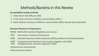
Antibiotic
Susceptibility Testing
for Aerobic Bacteria
2023 Update
Margie A. Morgan, PhD,
MT(ASCP), D(ABMM)
Antibiotic Classes
• Beta Lactams –contain a beta lactam
ring in molecular structure of antibiotic
• Penicillins
• Penicillin
• Amoxicillin
• Ampicillin
• Amp/Clavulanate
• Amp/Sulbactam
• Anti-Pseudomonal Penicillins
• Piperacillin/Tazobactam
• Ticarcillin/Clavulanate
• Anti- Staphylococcal Penicillins
• Nafcillin
• Oxacillin
• Cloxacillin
• Dicloxacillin
Antibiotic Classes (2)
• Beta lactam antibiotics
• Carbapenems
• Imipenem
• Meropenem
• Ertapenem
• Doripenem
• Monobactam
• Aztreonam
• Cephalosporins
• First generation
• Cefazolin
• Second generation
• Cefotetan
• Cefoxitin Cefuroxime
• Third generation
• Ceftriaxone
• Ceftazidime Cefpodoxime
• Fourth generation
• Cefepime Ceftaroline
Antibiotic Classes (3)
• Fluoroquinolones
• Ciprofloxacin
• Levofloxacin
• Moxifloxacin
• Aminoglycosides
• Gentamicin
• Tobramycin
• Amikacin
• Sulfonamide
• Trimethoprim/Sulfamethozaxole
• Macrolides
• Azithromycin
• Clarithromycin
• Erythromycin
• Tetracyclines
• Tetracycline
• Minocycline
• Doxycycline
• Lincosamide
• Clindamycin
• Nitrofurans
• Nitrofurantoin
Newer antibiotics for Multi-drug resistant Gram-negative
rod therapy
• Combination Beta-lactams
• Ceftolozane-Tazobactam
• Ceftazidime-Avibactam
• Imipenem-relebactam
• Meropenem-vaborbactam
• Siderophore Beta-lactam
• Cefiderocol
Major mechanisms of antibiotic resistance
•Enzymatic cleavage leads to inactivation of an antibiotic
• Active Beta lactamases and amino-glycoside modifying enzymes cleave and destroy an
antibiotic, such as CTX-M beta lactamase enzyme of ESBL producing gram negative rod
destroying Cefotaxime. Can be detected using molecular analysis.
•Altered receptors and binding proteins prevent attachment of
antibiotics to the bacterial surface so antibiotics cannot penetrate
• Penicillin binding proteins (PBPs). Can be detected using molecular assays.
• Mechanism for Strep pneumoniae resistance to penicillin and MRSA resistance to methicillin
•Altered permeability/influx and efflux pumps stop passage of
antibiotic through cell membrane porins – gram negative bacilli
• Pseudomonas aeruginosa resistance to amino-glycosides
• Resistance best detected by susceptibility testing using proper breakpoints.
•Bypass of a metabolic block/metabolic block imposed by antibiotic
• Enterococcus resistance to TMP/SXT
How are rules established for susceptibility testing?
CLSI – Clinical and Laboratory Standards Institute publish yearly
approved standards for the proper testing and reporting of
susceptibility results within the United States
1. Documents advising laboratories appropriate antibiotics to test
for each organism and infection site
2. Proper testing procedures for quality control of media,
antibiotics, and automated instruments
3. Charts outlining interpretation of susceptibility results
Methods/Bacteria in this Review
Susceptibility testing methods
1. Kirby Bauer disk diffusion (KB)
2. E Test Strip minimum inhibitory concentration (MIC )
3. Broth dilution minimum inhibitory concentration (MIC) manual and automated
Resistant Bacteria of Importance:
•MRSA Methicillin resistant Staphylococcus aureus
•VRE Vancomycin resistant Enterococcus
•ESBL Extended Spectrum Beta Lactamase (ESBL) producers (Enterobacterales)
•CRE/ Carbapenamase Resistant Enteric gram-negative rods (CRE)
CRO Carbapenamase resistant organisms (CRO)
•Streptococcus pneumoniae
•Neisseria gonorrhoeae
Quality Control for Susceptibility Testing
Verification: Before testing patient organisms, laboratories must correctly test
multiple QC organisms using the test method to assure you are performing tests
correctly.
Use ATCC strains of bacteria (American Type Culture Collection) with known activity
for testing QC. If QC strains are tested and within appropriate values, you can
develop a quality control plan for weekly testing on all antibiotic cards, disks, media,
and automated systems.
• Data must be recorded and reviewed monthly by supervisor
•If weekly QC results are out of control:
• Immediately repeat the test that failed QC / inform supervisor
• If repeat is OK and you know the source of your error – document and continue routine testing
• If repeat is NOT OK or if you do not know why the test failed – must investigate/document/ repeat
5 times to go back to routine testing. All repeat tests must be in control
• Must not report results of an out-of-control antibiotic until back in control
Preparation of Bacteria for Susceptibility Methods
• Susceptibility methods test for stasis not killing of bacteria
• Pure culture of one organism is tested / never test a mixed culture
• Log phase growth of bacteria grown on agar for 16-24 hours
• Standardized suspension of bacteria used for testing. This is
accomplished by using a O.5 McFarland standard, a barium sulfate
solution prepared in an acid pH that equals the turbidity of 10⁸
bacteria/ml
•Alternative method – use spectrophotometer measurement of the
suspension (more precise)
•Read tests after incubation at 35 °C in room air, or CO² for fastidious
bacterial species. Incubation usually 16- 24 hours
•Must control inoculum amount and incubation time or false resistance or
susceptible results could occur
Agar Disk Diffusion (Kirby Bauer)
Qualitative Susceptibility Method
•Use Mueller Hinton agar with or without addition of blood
• 150 mm plate diameter
• 4mm in depth
• Agar specifically balanced in Ca+ and Mg+,
• if the ions are too high % amino-glycosides test falsely resistant,
• if the ions too low % falsely susceptible amino-glycoside results
•Streak bacteria suspension on plate with cotton tipped
swab
•Apply up to 12 antibiotic disks onto plate/ each disk is a
different antibiotic. Paper disks 6mm in diameter
•Incubate for 16-24 hrs at 35*C
•Measure zone of diameter of inhibition of growth (mm) –
CLSI charts have interpretive guidelines for sensitive,
intermediate and resistant results
•Not all antibiotic / organism combinations can be tested
using disk diffusion for results will not be reliable
Kirby Bauer Disk Dispenser
for application of disks onto
agar surface
Each cartridge cylinder
contains a separate antibiotic
disk
Any Growth of colonies inside
a zone is considered resistant to
that antibiotic
Measure the diameter
of the zone of inhibition
Watch out for double
zones /possibly contaminated
E Test
Quantitative MIC Susceptibility Method
•Calibrated plastic strip impregnated with
concentration gradient (mcg/ml) of one antibiotic
placed on a lawn of bacteria (10⁸/ml)
•Diffusion gradient created as antibiotic diffuses into
agar in an elliptical shape (E)
•MIC (minimum inhibitory concentration) of
antibiotic is where the ellipse ends on the plastic
strip (blue arrow)
•Good method for slower growing fastidious
organisms that do not produce consistent results on
Kirby Bauer disk diffusion testing
.
Broth Dilution Minimum Inhibitory Concentration/MIC
Quantitative Susceptibility Method
• Bacteria inoculum: Adjusted to 0.5 McFarland standard then
further diluted to 5x10⁵ organisms /ml in sterile saline or broth
• Suspension of bacteria is inoculated into microtiter trays
containing known 2-fold dilutions (mcg/ml) of individual
antibiotics
• In the plate pictured, each horizontal row is a unique
antibiotic
• Growth produces turbidity in the broth which indicates
resistance to the antibiotic. Some systems include color
indicators to indicate growth (picture)
• Lowest concentration of an antibiotic with no growth is the
MIC value.
• CLSI charts have interpretive guidelines for sensitive,
intermediate, and resistant MIC values
MIC Related Definitions from the Past
MIC = Lowest concentration of an antibiotic inhibiting growth
MBC = Lowest concentration of an antibiotic killing the organism (99.9%)
• Antibiotic tolerance – calculated by the ratio of MBC/MIC
• If the ratio is >=32 this antibiotic/organism combination does not kill the organism
• MBC = 16 MIC= 8 16/8= 2 No Tolerance, bacteria killed
• MBC = 128 MIC = 8 128/2 = 64 Tolerance, the bacteria is damaged but not
killed. This could lead to lingering infections that continue to damage tissue
and possibly lead to antimicrobial resistance, do not treat with this antibiotic
Becton-Coulter MicroScan walkaway
AST system
BD Phoenix AST system
Biomerieux Vitek2 AST
Most used Automated Identification &
Susceptibility Testing Systems (AST)
Broth MIC two-fold dilutions
Calculated MIC value Broth MIC two-fold dilution MIC
Methicillin Resistant Staphylococcus aureus (MRSA)
•OLD WAY/ Test for oxacillin (OX) resistance to detect methicillin resistance – OX is
more stable for testing in the laboratory than methicillin
•If OX is resistant - S. aureus is reported as MRSA
•Improved way – Test antibiotic cefoxitin, if cefoxitin is resistant report as MRSA
•Cefoxitin testing is more sensitive than OX and the preferred way to detect MRSA
•Cephalosporin antibiotics are reported resistant for a MRSA and should not used
for therapy
•Resistance mechanism penicillin binding proteins (PBPs/PBP2a)
• PBPs are proteins that bind penicillin and related antibiotics and prevent their action
• The binding proteins prevent antibiotic disruption of the peptidoglycan synthesis in the cell wall
• PBPs are produced most often by the mecA gene
• Note: New emerging strain of MRSA produced by mecC gene
1. Cefoxitin KB disk testing is a sensitive
screen for MRSA using organism growth.
2. Immunochromatographic membrane
assays on Staph aureus colony. This
method uses sensitive recombinant
monoclonal antibody fragments to detect
the PBP2a protein directly from Staph
aureus isolates. The antibody fragments
are immobilized onto a nitrocellulose
membrane strip and form distinct lines if the
PBP2a is present (MRSA)
Methods to Detect MRSA
Cefoxitin = susceptible
Staph aureus
Cefoxitin = resistant
MRSA
3. Molecular assays are
also available to detect
mecA and mecC gene
resistance markers. This
can greatly decrease the
time to detection of MRSA.
Clindamycin Induction Test –
The D test
•The D test determines if Staph aureus is truly susceptible to
Clindamycin. It is more reliable than just testing Clindamycin
resistance by KB or MIC susceptibility methods.
•During antimicrobial therapy, Staph aureus isolates resistant to
Erythromycin possess enzymes that can be induced to make the
S. aureus resistant to Clindamycin, so called inducible resistance
•Clindamycin can be used to treat serious soft tissue infections
with MRSA – so reliable testing is necessary for appropriate
therapy.
D test Negative-
round Clindamycin zone
Kirby Bauer zone around Clindamycin is blunted to
form a “D” if Clindamycin can be induced by
Erythromycin to be resistant – so called INDUCIBLE
RESISTANCE.
Clindamycin should be reported as “resistant by
clindamycin induction” and not used for therapy
The D Test
Clindamycin susceptible
Enterococcus
•All Enterococcus are intrinsically resistant to:
• Cephalosporins
• Clindamycin
• Trimethoprim/sulfamethoxazole
•Synergistic antibiotic therapy can be important for the treatment of serious
Enterococcus infections
• Ampicillin plus Gentamicin is synergistic, that is increased bacterial killing
potential with combination of two antibiotics that kill by different mechanisms
• Particularly important for therapy of Enterococcus infective endocarditis
•Acquired resistance to vancomycin creating “vancomycin resistant enterococcus”
• Plasmid mediated vanA associated with E. faecium resistance
• Plasmid mediated vanB associated with E. faecalis resistance
Organism with intrinsic resistance to select
antibiotics
• Intrinsic resistance is when a bacterial species is naturally resistant to a certain
antibiotic or family of antibiotics, without the need for mutation or gain of
further genes. These antibiotics should never be used to treat infections caused
by that species of bacteria. A few important ones include:
• Proteus mirabilis - intrinsically resistant to Colistin due to epfB gene expression
• Stenotrophomonas maltophilia - intrinsically resistant to carbapenem, beta
lactams, and quinolone antibiotic classes
• Listeria monocytogenes – intrinsically resistant to cephalosporins
• Pseudomonas aeruginosa – intrinsically resistant to
Trimethoprim/sulfamethoxazole
• Enterococcus– intrinsically resistant to cephalosporins and aminoglycosides
Extended Spectrum Beta Lactamase [ESBL]
•Enzymes produced by Enteric gram-negative bacilli
• Beta lactamase enzymes confer resistance to Cephalosporins, Penicillins
and Monobactam (Aztreonam) antibiotics by opening the beta lactam
ring and inactivating the antibiotic
• ESBLs do not attack Cephamycin (cefoxitin, cefotetan) or the Carbapenem
antibiotic classes
• Most common in Escherichia coli, Klebsiella spp and Proteus mirabilis
•Plasmid mediated CTX-M beta lactamases are the most common ESBL
enzymes produced in the US currently, but many more ESBL types can be
found worldwide
•Therapy for ESBL producing gram-negative rods include the Carbapenems:
Imipenem, Meropenem, Doripenem, and Ertapenem
Detecting ESBL in the laboratory
•Phenotypic susceptibility testing, KB, Etest, and MIC testing
• Cephalosporin MIC values and KB zone breakpoints to “not miss”
the detection of ESBL organisms
• Newer MIC breakpoints are one to three doubling dilutions lower
than previously established breakpoints
•Molecular resistance marker testing is necessary to detect the type
of ESBL enzyme present, such as CTX-M
Why all the fuss about ESBLs?
• Gram negative rods (GNRs) with ESBL phenotypes >=10%
and increasing in US
• Limited treatment options for ESBL producers
• Carbapenems most used: meropenem, imipenem, ertapenem
• Risk factors:
• Long hospital stay – particularly in the ICU
• Central lines
• Issues with the intestine
• Long term care facility
• Ventilator assistance
Amp C beta lactamase activity
• AmpC β-lactamases are clinically important cephalosporinases encoded on the
chromosomes of many of the Enterobacteriales and a few other organisms, where
they mediate resistance to cephalothin, cefazolin, cefoxitin, most penicillins, and
β-lactamase inhibitor-β-lactam combinations.
• Transmissible plasmids have acquired genes for AmpC beta lactamase enzymes
and can be passed to a variety of different genera of GNRs
• Klebsiella (Enterobacter) aerogenes and Enterobacter cloacae are two of the
most common GNRs associated with this type of resistance
• Resistance due to plasmid-mediated AmpC enzymes is less common than
extended-spectrum β-lactamase (ESBL) production
• Treatment is successful with Carbapenemase antibiotics
Carbapenemase Producers
•CRE = Carbapenamase producing Enterobacteriales
•CRO= Carbapenamase producing gram-negative bacilli (not Enterobacteriales)
•Carbapenem antibiotics are commonly used antibiotics and have the high-level
activity against multi-drug resistant GNRs
•However, the appearance of carbapenem-hydrolyzing-beta-lactamases which
confer resistance to a broad spectrum of beta lactam antibiotics including
carbapenems – created GNRs resistant to most if not all antibiotics
•Two CREs get the most attention:
• KPC – “Klebsiella pneumoniae carbapenemase” most common in the US
• NDM-1 – New Delhi metallo-beta-lactamase.
• Many others including IMP and VIM resistance markers
•Infections with CRE/CRO producing GNRs associated with a 50% fatality rate
Carbapenem antibiotics (ertapenem, meropenem, imipenem) must
have elevated MICs in the resistance range to declare the
organism a carbapenamase producer. If any Carbapenem is
resistant the gram negative is a declared as CRE/CRO.
Treatment of CRE/CRO is determined by testing antibiotics to
determine the best choice. Choices include:
Polymyxins (Colistin and Polymyxin B)
Cefiderocol
Combination antibiotics:
Ceftazidime/avibactam
Ceftolozane/tazobactam
Meropenem/vabomere
Imipenem/relabactam
Carbapenemase CRE/CRO Testing
Streptococcus pneumoniae and
resistance to Penicillin (PEN)
• Perform a Minimum Inhibitory Concentration (MIC) using Penicillin
E Test or broth microdilution susceptibility method
•Kirby Bauer method should not be used to test Strep pneumoniae
because KB testing could under predict resistance to Penicillin (PCN)
•High level PCN resistance is found in <=10% of Strep pneumoniae in
the US
• If PCN resistant - antibiotic of choice becomes a 3rd gen
Cephalosporin, vancomycin, or a quinolone
Neisseria gonorrhoeae
• Increasing resistance over time
• 1980’s beta lactamase producing strains were detected/ making PCN no longer useful for
therapy
• By 2000, the quinolones were resistant due to the acquisition of mutations that altered the
binding sites, preventing quinolone activity
• Currently Cephalosporins (Ceftriaxone & Cefixime) are the mainstay for therapy in the US,
however resistance to these antibiotics are becoming more common in Asia.
• This is due to production of Penicillin Binding Proteins (PBPs) and over production of
efflux pump mechanisms
• Currently susceptibility testing is not routinely performed in most labs
• Detection of resistant strains in the US is problematic due to reliance on
amplification testing for GC diagnosis that only tests for the presence of GC
genes and do not detect antibiotic resistance markers
• Patients failing therapy should be cultured, and isolates sent to a public health
laboratory for susceptibility testing

Susceptibility 2023

  • 1.
    Antibiotic Susceptibility Testing for AerobicBacteria 2023 Update Margie A. Morgan, PhD, MT(ASCP), D(ABMM)
  • 2.
    Antibiotic Classes • BetaLactams –contain a beta lactam ring in molecular structure of antibiotic • Penicillins • Penicillin • Amoxicillin • Ampicillin • Amp/Clavulanate • Amp/Sulbactam • Anti-Pseudomonal Penicillins • Piperacillin/Tazobactam • Ticarcillin/Clavulanate • Anti- Staphylococcal Penicillins • Nafcillin • Oxacillin • Cloxacillin • Dicloxacillin
  • 3.
    Antibiotic Classes (2) •Beta lactam antibiotics • Carbapenems • Imipenem • Meropenem • Ertapenem • Doripenem • Monobactam • Aztreonam • Cephalosporins • First generation • Cefazolin • Second generation • Cefotetan • Cefoxitin Cefuroxime • Third generation • Ceftriaxone • Ceftazidime Cefpodoxime • Fourth generation • Cefepime Ceftaroline
  • 4.
    Antibiotic Classes (3) •Fluoroquinolones • Ciprofloxacin • Levofloxacin • Moxifloxacin • Aminoglycosides • Gentamicin • Tobramycin • Amikacin • Sulfonamide • Trimethoprim/Sulfamethozaxole • Macrolides • Azithromycin • Clarithromycin • Erythromycin • Tetracyclines • Tetracycline • Minocycline • Doxycycline • Lincosamide • Clindamycin • Nitrofurans • Nitrofurantoin
  • 5.
    Newer antibiotics forMulti-drug resistant Gram-negative rod therapy • Combination Beta-lactams • Ceftolozane-Tazobactam • Ceftazidime-Avibactam • Imipenem-relebactam • Meropenem-vaborbactam • Siderophore Beta-lactam • Cefiderocol
  • 6.
    Major mechanisms ofantibiotic resistance •Enzymatic cleavage leads to inactivation of an antibiotic • Active Beta lactamases and amino-glycoside modifying enzymes cleave and destroy an antibiotic, such as CTX-M beta lactamase enzyme of ESBL producing gram negative rod destroying Cefotaxime. Can be detected using molecular analysis. •Altered receptors and binding proteins prevent attachment of antibiotics to the bacterial surface so antibiotics cannot penetrate • Penicillin binding proteins (PBPs). Can be detected using molecular assays. • Mechanism for Strep pneumoniae resistance to penicillin and MRSA resistance to methicillin •Altered permeability/influx and efflux pumps stop passage of antibiotic through cell membrane porins – gram negative bacilli • Pseudomonas aeruginosa resistance to amino-glycosides • Resistance best detected by susceptibility testing using proper breakpoints. •Bypass of a metabolic block/metabolic block imposed by antibiotic • Enterococcus resistance to TMP/SXT
  • 7.
    How are rulesestablished for susceptibility testing? CLSI – Clinical and Laboratory Standards Institute publish yearly approved standards for the proper testing and reporting of susceptibility results within the United States 1. Documents advising laboratories appropriate antibiotics to test for each organism and infection site 2. Proper testing procedures for quality control of media, antibiotics, and automated instruments 3. Charts outlining interpretation of susceptibility results
  • 8.
    Methods/Bacteria in thisReview Susceptibility testing methods 1. Kirby Bauer disk diffusion (KB) 2. E Test Strip minimum inhibitory concentration (MIC ) 3. Broth dilution minimum inhibitory concentration (MIC) manual and automated Resistant Bacteria of Importance: •MRSA Methicillin resistant Staphylococcus aureus •VRE Vancomycin resistant Enterococcus •ESBL Extended Spectrum Beta Lactamase (ESBL) producers (Enterobacterales) •CRE/ Carbapenamase Resistant Enteric gram-negative rods (CRE) CRO Carbapenamase resistant organisms (CRO) •Streptococcus pneumoniae •Neisseria gonorrhoeae
  • 9.
    Quality Control forSusceptibility Testing Verification: Before testing patient organisms, laboratories must correctly test multiple QC organisms using the test method to assure you are performing tests correctly. Use ATCC strains of bacteria (American Type Culture Collection) with known activity for testing QC. If QC strains are tested and within appropriate values, you can develop a quality control plan for weekly testing on all antibiotic cards, disks, media, and automated systems. • Data must be recorded and reviewed monthly by supervisor •If weekly QC results are out of control: • Immediately repeat the test that failed QC / inform supervisor • If repeat is OK and you know the source of your error – document and continue routine testing • If repeat is NOT OK or if you do not know why the test failed – must investigate/document/ repeat 5 times to go back to routine testing. All repeat tests must be in control • Must not report results of an out-of-control antibiotic until back in control
  • 10.
    Preparation of Bacteriafor Susceptibility Methods • Susceptibility methods test for stasis not killing of bacteria • Pure culture of one organism is tested / never test a mixed culture • Log phase growth of bacteria grown on agar for 16-24 hours • Standardized suspension of bacteria used for testing. This is accomplished by using a O.5 McFarland standard, a barium sulfate solution prepared in an acid pH that equals the turbidity of 10⁸ bacteria/ml •Alternative method – use spectrophotometer measurement of the suspension (more precise) •Read tests after incubation at 35 °C in room air, or CO² for fastidious bacterial species. Incubation usually 16- 24 hours •Must control inoculum amount and incubation time or false resistance or susceptible results could occur
  • 11.
    Agar Disk Diffusion(Kirby Bauer) Qualitative Susceptibility Method •Use Mueller Hinton agar with or without addition of blood • 150 mm plate diameter • 4mm in depth • Agar specifically balanced in Ca+ and Mg+, • if the ions are too high % amino-glycosides test falsely resistant, • if the ions too low % falsely susceptible amino-glycoside results •Streak bacteria suspension on plate with cotton tipped swab •Apply up to 12 antibiotic disks onto plate/ each disk is a different antibiotic. Paper disks 6mm in diameter •Incubate for 16-24 hrs at 35*C •Measure zone of diameter of inhibition of growth (mm) – CLSI charts have interpretive guidelines for sensitive, intermediate and resistant results •Not all antibiotic / organism combinations can be tested using disk diffusion for results will not be reliable
  • 12.
    Kirby Bauer DiskDispenser for application of disks onto agar surface Each cartridge cylinder contains a separate antibiotic disk Any Growth of colonies inside a zone is considered resistant to that antibiotic Measure the diameter of the zone of inhibition Watch out for double zones /possibly contaminated
  • 13.
    E Test Quantitative MICSusceptibility Method •Calibrated plastic strip impregnated with concentration gradient (mcg/ml) of one antibiotic placed on a lawn of bacteria (10⁸/ml) •Diffusion gradient created as antibiotic diffuses into agar in an elliptical shape (E) •MIC (minimum inhibitory concentration) of antibiotic is where the ellipse ends on the plastic strip (blue arrow) •Good method for slower growing fastidious organisms that do not produce consistent results on Kirby Bauer disk diffusion testing .
  • 14.
    Broth Dilution MinimumInhibitory Concentration/MIC Quantitative Susceptibility Method • Bacteria inoculum: Adjusted to 0.5 McFarland standard then further diluted to 5x10⁵ organisms /ml in sterile saline or broth • Suspension of bacteria is inoculated into microtiter trays containing known 2-fold dilutions (mcg/ml) of individual antibiotics • In the plate pictured, each horizontal row is a unique antibiotic • Growth produces turbidity in the broth which indicates resistance to the antibiotic. Some systems include color indicators to indicate growth (picture) • Lowest concentration of an antibiotic with no growth is the MIC value. • CLSI charts have interpretive guidelines for sensitive, intermediate, and resistant MIC values
  • 15.
    MIC Related Definitionsfrom the Past MIC = Lowest concentration of an antibiotic inhibiting growth MBC = Lowest concentration of an antibiotic killing the organism (99.9%) • Antibiotic tolerance – calculated by the ratio of MBC/MIC • If the ratio is >=32 this antibiotic/organism combination does not kill the organism • MBC = 16 MIC= 8 16/8= 2 No Tolerance, bacteria killed • MBC = 128 MIC = 8 128/2 = 64 Tolerance, the bacteria is damaged but not killed. This could lead to lingering infections that continue to damage tissue and possibly lead to antimicrobial resistance, do not treat with this antibiotic
  • 16.
    Becton-Coulter MicroScan walkaway ASTsystem BD Phoenix AST system Biomerieux Vitek2 AST Most used Automated Identification & Susceptibility Testing Systems (AST) Broth MIC two-fold dilutions Calculated MIC value Broth MIC two-fold dilution MIC
  • 17.
    Methicillin Resistant Staphylococcusaureus (MRSA) •OLD WAY/ Test for oxacillin (OX) resistance to detect methicillin resistance – OX is more stable for testing in the laboratory than methicillin •If OX is resistant - S. aureus is reported as MRSA •Improved way – Test antibiotic cefoxitin, if cefoxitin is resistant report as MRSA •Cefoxitin testing is more sensitive than OX and the preferred way to detect MRSA •Cephalosporin antibiotics are reported resistant for a MRSA and should not used for therapy •Resistance mechanism penicillin binding proteins (PBPs/PBP2a) • PBPs are proteins that bind penicillin and related antibiotics and prevent their action • The binding proteins prevent antibiotic disruption of the peptidoglycan synthesis in the cell wall • PBPs are produced most often by the mecA gene • Note: New emerging strain of MRSA produced by mecC gene
  • 18.
    1. Cefoxitin KBdisk testing is a sensitive screen for MRSA using organism growth. 2. Immunochromatographic membrane assays on Staph aureus colony. This method uses sensitive recombinant monoclonal antibody fragments to detect the PBP2a protein directly from Staph aureus isolates. The antibody fragments are immobilized onto a nitrocellulose membrane strip and form distinct lines if the PBP2a is present (MRSA) Methods to Detect MRSA Cefoxitin = susceptible Staph aureus Cefoxitin = resistant MRSA 3. Molecular assays are also available to detect mecA and mecC gene resistance markers. This can greatly decrease the time to detection of MRSA.
  • 19.
    Clindamycin Induction Test– The D test •The D test determines if Staph aureus is truly susceptible to Clindamycin. It is more reliable than just testing Clindamycin resistance by KB or MIC susceptibility methods. •During antimicrobial therapy, Staph aureus isolates resistant to Erythromycin possess enzymes that can be induced to make the S. aureus resistant to Clindamycin, so called inducible resistance •Clindamycin can be used to treat serious soft tissue infections with MRSA – so reliable testing is necessary for appropriate therapy.
  • 20.
    D test Negative- roundClindamycin zone Kirby Bauer zone around Clindamycin is blunted to form a “D” if Clindamycin can be induced by Erythromycin to be resistant – so called INDUCIBLE RESISTANCE. Clindamycin should be reported as “resistant by clindamycin induction” and not used for therapy The D Test Clindamycin susceptible
  • 21.
    Enterococcus •All Enterococcus areintrinsically resistant to: • Cephalosporins • Clindamycin • Trimethoprim/sulfamethoxazole •Synergistic antibiotic therapy can be important for the treatment of serious Enterococcus infections • Ampicillin plus Gentamicin is synergistic, that is increased bacterial killing potential with combination of two antibiotics that kill by different mechanisms • Particularly important for therapy of Enterococcus infective endocarditis •Acquired resistance to vancomycin creating “vancomycin resistant enterococcus” • Plasmid mediated vanA associated with E. faecium resistance • Plasmid mediated vanB associated with E. faecalis resistance
  • 22.
    Organism with intrinsicresistance to select antibiotics • Intrinsic resistance is when a bacterial species is naturally resistant to a certain antibiotic or family of antibiotics, without the need for mutation or gain of further genes. These antibiotics should never be used to treat infections caused by that species of bacteria. A few important ones include: • Proteus mirabilis - intrinsically resistant to Colistin due to epfB gene expression • Stenotrophomonas maltophilia - intrinsically resistant to carbapenem, beta lactams, and quinolone antibiotic classes • Listeria monocytogenes – intrinsically resistant to cephalosporins • Pseudomonas aeruginosa – intrinsically resistant to Trimethoprim/sulfamethoxazole • Enterococcus– intrinsically resistant to cephalosporins and aminoglycosides
  • 23.
    Extended Spectrum BetaLactamase [ESBL] •Enzymes produced by Enteric gram-negative bacilli • Beta lactamase enzymes confer resistance to Cephalosporins, Penicillins and Monobactam (Aztreonam) antibiotics by opening the beta lactam ring and inactivating the antibiotic • ESBLs do not attack Cephamycin (cefoxitin, cefotetan) or the Carbapenem antibiotic classes • Most common in Escherichia coli, Klebsiella spp and Proteus mirabilis •Plasmid mediated CTX-M beta lactamases are the most common ESBL enzymes produced in the US currently, but many more ESBL types can be found worldwide •Therapy for ESBL producing gram-negative rods include the Carbapenems: Imipenem, Meropenem, Doripenem, and Ertapenem
  • 24.
    Detecting ESBL inthe laboratory •Phenotypic susceptibility testing, KB, Etest, and MIC testing • Cephalosporin MIC values and KB zone breakpoints to “not miss” the detection of ESBL organisms • Newer MIC breakpoints are one to three doubling dilutions lower than previously established breakpoints •Molecular resistance marker testing is necessary to detect the type of ESBL enzyme present, such as CTX-M
  • 25.
    Why all thefuss about ESBLs? • Gram negative rods (GNRs) with ESBL phenotypes >=10% and increasing in US • Limited treatment options for ESBL producers • Carbapenems most used: meropenem, imipenem, ertapenem • Risk factors: • Long hospital stay – particularly in the ICU • Central lines • Issues with the intestine • Long term care facility • Ventilator assistance
  • 26.
    Amp C betalactamase activity • AmpC β-lactamases are clinically important cephalosporinases encoded on the chromosomes of many of the Enterobacteriales and a few other organisms, where they mediate resistance to cephalothin, cefazolin, cefoxitin, most penicillins, and β-lactamase inhibitor-β-lactam combinations. • Transmissible plasmids have acquired genes for AmpC beta lactamase enzymes and can be passed to a variety of different genera of GNRs • Klebsiella (Enterobacter) aerogenes and Enterobacter cloacae are two of the most common GNRs associated with this type of resistance • Resistance due to plasmid-mediated AmpC enzymes is less common than extended-spectrum β-lactamase (ESBL) production • Treatment is successful with Carbapenemase antibiotics
  • 27.
    Carbapenemase Producers •CRE =Carbapenamase producing Enterobacteriales •CRO= Carbapenamase producing gram-negative bacilli (not Enterobacteriales) •Carbapenem antibiotics are commonly used antibiotics and have the high-level activity against multi-drug resistant GNRs •However, the appearance of carbapenem-hydrolyzing-beta-lactamases which confer resistance to a broad spectrum of beta lactam antibiotics including carbapenems – created GNRs resistant to most if not all antibiotics •Two CREs get the most attention: • KPC – “Klebsiella pneumoniae carbapenemase” most common in the US • NDM-1 – New Delhi metallo-beta-lactamase. • Many others including IMP and VIM resistance markers •Infections with CRE/CRO producing GNRs associated with a 50% fatality rate
  • 28.
    Carbapenem antibiotics (ertapenem,meropenem, imipenem) must have elevated MICs in the resistance range to declare the organism a carbapenamase producer. If any Carbapenem is resistant the gram negative is a declared as CRE/CRO. Treatment of CRE/CRO is determined by testing antibiotics to determine the best choice. Choices include: Polymyxins (Colistin and Polymyxin B) Cefiderocol Combination antibiotics: Ceftazidime/avibactam Ceftolozane/tazobactam Meropenem/vabomere Imipenem/relabactam Carbapenemase CRE/CRO Testing
  • 29.
    Streptococcus pneumoniae and resistanceto Penicillin (PEN) • Perform a Minimum Inhibitory Concentration (MIC) using Penicillin E Test or broth microdilution susceptibility method •Kirby Bauer method should not be used to test Strep pneumoniae because KB testing could under predict resistance to Penicillin (PCN) •High level PCN resistance is found in <=10% of Strep pneumoniae in the US • If PCN resistant - antibiotic of choice becomes a 3rd gen Cephalosporin, vancomycin, or a quinolone
  • 30.
    Neisseria gonorrhoeae • Increasingresistance over time • 1980’s beta lactamase producing strains were detected/ making PCN no longer useful for therapy • By 2000, the quinolones were resistant due to the acquisition of mutations that altered the binding sites, preventing quinolone activity • Currently Cephalosporins (Ceftriaxone & Cefixime) are the mainstay for therapy in the US, however resistance to these antibiotics are becoming more common in Asia. • This is due to production of Penicillin Binding Proteins (PBPs) and over production of efflux pump mechanisms • Currently susceptibility testing is not routinely performed in most labs • Detection of resistant strains in the US is problematic due to reliance on amplification testing for GC diagnosis that only tests for the presence of GC genes and do not detect antibiotic resistance markers • Patients failing therapy should be cultured, and isolates sent to a public health laboratory for susceptibility testing