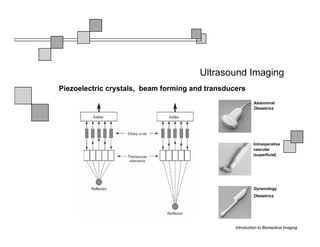
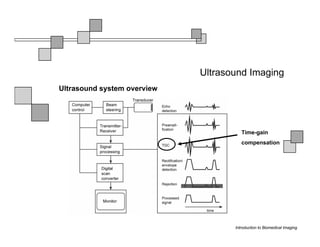
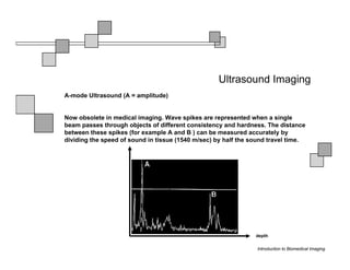
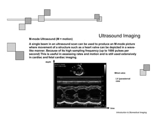
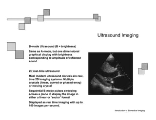
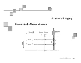
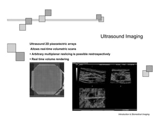
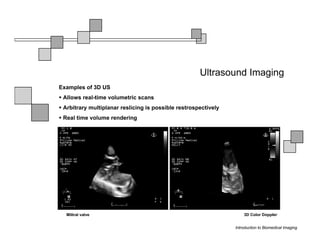
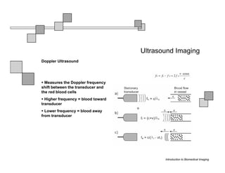
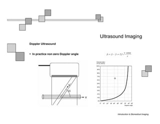
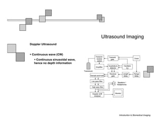
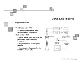
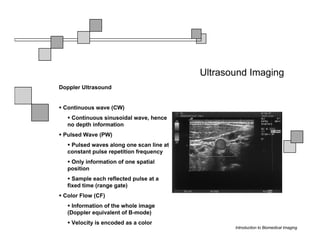
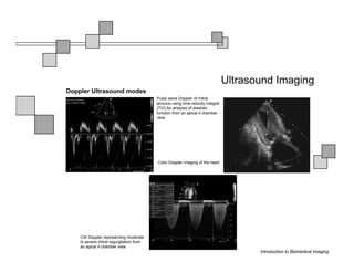
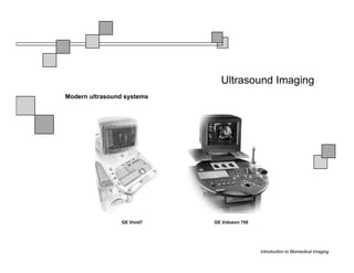
Introduction to Biomedical Imaging


                Alejandro Frangi, PhD
                Computational Imaging Lab
  Department of Information & Communication Technology
                 Pompeu Fabra University
                     www.cilab.upf.edu
Ultrasound Imaging


                     Introduction to Biomedical Imaging
Ultrasound Imaging
Basic principles. Comparison to X-rays


 Ultrasound > 20kHz
 Medical/Diagnostic Ultrasound 1-15 MHz
 Sound spreads in all directions
 Ultrasound can be formed into a narrow beam (it is more “light-like”)
 Periodic motion yields pressure waves
 Speed of sound vs. speed of light
 Ultrasound requires a medium to propagate (no sound in vacuum)
 X-rays can be scattered. US can be reflected, refracted and focused


                                                         Introduction to Biomedical Imaging
Ultrasound Imaging
Ultrasound pulse/continuous wave modes


 Pulsed Wave Mode
   Short DC pulse is applied to the crystal producing its instantaneous
  expansion an tissue compression
    Due to elastic tissue properties: high pressure wave front (compression)
  travels through the body at speed ν followed by a low wave front
  (decompression or rarefaction)
    Multiple frequencies are present in the signal and echo (bandwidth)
 Continuous Wave Mode
    AC voltage applied to crystal
   Periodic pattern of compression and rarefaction will travel across the
  body with speed ν and wavelength λ
   The frequency is given by the AC voltage applied to the transducer
  crystal: ν = λ f

                                                                Introduction to Biomedical Imaging
Ultrasound Imaging
Ultrasound pulse/wave
generation:
transmission


Ultrasound signal
detection


                               Introduction to Biomedical Imaging
Ultrasound Imaging
Ultrasound transducer frequency vs. resolution


                                                 Introduction to Biomedical Imaging
Ultrasound Imaging
Piezoelectric crystals, beam forming and transducers


                                                 Introduction to Biomedical Imaging
Ultrasound Imaging
Piezoelectric crystals, beam forming and transducers


                                                 Introduction to Biomedical Imaging
Ultrasound Imaging
Piezoelectric crystals, beam forming and transducers
                                                           Abdominal
                                                           Obstetrics


                                                          Intraoperative
                                                          vascular
                                                          (superficial)


                                                           Gynecology
                                                           Obstetrics


                                                 Introduction to Biomedical Imaging
Ultrasound Imaging
Ultrasound system overview


                                       Time-gain
                                       compensation


                                    Introduction to Biomedical Imaging
Ultrasound Imaging
A-mode Ultrasound (A = amplitude)


Now obsolete in medical imaging. Wave spikes are represented when a single
beam passes through objects of different consistency and hardness. The distance
between these spikes (for example A and B ) can be measured accurately by
dividing the speed of sound in tissue (1540 m/sec) by half the sound travel time.


                                                                depth

                                                                Introduction to Biomedical Imaging
Ultrasound Imaging
M-mode Ultrasound (M = motion)
A single beam in an ultrasound scan can be used to produce an M-mode picture
where movement of a structure such as a heart valve can be depicted in a wave-
like manner. Because of its high sampling frequency (up to 1000 pulses per
second) This is useful in assessing rates and motion and is still used extensively
in cardiac and fetal cardiac imaging.
            depth


                                                                 Mitral valve

                                                                     LV (parasternal
                                                                     view


                                                              time

                                                                     Introduction to Biomedical Imaging
Ultrasound Imaging

B-mode Ultrasound (B = brightness)
Same as A-mode, but one dimensional
graphical display with brightness
corresponding to amplitude of reflected
sound


2D real-time ultrasound
Most modern ultrasound devices are real-
time 2D imaging systems. Multiple
crystals (linear, curved or phased-array)
or moving crystal
Sequential B-mode pulses sweeping
across a plane to display the image in
either a linear or ‘sector’ format
Displayed as real time imaging with up to
100 images per second.
                                                   Introduction to Biomedical Imaging
Ultrasound Imaging

Summary A-, B-, M-mode ultrasound


                                           Introduction to Biomedical Imaging
Ultrasound Imaging
Ultrasound 2D piezoelectric arrays
Allows real-time volumetric scans
 Arbitrary multiplanar reslicing is possible restrospectively
 Real time volume rendering


                                                                   Brest biopsy


                                                                Introduction to Biomedical Imaging
Ultrasound Imaging
Examples of 3D US
 Allows real-time volumetric scans
 Arbitrary multiplanar reslicing is possible restrospectively
 Real time volume rendering


  Miltral valve                                                     3D Color Doppler


                                                                Introduction to Biomedical Imaging
Ultrasound Imaging
Doppler Ultrasound


  Measures the Doppler frequency
shift between the transducer and
the red blood cells
  Higher frequency = blood toward
transducer
  Lower frequency = blood away
from transducer


                                           Introduction to Biomedical Imaging
Ultrasound Imaging
Doppler Ultrasound


 In practice non zero Doppler angle


                                             Introduction to Biomedical Imaging
Ultrasound Imaging
Doppler Ultrasound


 Continuous wave (CW)
   Continuous sinusoidal wave,
  hence no depth information


                                        Introduction to Biomedical Imaging
Ultrasound Imaging
Doppler Ultrasound


 Continuous wave (CW)
   Continuous sinusoidal wave,
  hence no depth information
 Pulsed Wave (PW)
    Pulsed waves along one scan line
  at constant pulse repetition
  frequency
   Only information of one spatial
  position
    Sample each reflected pulse at a
  fixed time (range gate)


                                              Introduction to Biomedical Imaging
Ultrasound Imaging
Doppler Ultrasound


 Continuous wave (CW)
   Continuous sinusoidal wave, hence
  no depth information
 Pulsed Wave (PW)
   Pulsed waves along one scan line at
  constant pulse repetition frequency
   Only information of one spatial
  position
    Sample each reflected pulse at a
  fixed time (range gate)
 Color Flow (CF)
    Information of the whole image
  (Doppler equivalent of B-mode)
    Velocity is encoded as a color
                                                Introduction to Biomedical Imaging
Ultrasound Imaging
Doppler Ultrasound modes
                                          Pulse wave Doppler of mitral
                                          annulus using time-velocity integral
                                          (TVI) for analysis of diastolic
                                          function from an apical 4 chamber
                                          view.


                                          Color Doppler imaging of the heart


    CW Doppler representing moderate
    to severe mitral regurgitation from
    an apical 4 chamber view.
                                                                                        Introduction to Biomedical Imaging
Ultrasound Imaging
Modern ultrasound systems


                 GE Vivid7   GE Voluson 750


                                              Introduction to Biomedical Imaging

Introduction to Biomedical Imaging

  • 1.
    Introduction to BiomedicalImaging Alejandro Frangi, PhD Computational Imaging Lab Department of Information & Communication Technology Pompeu Fabra University www.cilab.upf.edu
  • 2.
    Ultrasound Imaging Introduction to Biomedical Imaging
  • 3.
    Ultrasound Imaging Basic principles.Comparison to X-rays Ultrasound > 20kHz Medical/Diagnostic Ultrasound 1-15 MHz Sound spreads in all directions Ultrasound can be formed into a narrow beam (it is more “light-like”) Periodic motion yields pressure waves Speed of sound vs. speed of light Ultrasound requires a medium to propagate (no sound in vacuum) X-rays can be scattered. US can be reflected, refracted and focused Introduction to Biomedical Imaging
  • 4.
    Ultrasound Imaging Ultrasound pulse/continuouswave modes Pulsed Wave Mode Short DC pulse is applied to the crystal producing its instantaneous expansion an tissue compression Due to elastic tissue properties: high pressure wave front (compression) travels through the body at speed ν followed by a low wave front (decompression or rarefaction) Multiple frequencies are present in the signal and echo (bandwidth) Continuous Wave Mode AC voltage applied to crystal Periodic pattern of compression and rarefaction will travel across the body with speed ν and wavelength λ The frequency is given by the AC voltage applied to the transducer crystal: ν = λ f Introduction to Biomedical Imaging
  • 5.
    Ultrasound Imaging Ultrasound pulse/wave generation: transmission Ultrasoundsignal detection Introduction to Biomedical Imaging
  • 6.
    Ultrasound Imaging Ultrasound transducerfrequency vs. resolution Introduction to Biomedical Imaging
  • 7.
    Ultrasound Imaging Piezoelectric crystals,beam forming and transducers Introduction to Biomedical Imaging
  • 8.
    Ultrasound Imaging Piezoelectric crystals,beam forming and transducers Introduction to Biomedical Imaging
  • 9.
    Ultrasound Imaging Piezoelectric crystals,beam forming and transducers Abdominal Obstetrics Intraoperative vascular (superficial) Gynecology Obstetrics Introduction to Biomedical Imaging
  • 10.
    Ultrasound Imaging Ultrasound systemoverview Time-gain compensation Introduction to Biomedical Imaging
  • 11.
    Ultrasound Imaging A-mode Ultrasound(A = amplitude) Now obsolete in medical imaging. Wave spikes are represented when a single beam passes through objects of different consistency and hardness. The distance between these spikes (for example A and B ) can be measured accurately by dividing the speed of sound in tissue (1540 m/sec) by half the sound travel time. depth Introduction to Biomedical Imaging
  • 12.
    Ultrasound Imaging M-mode Ultrasound(M = motion) A single beam in an ultrasound scan can be used to produce an M-mode picture where movement of a structure such as a heart valve can be depicted in a wave- like manner. Because of its high sampling frequency (up to 1000 pulses per second) This is useful in assessing rates and motion and is still used extensively in cardiac and fetal cardiac imaging. depth Mitral valve LV (parasternal view time Introduction to Biomedical Imaging
  • 13.
    Ultrasound Imaging B-mode Ultrasound(B = brightness) Same as A-mode, but one dimensional graphical display with brightness corresponding to amplitude of reflected sound 2D real-time ultrasound Most modern ultrasound devices are real- time 2D imaging systems. Multiple crystals (linear, curved or phased-array) or moving crystal Sequential B-mode pulses sweeping across a plane to display the image in either a linear or ‘sector’ format Displayed as real time imaging with up to 100 images per second. Introduction to Biomedical Imaging
  • 14.
    Ultrasound Imaging Summary A-,B-, M-mode ultrasound Introduction to Biomedical Imaging
  • 15.
    Ultrasound Imaging Ultrasound 2Dpiezoelectric arrays Allows real-time volumetric scans Arbitrary multiplanar reslicing is possible restrospectively Real time volume rendering Brest biopsy Introduction to Biomedical Imaging
  • 16.
    Ultrasound Imaging Examples of3D US Allows real-time volumetric scans Arbitrary multiplanar reslicing is possible restrospectively Real time volume rendering Miltral valve 3D Color Doppler Introduction to Biomedical Imaging
  • 17.
    Ultrasound Imaging Doppler Ultrasound Measures the Doppler frequency shift between the transducer and the red blood cells Higher frequency = blood toward transducer Lower frequency = blood away from transducer Introduction to Biomedical Imaging
  • 18.
    Ultrasound Imaging Doppler Ultrasound In practice non zero Doppler angle Introduction to Biomedical Imaging
  • 19.
    Ultrasound Imaging Doppler Ultrasound Continuous wave (CW) Continuous sinusoidal wave, hence no depth information Introduction to Biomedical Imaging
  • 20.
    Ultrasound Imaging Doppler Ultrasound Continuous wave (CW) Continuous sinusoidal wave, hence no depth information Pulsed Wave (PW) Pulsed waves along one scan line at constant pulse repetition frequency Only information of one spatial position Sample each reflected pulse at a fixed time (range gate) Introduction to Biomedical Imaging
  • 21.
    Ultrasound Imaging Doppler Ultrasound Continuous wave (CW) Continuous sinusoidal wave, hence no depth information Pulsed Wave (PW) Pulsed waves along one scan line at constant pulse repetition frequency Only information of one spatial position Sample each reflected pulse at a fixed time (range gate) Color Flow (CF) Information of the whole image (Doppler equivalent of B-mode) Velocity is encoded as a color Introduction to Biomedical Imaging
  • 22.
    Ultrasound Imaging Doppler Ultrasoundmodes Pulse wave Doppler of mitral annulus using time-velocity integral (TVI) for analysis of diastolic function from an apical 4 chamber view. Color Doppler imaging of the heart CW Doppler representing moderate to severe mitral regurgitation from an apical 4 chamber view. Introduction to Biomedical Imaging
  • 23.
    Ultrasound Imaging Modern ultrasoundsystems GE Vivid7 GE Voluson 750 Introduction to Biomedical Imaging