This document discusses the use of surgeon-performed ultrasound in proctological practice. It provides an overview of how endoluminal ultrasonography can be used to evaluate perirectal sepsis, faecal incontinence, and rectal and anal cancers. It describes the principles, techniques, and applications of endoanal, endorectal, 3D, and transperineal ultrasound. Dynamic transperineal ultrasound is presented as a non-invasive alternative to defaecography for assessing pelvic floor disorders and functional conditions.
Una nuova variante di cheratoplastica perforante: la PK decagonale con laser ...Merqurio
This document describes a prospective study that assesses the use of a decagonal (10-sided polygon) trephination pattern for penetrating keratoplasty (PK) assisted by femtosecond laser. Sixteen eyes underwent the procedure. Short-term results showed that the decagonal trephination profile was effective and safe for PK. Visual acuity improved and astigmatism was reduced compared to conventional PK. However, longer follow-up is needed to determine precise advantages of this technique over conventional PK.
This document provides an overview of magnetic resonance imaging (MRI) physics and the MRI appearance of normal anal canal anatomy and various grades of anal fistulas. Key points include:
- MRI uses strong magnetic fields and radio waves to produce detailed images based on the magnetic properties of hydrogen protons in tissues.
- Normal anatomy includes identification of anal sphincter muscles and mesorectal fascia on MRI images.
- Anal fistulas are classified using the Parks or St James's University Hospital system, grading complexity from intersphincteric to transsphincteric or supralevator types.
- Examples of each grade are shown with drawings and MRI images, demonstrating internal openings, tracts,
The anal canal extends from the anorectal ring to the anal verge. It is supplied by branches from the internal iliac and pudendal arteries and drained by veins that connect to the internal iliac and inferior mesenteric veins. It contains two sphincters - the internal involuntary sphincter and external voluntary sphincter - that help maintain fecal continence. Diseases that can affect the anal canal include hemorrhoids, anal fissures, abscesses, fistulas, and various cancers. Evaluation and management involve examination, imaging, and treatments ranging from nonsurgical options to procedures like sphincterotomy or fistulotomy.
This document summarizes key anatomical features of the rectum and anal canal. It describes the rectum's length, curvature, blood supply, nerve supply and relations to surrounding structures. It discusses the anal canal's measurements and divisions. It also outlines the musculature including the internal and external anal sphincters. Clinical conditions involving the rectum and anal canal like hemorrhoids, anal fissures and fistulas are briefly mentioned.
Evaluation of Complex Anal Fistula - EUA, EUAS OR MRI?ensteve
1. The document discusses different imaging modalities for evaluating complex anal fistulas, including endoanal ultrasound (EAUS), MRI, and their ability to classify fistulas and identify secondary extensions compared to clinical exam alone.
2. Several studies found EAUS and MRI to have higher sensitivity than clinical exam alone in identifying primary tracts, abscesses, horseshoe extensions, and internal openings.
3. Pre-operative MRI can alter surgical management by identifying complex tracts and internal openings and reducing recurrence rates when surgery follows the MRI findings.
Natural history of caesarean scar pregnancy on prenatal ultrasound the cross...Võ Tá Sơn
Natural history of caesarean scar pregnancy on prenatal ultrasound the cross-over sign Cali 2016
Dấu hiệu cross-over trong siêu âm tiền sản tiên lượng tiến triển của thai ở sẹo mổ lấy thai, võ tá sơn
This document provides information about a case presentation of a 19-year-old male student named Ahmad who presented with rectal bleeding and anal pain and swelling. On examination, he was found to have hemorrhoids and signs of anemia. Sigmoidoscopy revealed hemorrhoids that were banded. He received a blood transfusion and IV fluids and was diagnosed with anemia secondary to bleeding hemorrhoids. The document also provides background information on hemorrhoids, appendicitis, colorectal polyps, and familial adenomatous polyposis.
The anatomy and physiology of normal anorectumRavi Kanojia
This document discusses the anatomy related to pediatric anorectal surgery and continence. It covers the anatomy of the rectum, anal canal, levator ani muscles, internal and external anal sphincters, and their roles in continence. It also discusses techniques for evaluating postoperative continence, including subjective and objective scoring systems that assess various anorectal properties and functions.
The document provides detailed anatomical information about the anal canal. It describes the length and divisions of the anal canal. It discusses the muscles including the internal and external sphincters. It covers the blood supply, lymphatic drainage and innervation of the anal canal. It also describes anal gland anatomy and various congenital anomalies that can occur in the anal region.
The anal canal begins at the ano-rectal junction and extends downwards for approximately 4 cm, ending at the anal orifice. It is surrounded by two sphincter muscles - the internal and external sphincters. The pectinate line divides the anal canal into upper and lower regions with different anatomical features. Blood supply comes from the superior, middle, and inferior rectal arteries while drainage is through internal and external hemorrhoidal veins. Nerve supply is from the inferior hypogastric plexus and pudendal nerve. Anal fistulae are abnormal tracts that can develop connecting the anal canal to the perianal skin.
This document discusses intensity modulated radiation therapy (IMRT) and image guided radiation therapy (IGRT) for head and neck cancers. It provides details on contouring targets and organs at risk for treatment planning. It summarizes evidence from trials on reducing xerostomia with IMRT. It also discusses the benefits of daily imaging with IGRT for accurate treatment delivery and potentially reducing planning target volume margins. Adaptive planning is mentioned as an area that continues to be explored to account for anatomical changes over the course of radiation treatment.
The document discusses various approaches for managing chronic pancreatitis and pancreatic pseudocysts. It compares EUS, MRCP, and the combination for evaluating chronic pancreatitis. For pain relief in chronic pancreatitis, endotherapy using pancreatic stents is discussed as less invasive than surgery, with short-term relief rates of 70-94% and long-term relief of 52-82%. Early endoscopic treatment of pancreatic stones is recommended to help with long-term pain management. For pancreatic pseudocysts, the document discusses definitions, incidence, imaging with CT, and endoscopic drainage approaches including transmural versus transpapillary based on characteristics like size and communication with the pancreatic duct.
This document discusses endoscopic therapies for the management of variceal hemorrhage, specifically endoscopic sclerotherapy (EST) and endoscopic variceal ligation (EVL). It provides background on variceal bleeding and survival rates over time. It then describes the modalities and techniques of EST and EVL, including injection methods, sclerosing agents used, risks, and indications. Randomized controlled trials comparing EST and EVL are summarized, showing higher eradication rates with EVL. In conclusion, endoscopic therapies like EST and EVL are effective for controlling acute variceal bleeding and reducing recurrence when used for primary or secondary prophylaxis.
The document discusses laparoscopy procedures for various gynecological conditions such as infertility, chronic pelvic pain, ectopic pregnancy, and oncological issues. It notes that laparoscopy can be used for both diagnostic and operative purposes. It then discusses different techniques for laparoscopic access such as direct trocar insertion versus Verres needle insertion. It reviews studies comparing complication rates between different access techniques. The document emphasizes the importance of evidence-based medicine and following guidelines from organizations like NICE when determining appropriate diagnostic tests and treatments for conditions like infertility.
The document discusses laparoscopy procedures for various gynecological conditions. It begins by outlining conditions that can be diagnosed or treated via laparoscopy, including infertility, ectopic pregnancy, adhesions, endometriosis, ovarian masses, hysterectomy, uterine fibroids, and gynecological oncology issues. It then discusses different laparoscopy access techniques such as direct trocar insertion, open laparoscopy, and Verres needle insertion. It provides data on complication rates for different access methods. The document also discusses techniques for avoiding major vascular injuries during access. In summary, the document provides an overview of laparoscopy procedures and techniques for gynecological conditions.
Slides do curso avançado de atualização em implante de Anel de Ferrara, elaborado por Ferrara Ophtalmics. Para material completo, acesse www.aneldeferrara.com.br
The document discusses endoscopic and open neck conservative surgery options for laryngeal cancer preservation. It summarizes the epidemiology, diagnosis, and treatment of laryngeal tumors including transoral laser surgery for glottic and supraglottic tumors. Oncologic and functional results of endoscopic cordectomies, partial laryngectomies, and open neck conservative surgeries like vertical partial laryngectomy and supracricoid partial laryngectomy are presented based on level III evidence studies. The document emphasizes that endoscopic surgery and open neck conservative surgeries can achieve high laryngeal preservation rates with good oncologic control and functional outcomes for early laryngeal cancers.
Percutaneous endoscopic lumbar discectomy (PELD) is one of the less invasive treatments for lumbar disc herniation, and has 3 different operative approaches. Here, we will review the posterolateral approach (PLA) known as Exraforaminal PELD.
Value Of Ear Endoscopy In Cholesteatoma Surgery.Pptaliabbas07
This document discusses the value of ear endoscopy in cholesteatoma surgery. It finds that ear endoscopy can be used as an adjunct to the microscope to provide educational, diagnostic, and therapeutic benefits. A study of 942 patients undergoing cholesteatoma surgery found that endoscopy assisted in the identification and removal of residual cholesteatoma in 19% of primary cases and 14% of second look procedures. The conclusions are that endoscopy is a complementary tool that can help confirm thorough cholesteatoma removal, assess the adequacy of tympanoplasty, and aid in second look explorations when used alongside the microscope.
This document discusses the importance and implementation of continuous EEG monitoring in intensive care units. It notes that EEG can detect brain ischemia earlier than clinical signs, monitor for nonconvulsive seizures, and provide early prognostic information. Successful ICU/cEEG programs require collaboration between neurophysiologists, neurointensivists, ICU staff and others. Regular training of ICU staff is needed so they can recognize normal and abnormal waveforms and ensure monitoring is available 24/7. Teamwork and institutional support are essential for effective ICU/cEEG monitoring.
The document lists medical equipment inventory for different departments including quantities. It includes surgical vacuums, scales, hospital beds, monitors, defibrillators, stethoscopes, lights, incubators, and other neonatal equipment. Specifications are provided for some items like incubators and hospital beds.
This document summarizes a study on proptosis, or forward displacement of the eye, in pediatric patients. The study aimed to analyze the incidence, etiological factors, correlation with investigations, and management/outcomes of childhood proptosis. It found that the most common causes were orbital cellulitis secondary to sinusitis and cavernous sinus thrombosis. Aggressive antibiotic treatment and supportive care led to complete recovery in most cases. Early and prompt therapy was determined to lead to satisfying outcomes.
Unresolved Issues In Myocardial ViabilityMuhammad Ayub
This document discusses myocardial viability and techniques to detect viable myocardium. It begins by defining viable myocardium and explaining why detection is important for predicting prognosis and response to revascularization. It then reviews techniques to detect viability including those assessing contractile reserve using stress echocardiography, SPECT, or MRI as well as techniques evaluating preserved metabolism using PET tracers or cell membrane integrity using thallium or sestamibi SPECT. The document compares the sensitivity and specificity of different techniques and concludes that while techniques assessing contractile reserve are highly specific, nuclear techniques provide good sensitivity for viability assessment.
The document discusses the anatomy, physiology, and pathology of the cornea. It provides details on the layers of the cornea and describes corneal ulcers. Key points include:
1) The cornea has 5 layers - epithelium, Bowman's layer, stroma, Descemet's membrane, and endothelium. The stroma makes up 90% of the corneal thickness.
2) Corneal ulcers are localized areas of necrosis in the superficial stroma caused by trauma, infection, or other insults. Common causes of infection include bacteria like Staphylococcus and Pneumococcus.
3) Complications of corneal ulcers can include iritis, glaucoma, corneal opacity
This document analyzes the results of unicompartmental knee prostheses with mobile and fixed plates from the 1950s to the 2000s. It summarizes the history and development of these prostheses over time. Key findings from literature on survival rates of unicondylar prostheses show survival rates comparable to total knee replacements at 10 years and questions remain about survival rates at 15-20 years. The document advocates for unicondylar prostheses based on appropriate patient selection and surgical technique to achieve long prosthesis life expectancy.
The Use of Achieve Circular Mapping Catheter Significantly Reduces Procedure ...Jc Templar
Michaël Peyrol, Pascal Sbragia, Morgane Orabona, Anne-Claire Casalta, Amandine Quatre, Zinedine Zerrouk, Gilles Boccara, Maxime Guenoun, Samuel Lévy, Franck Paganelli
Department of Cardiology, Centre Hospitalier Universitaire Nord, Marseille, France
Full-endoscopic lumbar discectomy is an innovative, minimally invasive alternative to microdiscectomy for patients with symptomatic lumbar disc herniations. IELD and TELD offer two complementary surgical corridors to spinal pathology and allow for treatment of the vast majority of lumbar disc herniations. There is level one evidence suggesting that full-endoscopic spine surgery results in similar functional outcomes compared with microsurgical technique, and has a favorable rate of perioperative complications.
The document discusses fractures of the distal radius, including:
1) Distal radius fractures are an important epidemiological problem as the population ages and life expectancies increase.
2) Classification systems help compare fracture types for outcomes but do not guide treatment on their own. Common systems include the Frykman classification and AO classification.
3) Treatment depends on the fracture pattern and stability. Closed reduction with casting is appropriate for stable fractures, while unstable or complex fractures may require open reduction and fixation methods like K-wires, plates, or external fixation.
This document provides information on various types of colonic polyps. It discusses neoplastic polyps such as adenomas, which can be classified as exophytic or non-exophytic. Non-exophytic adenomas, previously called flat adenomas, occur more frequently with high-grade dysplasia. The document also covers non-neoplastic polyps such as hyperplastic polyps and hamartomas. Different classification systems are described including endoscopic, histopathological, and clinicopathological classifications. Specific polyposis syndromes associated with multiple polyp growth are mentioned.
Similar to Surgeon Performed Ultrasound In Proctological Practice (20)
A researcher from the University of Zaragoza proposed a new technique called "Watering the rectum" to image rectal tumors, which fills the rectum with water instead of using balloons or air as in conventional colonoscopy. Initial images using the new water technique showed similar or improved visibility of rectal tumors compared to traditional methods.
This document discusses ultrasound physics and how to optimize ultrasound images. It defines ultrasound and describes how it is produced using piezoelectric crystals. It explains pulse echo imaging and the factors that influence resolution such as frequency, pulse length, and focal zone. The document also covers ultrasound transmission, Doppler imaging, transducer types, scanning techniques, controls to optimize images, and common artifacts.
This document discusses ultrasound physics and principles. It covers the characteristics of sound waves including their need for a medium, compression and rarefaction, and propagation. It describes ultrasound wave properties like range, velocity in different media, and how velocity relates to compressibility, density, and intensity. Transducers are discussed including their piezoelectric crystal, electrode, and backing block components. Modes of ultrasound like continuous wave and pulse wave are summarized. Key interactions of ultrasound with matter like reflection, refraction, and absorption are covered. Principles of Doppler ultrasound for blood flow measurement are outlined.
Ultrasound uses piezoelectric crystals to both transmit and receive sound waves. It propagates through materials at different velocities depending on factors like density and modulus. Intensity decreases with distance due to attenuation from scattering and absorption. Safety depends on keeping intensity below thresholds for potential tissue heating. Scattering strength varies based on particle and wavelength size scales. Ultrasound finds medical use in diagnostics and therapy through techniques like lithotripsy.
The Epidemic of Thyroid Nodules: Which Should Undergo Fine Needle Aspiration?u.surgery
The document discusses thyroid nodules and guidelines for performing fine needle aspiration (FNA). It notes that while thyroid nodules are common, detected in up to 67% of ultrasound exams, only 5-15% of nodules are malignant. Sonographic features that increase the likelihood of malignancy include microcalcifications, irregular margins, hypoechogenicity, and intranodular flow. The document recommends FNA for nodules over 1 cm in size or if sonographic features suggest cancer risk. Strategic FNA based on risk factors aims to identify the minority of nodules that are cancer while avoiding unnecessary biopsies of predominantly benign nodules.
Sonographic Assessment of Blunt Abdominal Trauma in the Emergency Department:...u.surgery
The document discusses the Focused Assessment with Sonography for Trauma (FAST) exam used to diagnose blunt abdominal trauma. The FAST exam uses ultrasound to quickly check for fluid in four areas of the abdomen that could indicate internal bleeding. While fast and non-invasive, the FAST exam has limitations as it can miss some injuries and has variable accuracy depending on the operator. The document explores different trauma algorithms and how the FAST could be incorporated while accounting for its limitations.
Abdominal sonography is a non-invasive imaging technique that has several advantages over other modalities. It does not require contrast agents, radiation, or isotopes and can visualize organs and surrounding structures along with morphological abnormalities. While obesity, gas, or lack of patient cooperation can limit its effectiveness, sonography should be the first examination used to evaluate diseases of the liver, biliary system, pancreas, and urinary tract. It allows assessment of organ size, structure, lesions, and blood flow and can guide minimally invasive biopsies and procedures. Disadvantages include limited specificity requiring additional tests, but it provides real-time dynamic imaging without known health risks.
The document discusses the physics of ultrasound imaging, including an overview of acoustic waves, wave propagation equations, reflection and refraction of waves, Doppler effect, and the functioning of ultrasound transducers using piezoelectric crystals to generate and receive acoustic waves for medical imaging applications.
This document discusses the increasing use of ultrasound by surgeons. It provides examples of how ultrasound is used in trauma settings to detect internal bleeding and guide procedures. The author also shares their experience using ultrasound to diagnose acute cholecystitis and guide radiofrequency ablation of liver tumors. Percutaneous drainage of intra-abdominal collections under ultrasound guidance is described as well, with a high success rate.
Ultrasound Education In The American College Of Surgeons A Historyu.surgery
This document discusses the history of ultrasound education within the American College of Surgeons (ACS). It describes how early European experience with surgical ultrasound helped establish its value. In the 1990s, the ACS began developing an ultrasound educational program through initiatives like workshops at clinical meetings. This led to the formation of an Ultrasound Users Group and National Ultrasound Faculty to standardize ultrasound education for surgeons. Over years, the ACS program expanded to include multiple modules and courses, establishing ultrasound firmly within the surgical domain through education.
Thyroid Ultrasound For The Endocrine Surgeonu.surgery
This document discusses the value of ultrasound for endocrine surgeons. It summarizes a study of over 5,700 ultrasounds performed by an endocrine surgeon between 1999-2004. Key findings include that ultrasound enhanced the diagnosis of coexisting thyroid and parathyroid diseases, helped localize abnormal parathyroids, modified treatment in 62% of thyroid cancer cases, and improved resident education. The conclusion is that surgeon-performed neck ultrasound is a specific, accurate tool that enhances patient care and surgical decision making.
Surgeon Performed Ultrasound Privileges, Competency And Practiceu.surgery
1) The document discusses the results of an ACS survey on surgeon-performed ultrasound privileges, competency, and practice.
2) The survey found that over half of responding surgeons use ultrasound in multiple clinical areas and many perform ultrasound-guided procedures, most commonly for breast procedures.
3) In general, surgeons reported competency with less than 20 ultrasound examinations in most clinical areas, except for the acute care setting which required 21-30 examinations.
This document discusses the physical principles of ultrasound used in medical imaging. It defines key terms like frequency, wavelength, attenuation and resolution. It describes how piezoelectric transducers convert electrical pulses to ultrasound pulses and echoes. It explains how sector and linear array transducers work and the different display modes. It also discusses artifacts and the safety of diagnostic medical ultrasound.
International Guidelines And Regulations For The Safe Use Of Diagnostic Ultra...u.surgery
The document discusses international guidelines and regulations for the safe use of diagnostic ultrasound in medicine. It covers ultrasound biophysics and potential bioeffects, the development of safety guidelines and regulations, and balancing the benefits of ultrasound with potential risks. Key points addressed include thermal and non-thermal bioeffects, limits set by organizations like the FDA, and the importance of prudent and ALARA use of diagnostic ultrasound to minimize risks while optimizing medical benefits.
Latest Oncologic Strategies For Well Differentiated Thyroid Carcinomau.surgery
The presentation reviewed the appropriate surgical approach for papillary thyroid carcinoma, highlighting the importance of (1) total thyroidectomy with central lymph node dissection for tumors greater than 1 cm, (2) clearing all sonographically detectable lymph node metastases at initial surgery to prevent recurrence, and (3) obtaining high-quality pre-operative ultrasound to thoroughly stage the cancer. Radioiodine therapy has limited effectiveness for macroscopic lymph node disease and is not a substitute for complete surgical resection.
The document discusses emergency ultrasound in trauma patients. It describes how focused abdominal sonography for trauma (FAST) uses 4 views to detect free fluid in trauma patients in 3 minutes or less. Studies show FAST has a sensitivity of 81-98% and specificity of 88-100% for detecting intra-abdominal bleeding. While not showing specific injuries, FAST effectively detects patients needing surgery. The document also discusses using ultrasound to detect hemothorax, pericardial fluid, and penetrating cardiac wounds.
An Evaluation Of The Acs Ultrasound Education Programu.surgery
This study evaluated the effectiveness of the American College of Surgeons' ultrasound education program from 1998-2002. A survey of 873 surgeons found that after completing ACS courses, the percentage of surgeons performing ultrasound increased from 58% to 65%. On average, surgeons received 10 hours of total ultrasound instruction, with 5-8 hours dedicated to didactic lessons and 4-8 hours of hands-on training. The majority of surgeons rated the courses as good to excellent and reported applying the new skills and knowledge to their surgical practice. The study concluded the ACS ultrasound education program has been successful in formalizing ultrasound training for surgeons.
Staging and Surgical Management of Pancreatiic Canceru.surgery
This document discusses staging and surgical management of pancreatic cancer. It covers staging techniques like CT, endoscopic ultrasound, laparoscopy and biomarkers. CT is good for predicting resectability but not lymph node status. Endoscopic ultrasound is useful for diagnosis and staging. Laparoscopy can detect small volume disease and changes management in 10-15% of cases. Extended resections, portal vein resection and pylorus-preserving pancreaticoduodenectomy are discussed. Reconstruction techniques after resection include pancreaticojejunostomy. Drains and stents may not provide benefits while octreotide could help for high-risk anastomoses. Outcome depends on tumour type and quality of life is improved at high-volume
Medical uses of ultrasound can include imaging the inside of the body and detecting blood flow. Bats and dolphins use echolocation with ultrasound to navigate. We can apply this principle to medical ultrasound imaging by emitting ultrasound pulses and mapping the echoes to visualize internal structures. Doppler ultrasound further analyzes the ultrasound echoes to determine blood flow velocity within the body. While 2D ultrasound is considered safe for imaging fetuses, more advanced 4D ultrasound using higher energies requires further consideration regarding any potential risks from heating effects.
Does Over-Masturbation Contribute to Chronic Prostatitis.pptxwalterHu5
In some case, your chronic prostatitis may be related to over-masturbation. Generally, natural medicine Diuretic and Anti-inflammatory Pill can help mee get a cure.
Cell Therapy Expansion and Challenges in Autoimmune DiseaseHealth Advances
There is increasing confidence that cell therapies will soon play a role in the treatment of autoimmune disorders, but the extent of this impact remains to be seen. Early readouts on autologous CAR-Ts in lupus are encouraging, but manufacturing and cost limitations are likely to restrict access to highly refractory patients. Allogeneic CAR-Ts have the potential to broaden access to earlier lines of treatment due to their inherent cost benefits, however they will need to demonstrate comparable or improved efficacy to established modalities.
In addition to infrastructure and capacity constraints, CAR-Ts face a very different risk-benefit dynamic in autoimmune compared to oncology, highlighting the need for tolerable therapies with low adverse event risk. CAR-NK and Treg-based therapies are also being developed in certain autoimmune disorders and may demonstrate favorable safety profiles. Several novel non-cell therapies such as bispecific antibodies, nanobodies, and RNAi drugs, may also offer future alternative competitive solutions with variable value propositions.
Widespread adoption of cell therapies will not only require strong efficacy and safety data, but also adapted pricing and access strategies. At oncology-based price points, CAR-Ts are unlikely to achieve broad market access in autoimmune disorders, with eligible patient populations that are potentially orders of magnitude greater than the number of currently addressable cancer patients. Developers have made strides towards reducing cell therapy COGS while improving manufacturing efficiency, but payors will inevitably restrict access until more sustainable pricing is achieved.
Despite these headwinds, industry leaders and investors remain confident that cell therapies are poised to address significant unmet need in patients suffering from autoimmune disorders. However, the extent of this impact on the treatment landscape remains to be seen, as the industry rapidly approaches an inflection point.
Osteoporosis - Definition , Evaluation and Management .pdfJim Jacob Roy
Osteoporosis is an increasing cause of morbidity among the elderly.
In this document , a brief outline of osteoporosis is given , including the risk factors of osteoporosis fractures , the indications for testing bone mineral density and the management of osteoporosis
Local Advanced Lung Cancer: Artificial Intelligence, Synergetics, Complex Sys...Oleg Kshivets
Overall life span (LS) was 1671.7±1721.6 days and cumulative 5YS reached 62.4%, 10 years – 50.4%, 20 years – 44.6%. 94 LCP lived more than 5 years without cancer (LS=2958.6±1723.6 days), 22 – more than 10 years (LS=5571±1841.8 days). 67 LCP died because of LC (LS=471.9±344 days). AT significantly improved 5YS (68% vs. 53.7%) (P=0.028 by log-rank test). Cox modeling displayed that 5YS of LCP significantly depended on: N0-N12, T3-4, blood cell circuit, cell ratio factors (ratio between cancer cells-CC and blood cells subpopulations), LC cell dynamics, recalcification time, heparin tolerance, prothrombin index, protein, AT, procedure type (P=0.000-0.031). Neural networks, genetic algorithm selection and bootstrap simulation revealed relationships between 5YS and N0-12 (rank=1), thrombocytes/CC (rank=2), segmented neutrophils/CC (3), eosinophils/CC (4), erythrocytes/CC (5), healthy cells/CC (6), lymphocytes/CC (7), stick neutrophils/CC (8), leucocytes/CC (9), monocytes/CC (10). Correct prediction of 5YS was 100% by neural networks computing (error=0.000; area under ROC curve=1.0).
Histololgy of Female Reproductive System.pptxAyeshaZaid1
Dive into an in-depth exploration of the histological structure of female reproductive system with this comprehensive lecture. Presented by Dr. Ayesha Irfan, Assistant Professor of Anatomy, this presentation covers the Gross anatomy and functional histology of the female reproductive organs. Ideal for students, educators, and anyone interested in medical science, this lecture provides clear explanations, detailed diagrams, and valuable insights into female reproductive system. Enhance your knowledge and understanding of this essential aspect of human biology.
These lecture slides, by Dr Sidra Arshad, offer a quick overview of the physiological basis of a normal electrocardiogram.
Learning objectives:
1. Define an electrocardiogram (ECG) and electrocardiography
2. Describe how dipoles generated by the heart produce the waveforms of the ECG
3. Describe the components of a normal electrocardiogram of a typical bipolar lead (limb II)
4. Differentiate between intervals and segments
5. Enlist some common indications for obtaining an ECG
6. Describe the flow of current around the heart during the cardiac cycle
7. Discuss the placement and polarity of the leads of electrocardiograph
8. Describe the normal electrocardiograms recorded from the limb leads and explain the physiological basis of the different records that are obtained
9. Define mean electrical vector (axis) of the heart and give the normal range
10. Define the mean QRS vector
11. Describe the axes of leads (hexagonal reference system)
12. Comprehend the vectorial analysis of the normal ECG
13. Determine the mean electrical axis of the ventricular QRS and appreciate the mean axis deviation
14. Explain the concepts of current of injury, J point, and their significance
Study Resources:
1. Chapter 11, Guyton and Hall Textbook of Medical Physiology, 14th edition
2. Chapter 9, Human Physiology - From Cells to Systems, Lauralee Sherwood, 9th edition
3. Chapter 29, Ganong’s Review of Medical Physiology, 26th edition
4. Electrocardiogram, StatPearls - https://www.ncbi.nlm.nih.gov/books/NBK549803/
5. ECG in Medical Practice by ABM Abdullah, 4th edition
6. Chapter 3, Cardiology Explained, https://www.ncbi.nlm.nih.gov/books/NBK2214/
7. ECG Basics, http://www.nataliescasebook.com/tag/e-c-g-basics
Surgeon Performed Ultrasound In Proctological Practice
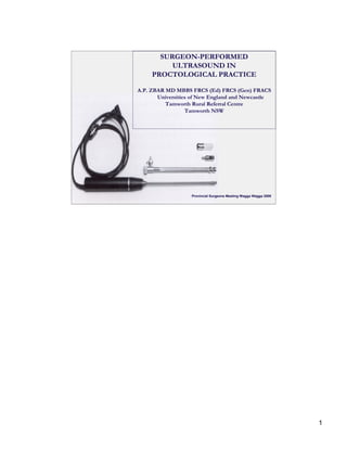
1. SURGEON-PERFORMED
ULTRASOUND IN
PROCTOLOGICAL PRACTICE
A.P. ZBAR MD MBBS FRCS (Ed) FRCS (Gen) FRACS
Universities of New England and Newcastle
Tamworth Rural Referral Centre
Tamworth NSW
Provincial Surgeons Meeting Wagga Wagga 2008
1
2. ENDOLUMINAL ULTRASONOGRAPHY
First introduced by Law and Bartram in 1989
Association with normal anatomy
Constitutive variations with age
Modifications – 3-dimensional, Contrast enhancement
and Transperineal technology
UTILIZATION
Perirectal Sepsis
Faecal Incontinence
Rectal and Anal Cancers
Functional Disorders
2
3. PRINCIPLES OF ULTRASOUND
Resolution depends on frequency (MHz)
and pulse repetition frequency
Lateral resolution is controlled by beam width
A standard BK 10 MHz transducer has an
axial resolution < 0.05 mm with a lateral
resolution of 0.5-1 mm and a focal range
of 5-45 mm.
3
8. TRANSPERINEAL SONOGRAPHY
Sensitivity for detection of trans-
sphincteric and extrasphincteric
fistulae = 100%
Sensitivity for internal opening
with TPUS is 90% and 80% for
EAUS
Sensitivity for horseshoeing with
TPUS is 28.6%
Sensitivity for ancillary abscess
and secondary tracks with TPUS
Zbar AP et al. Techniques in Coloproctol 2007
is 63.6%
8
28. DYNAMIC TRANSPERINEAL ULTRASONOGRAPHY (DTP-US)
Simple
Non-invasive
Avoids radiation in young patients
Repeatable
Portable
Simultaneously assesses the anterior and posterior perineum & pelvic floor
Avoids the need for multiorgan opacification
Dynamic transperineal ultrasound is accurate in
the diagnosis of specific pelvic floor conditions
It tends to diagnose multiple conditions in
functional disorders
Quantitative assessment of the pelvic floor is
comparable with defaecography for pelvic
floor disorders at rest and during straining
28