Downloaded 86 times


The document discusses analog and digital medical images. It notes that analog images are continuous representations of brightness or color, while digital images represent images as a matrix of pixels with numerical values. The advantages of digital images include easier processing, storage, and transfer by computer systems. It also discusses key aspects of digital medical images like pixel bit depth, resolution, matrix size, and compression.
Overview of the class and instructor details.
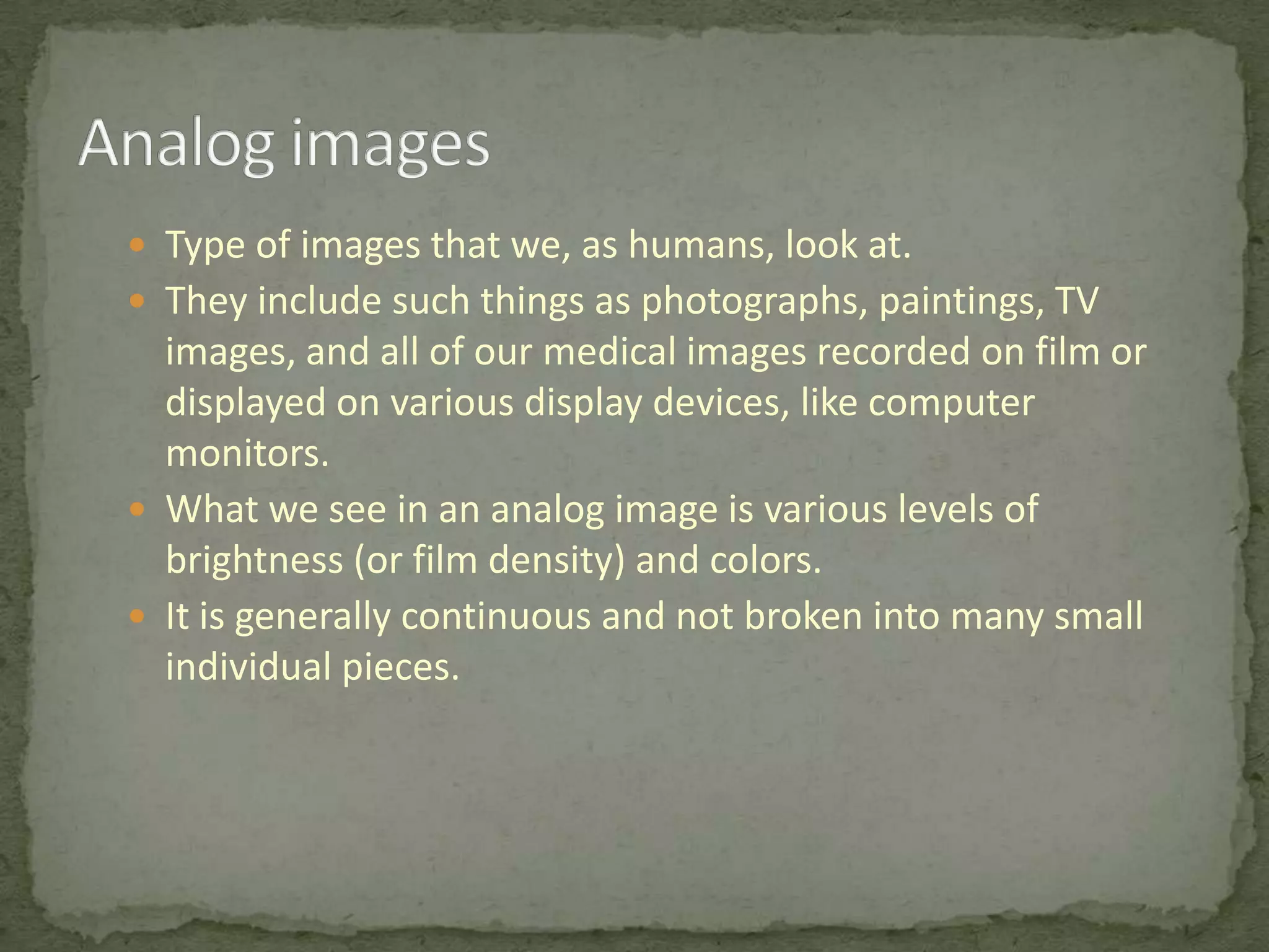
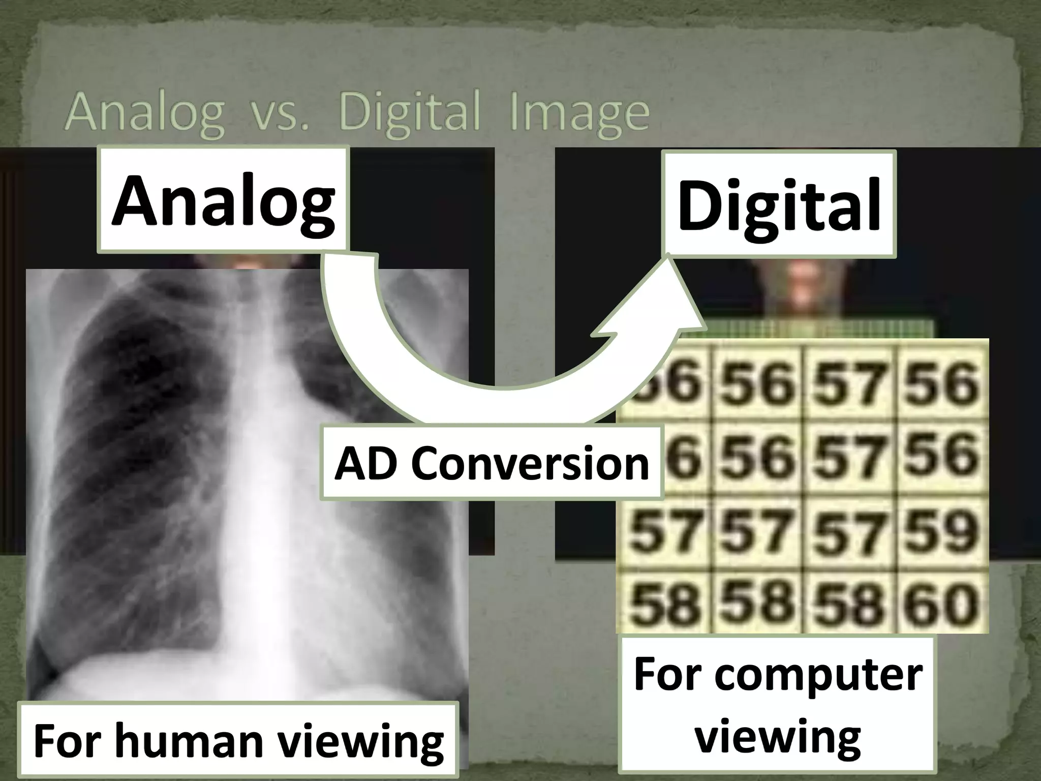
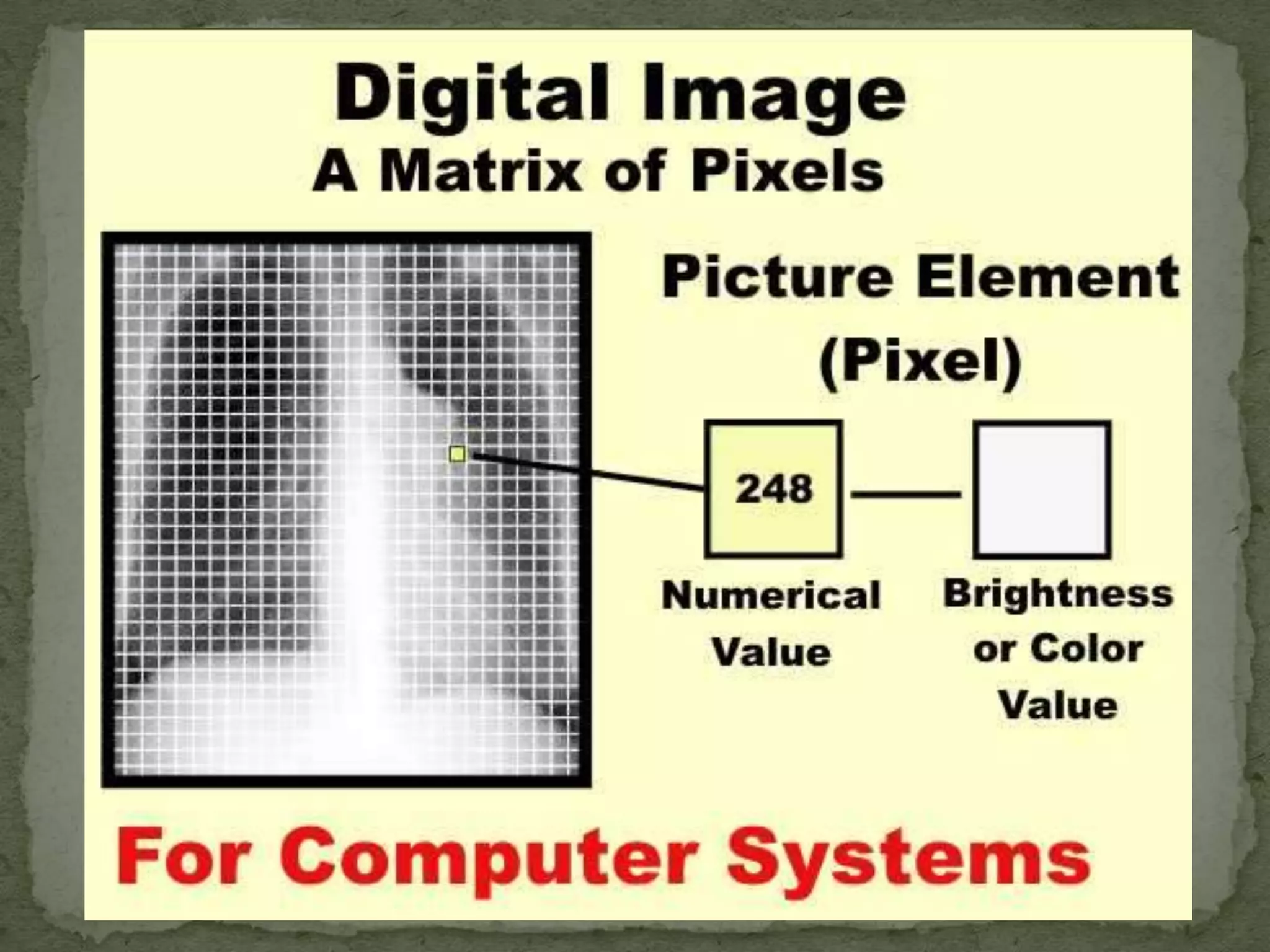
Contrast between analog and digital images; discussions on image properties and pixel representation.
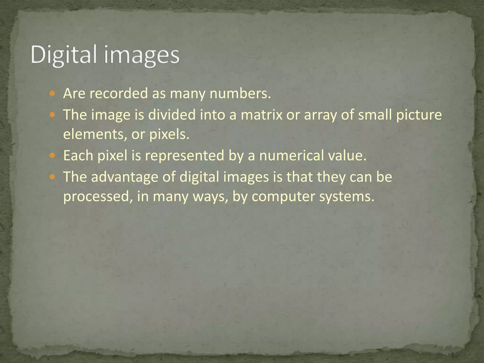
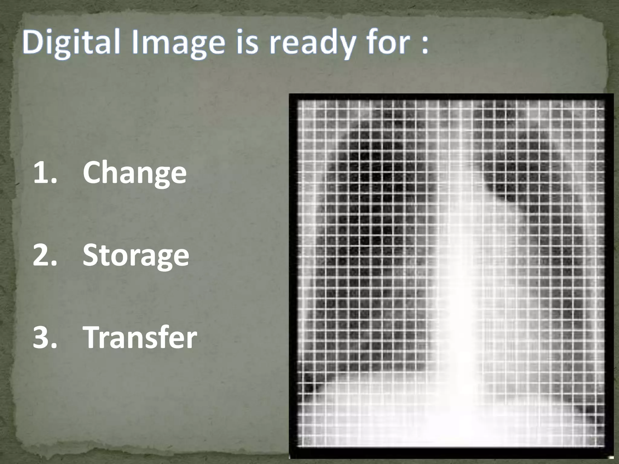
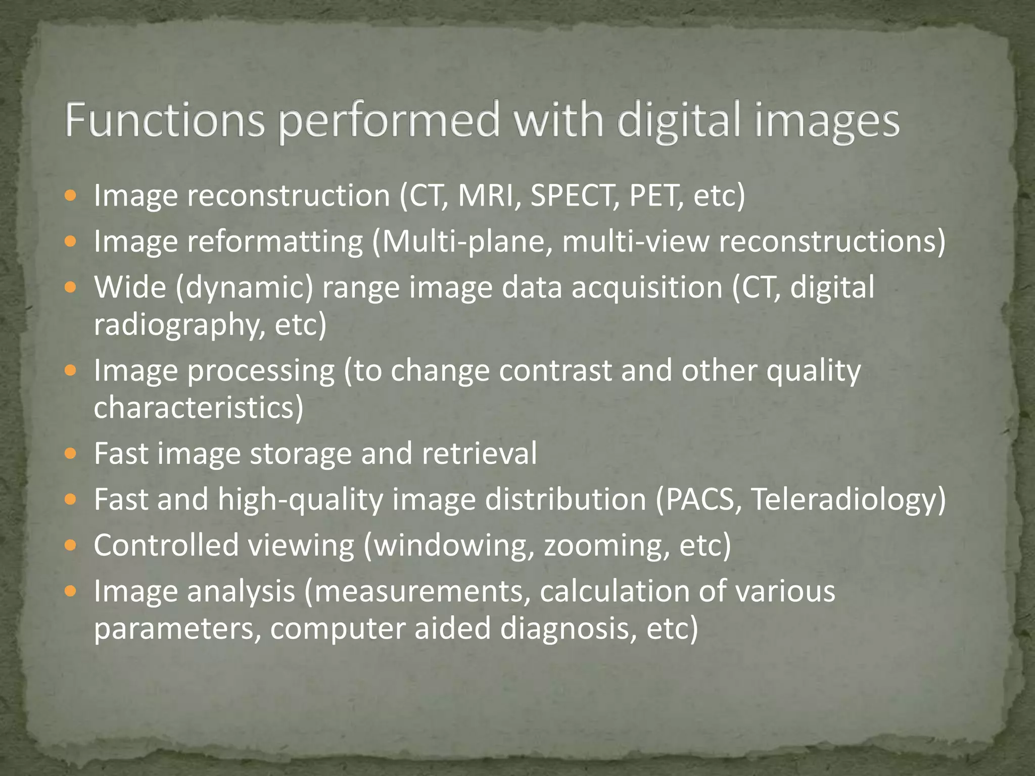
Key benefits of digital images; includes image reconstruction, storage, transfer, and analysis.
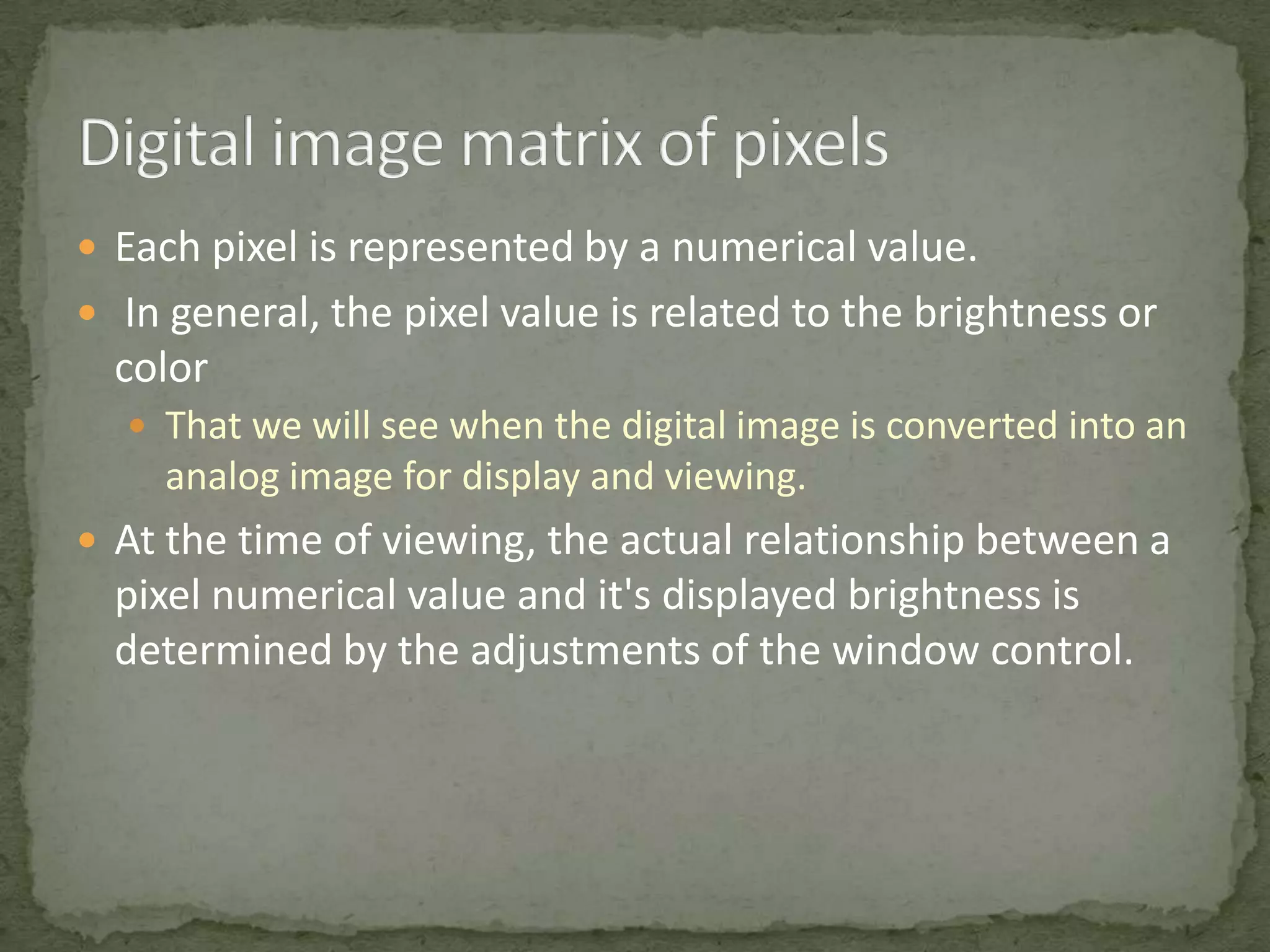
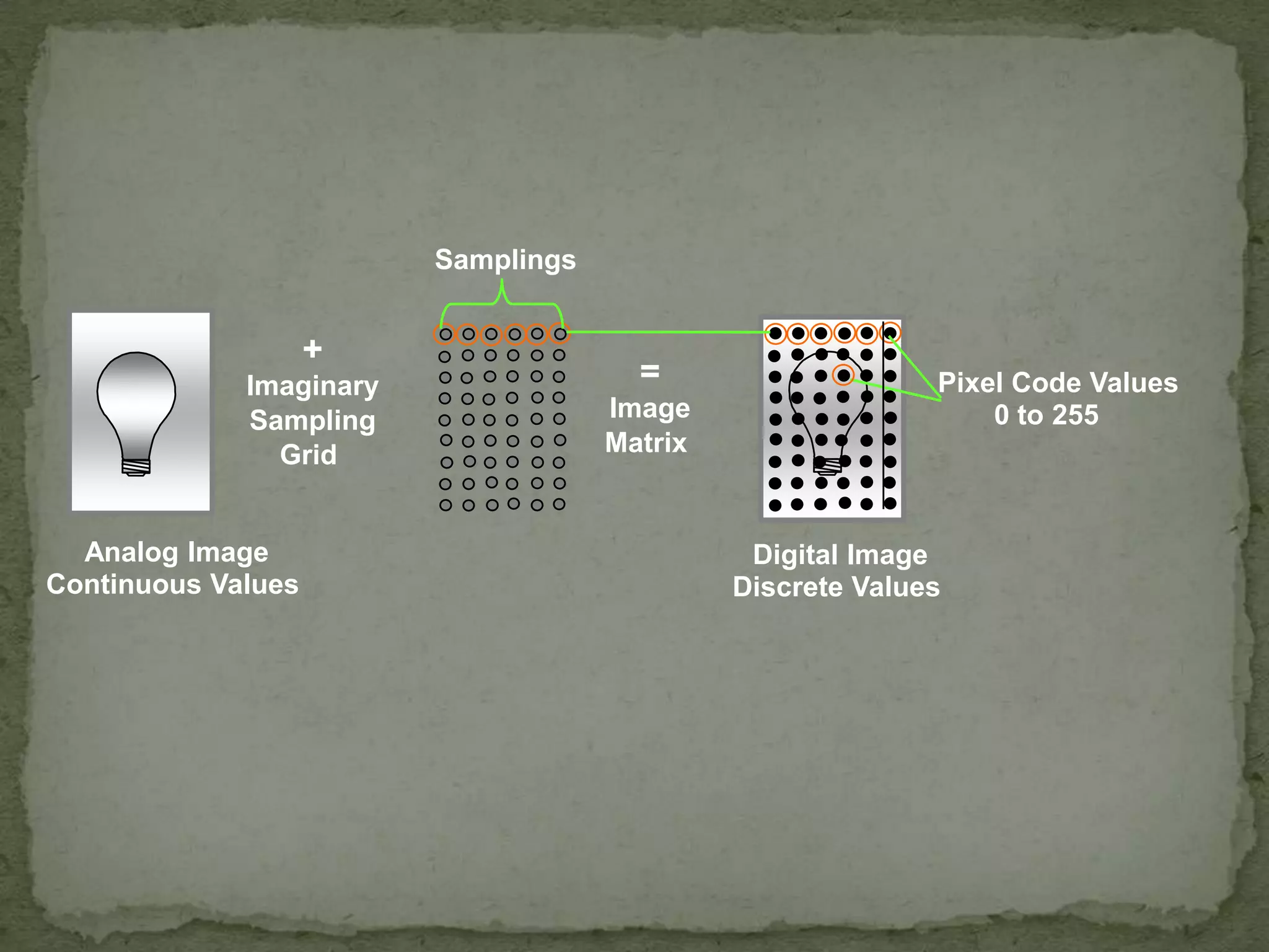
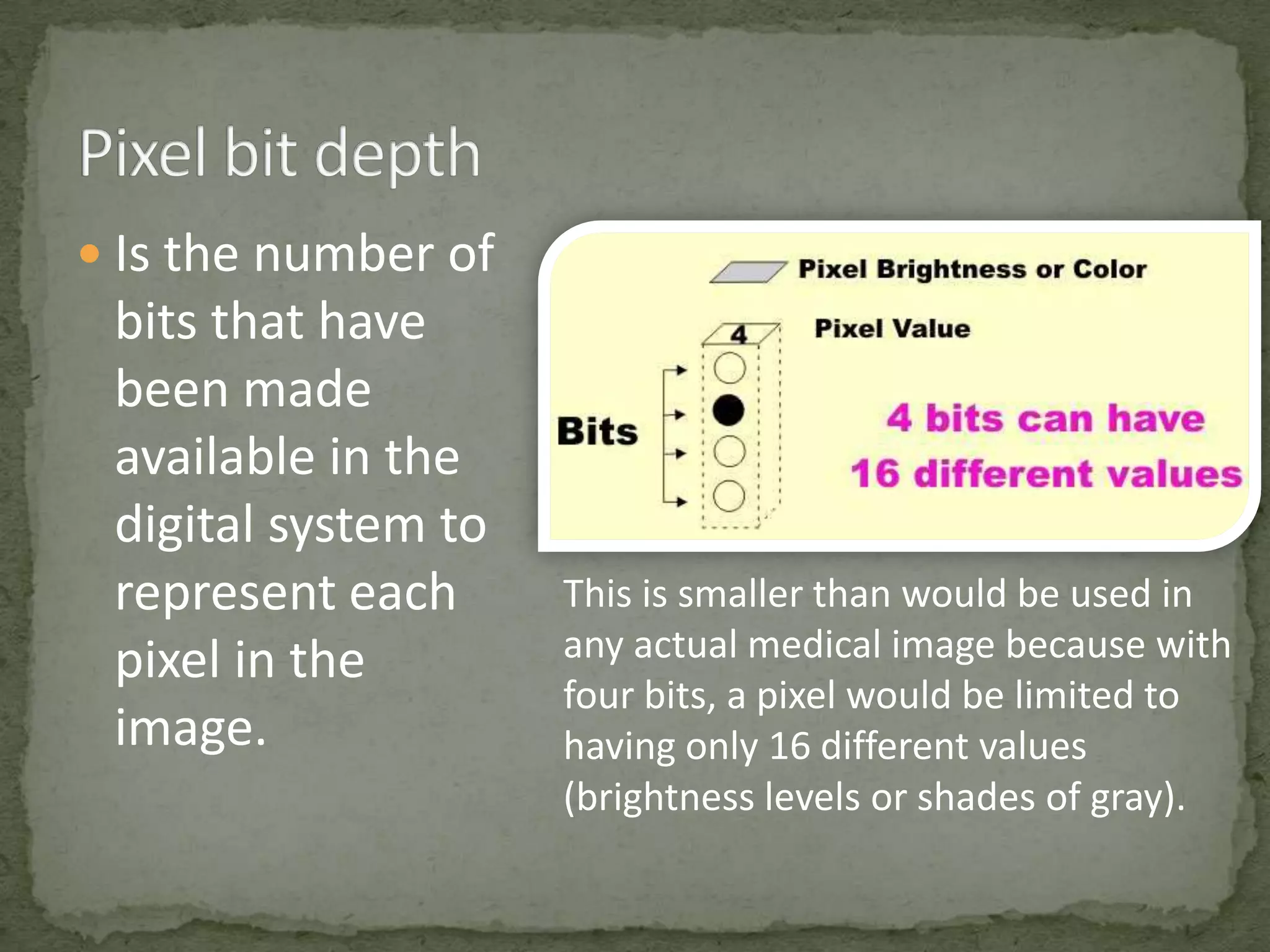
Description of pixel values in digital images and the AD conversion process for different viewing.
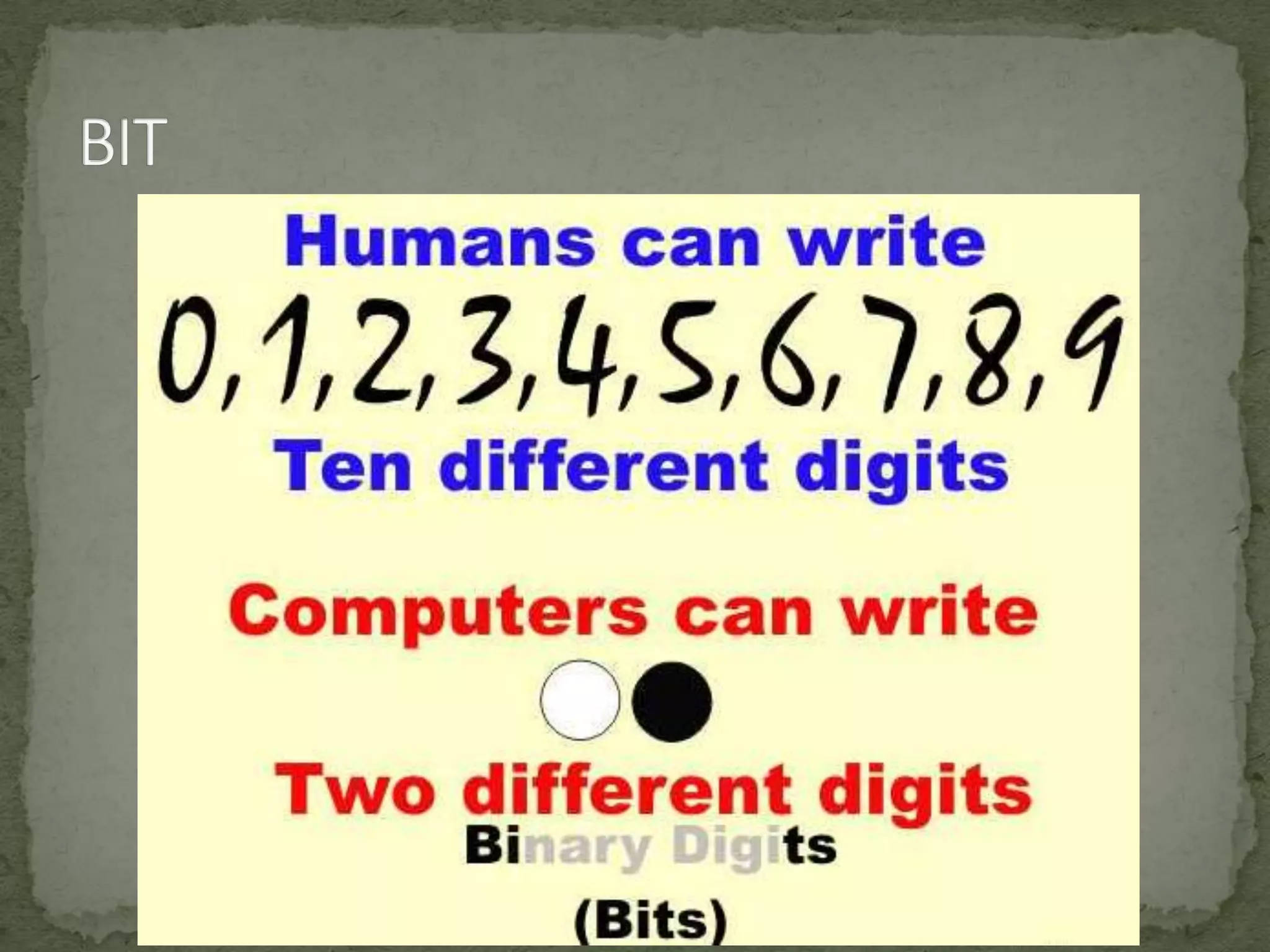
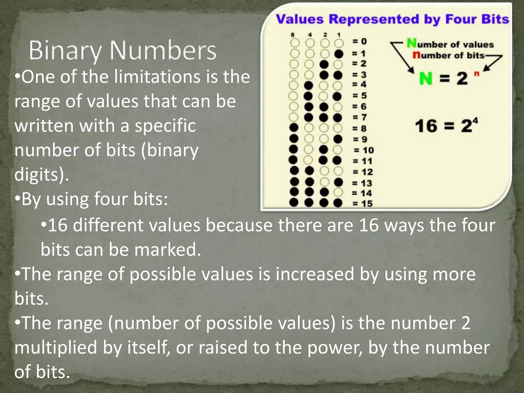
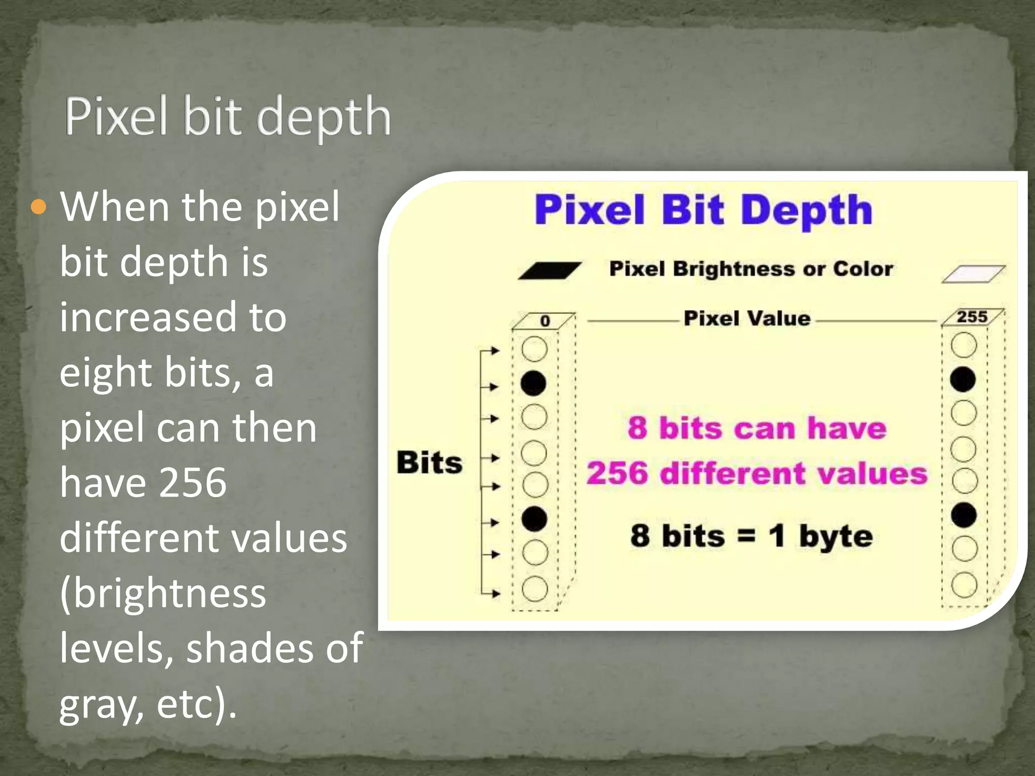
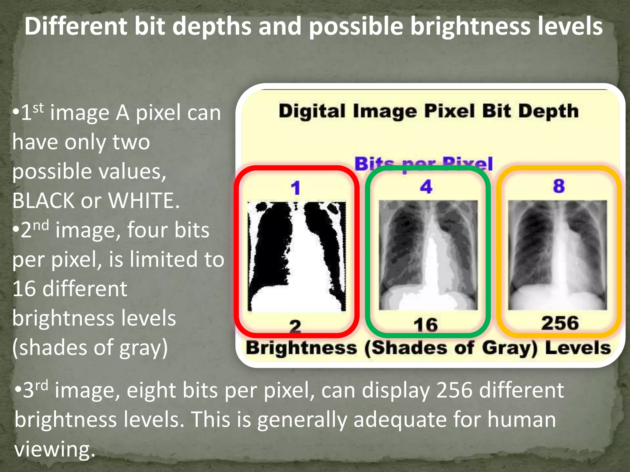
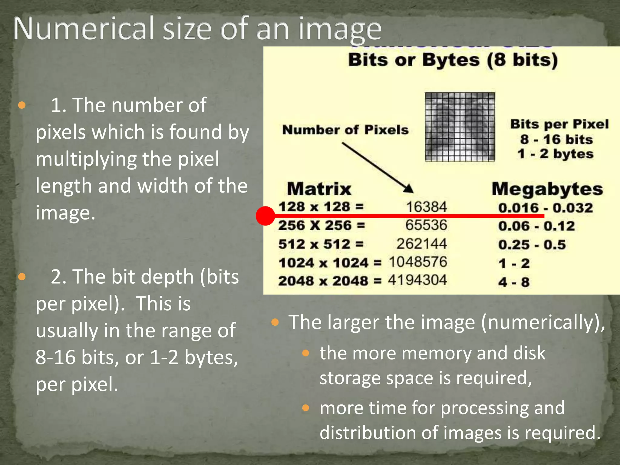
Impact of bit depth on image quality; explanations of pixel value ranges and their implications.
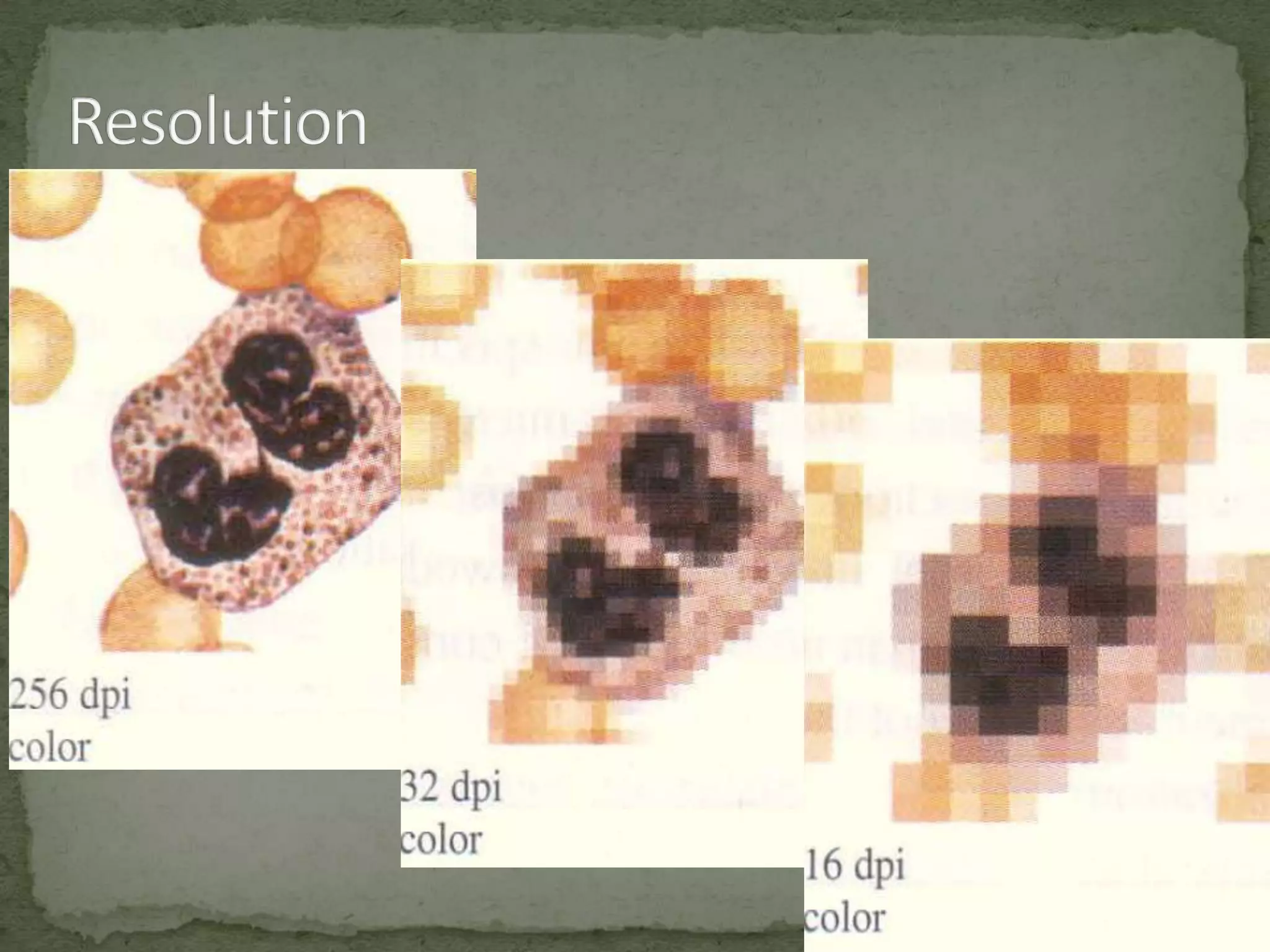
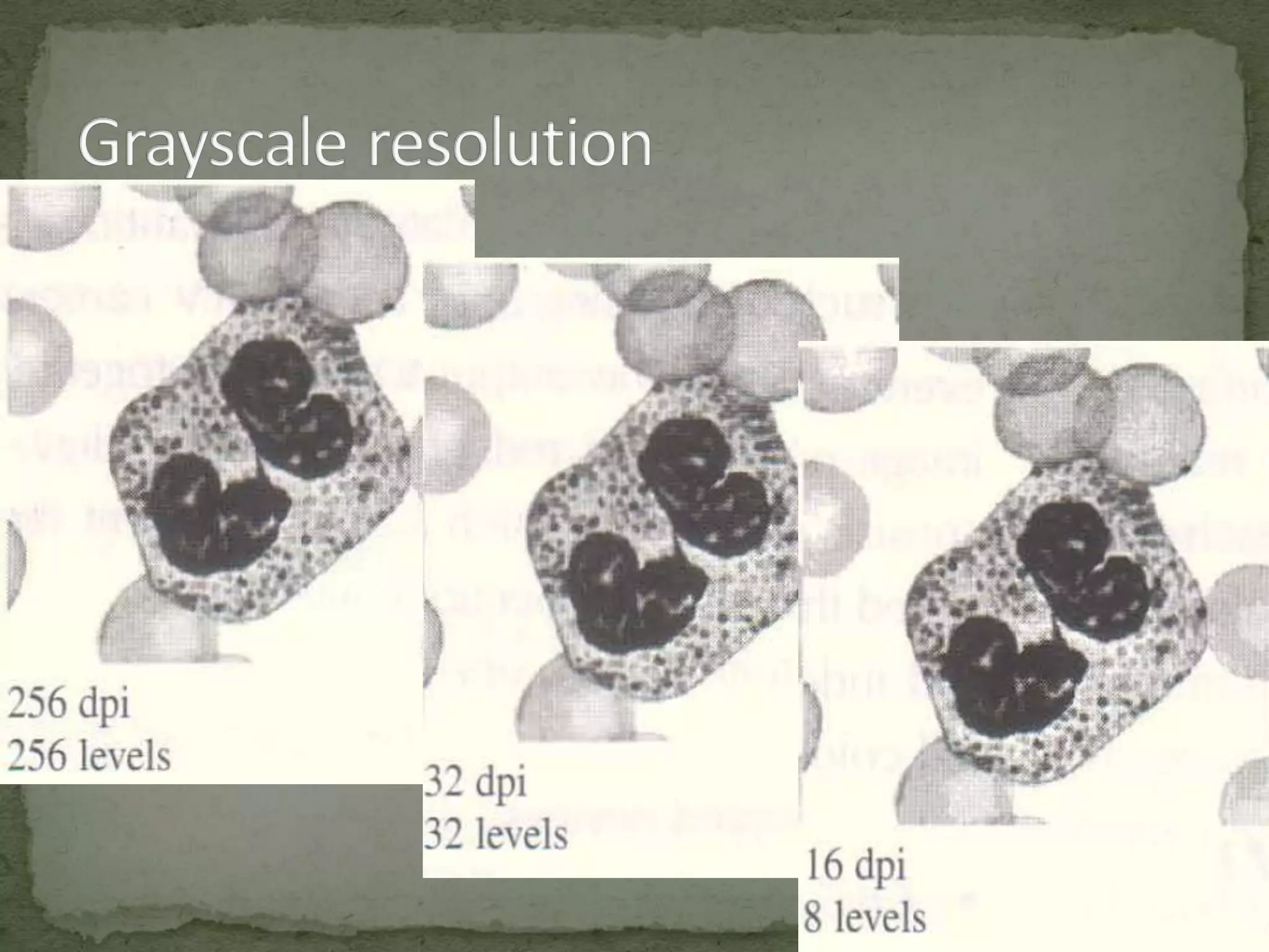
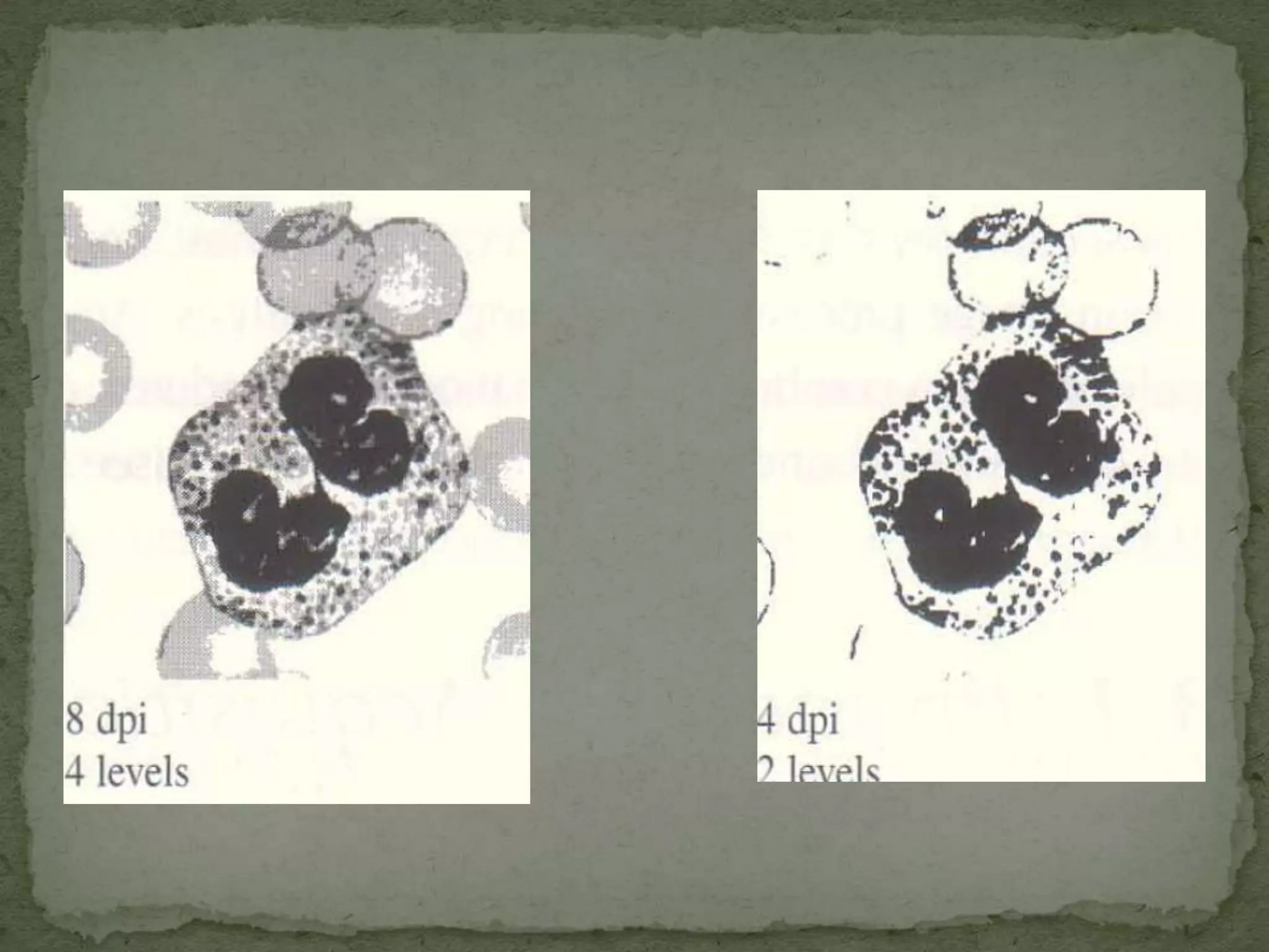
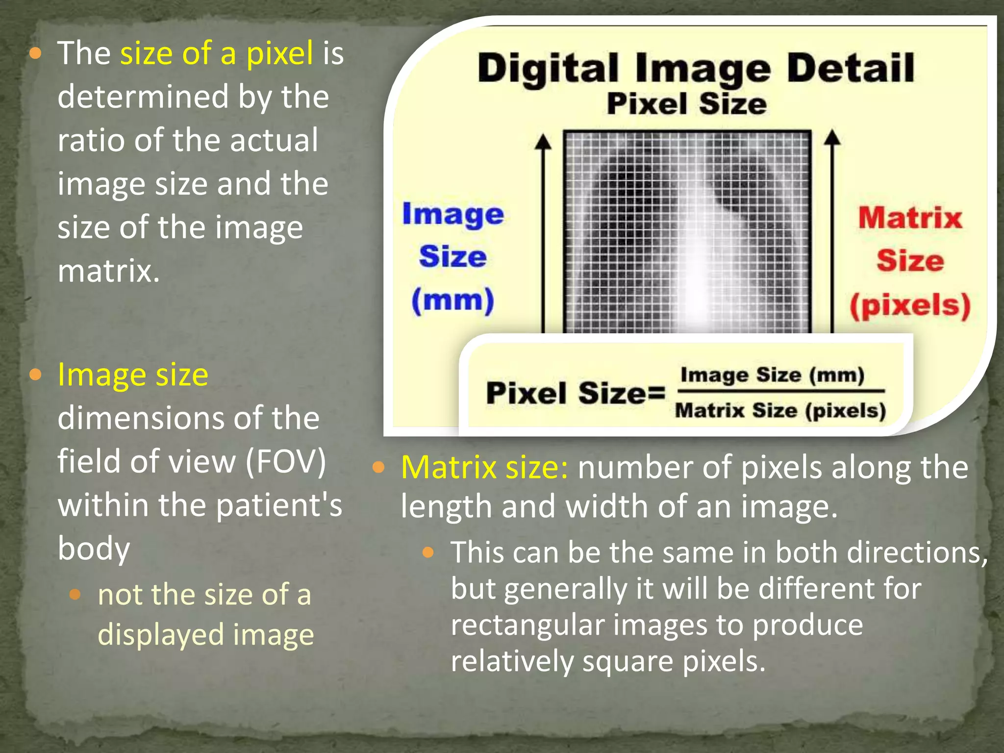
Definition of image resolution; the relationship between pixel count and image quality in detail.
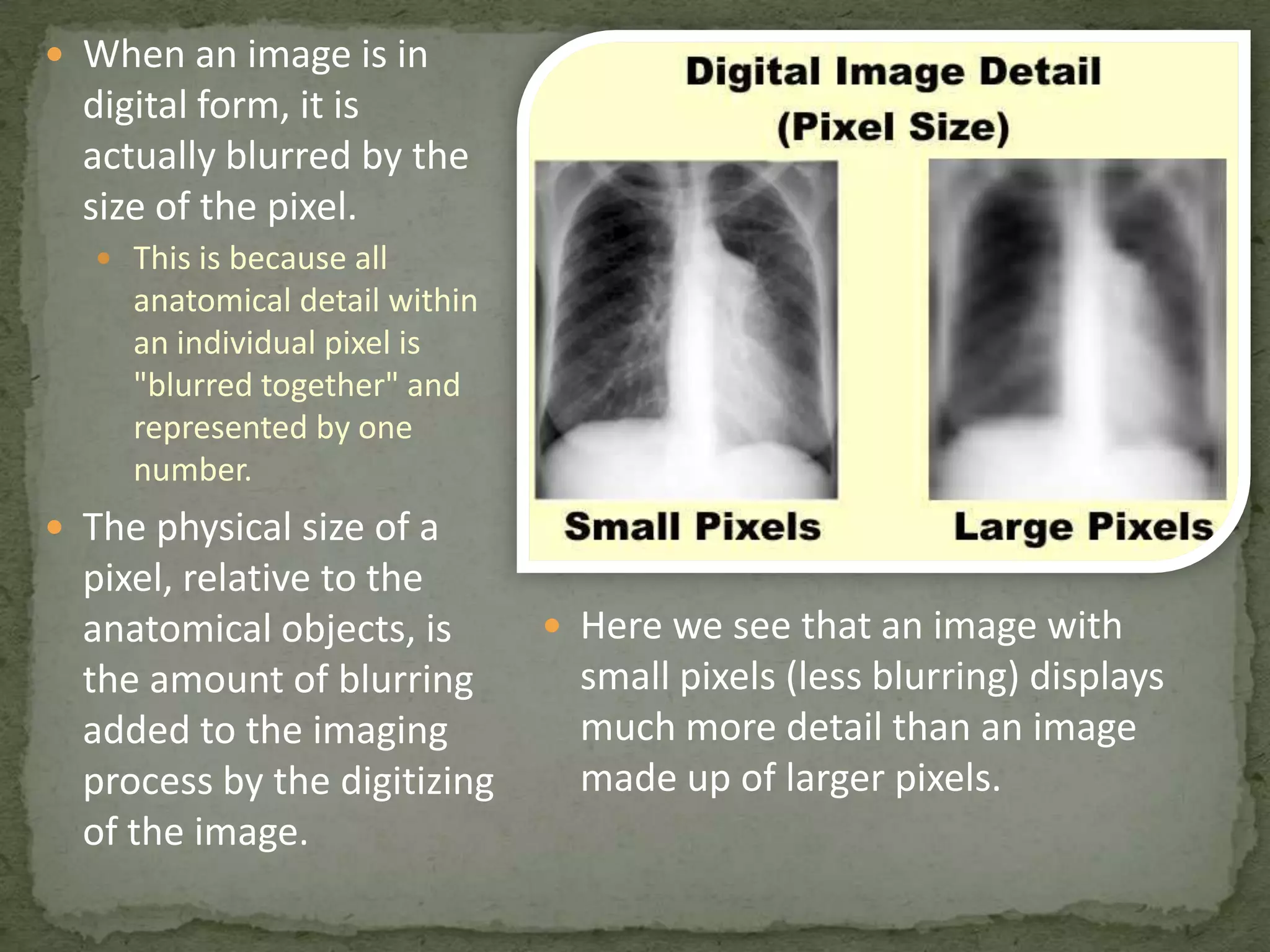
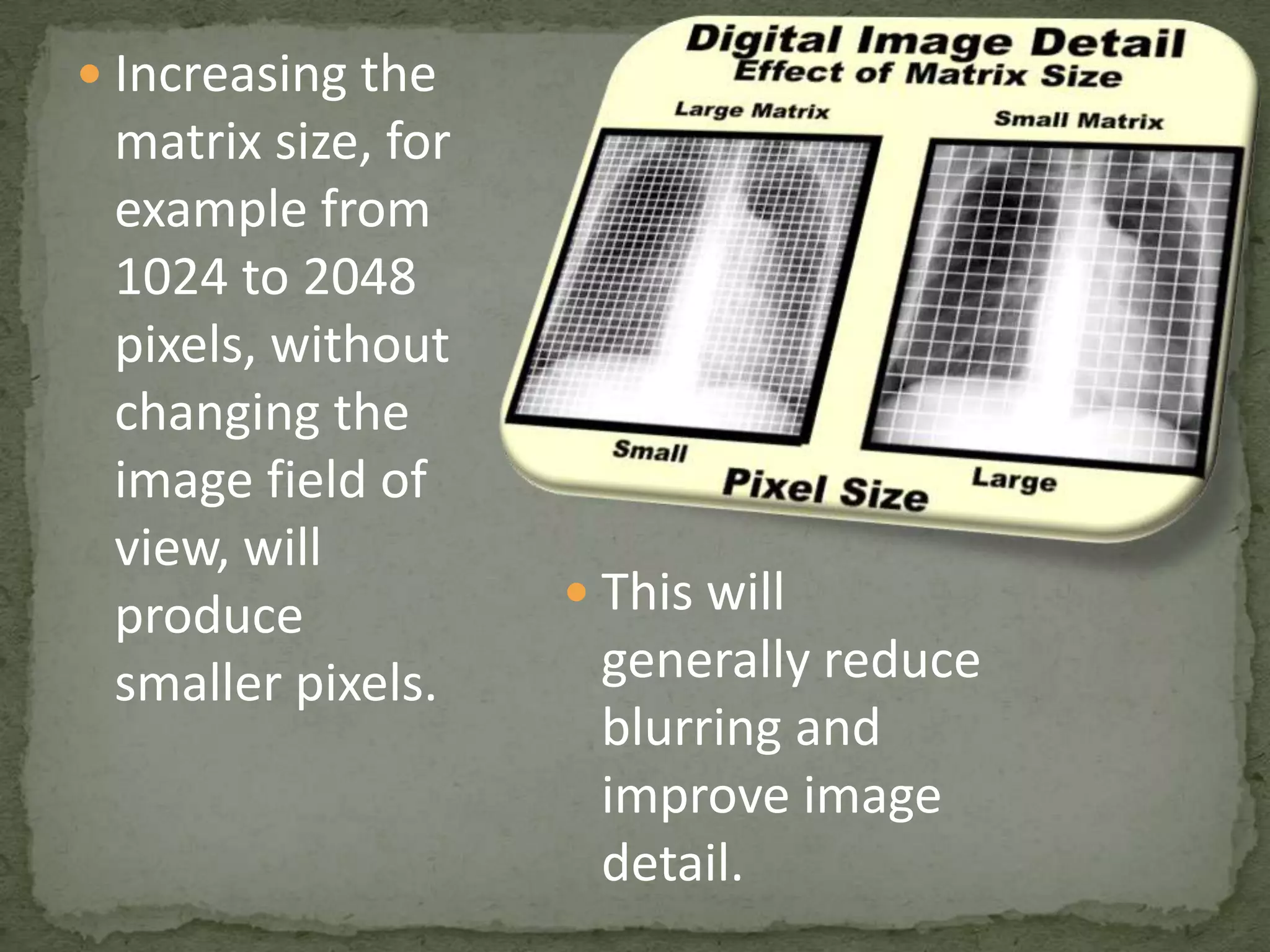
Effects of pixel size on image detail and blurriness; discussion on matrix size increases.
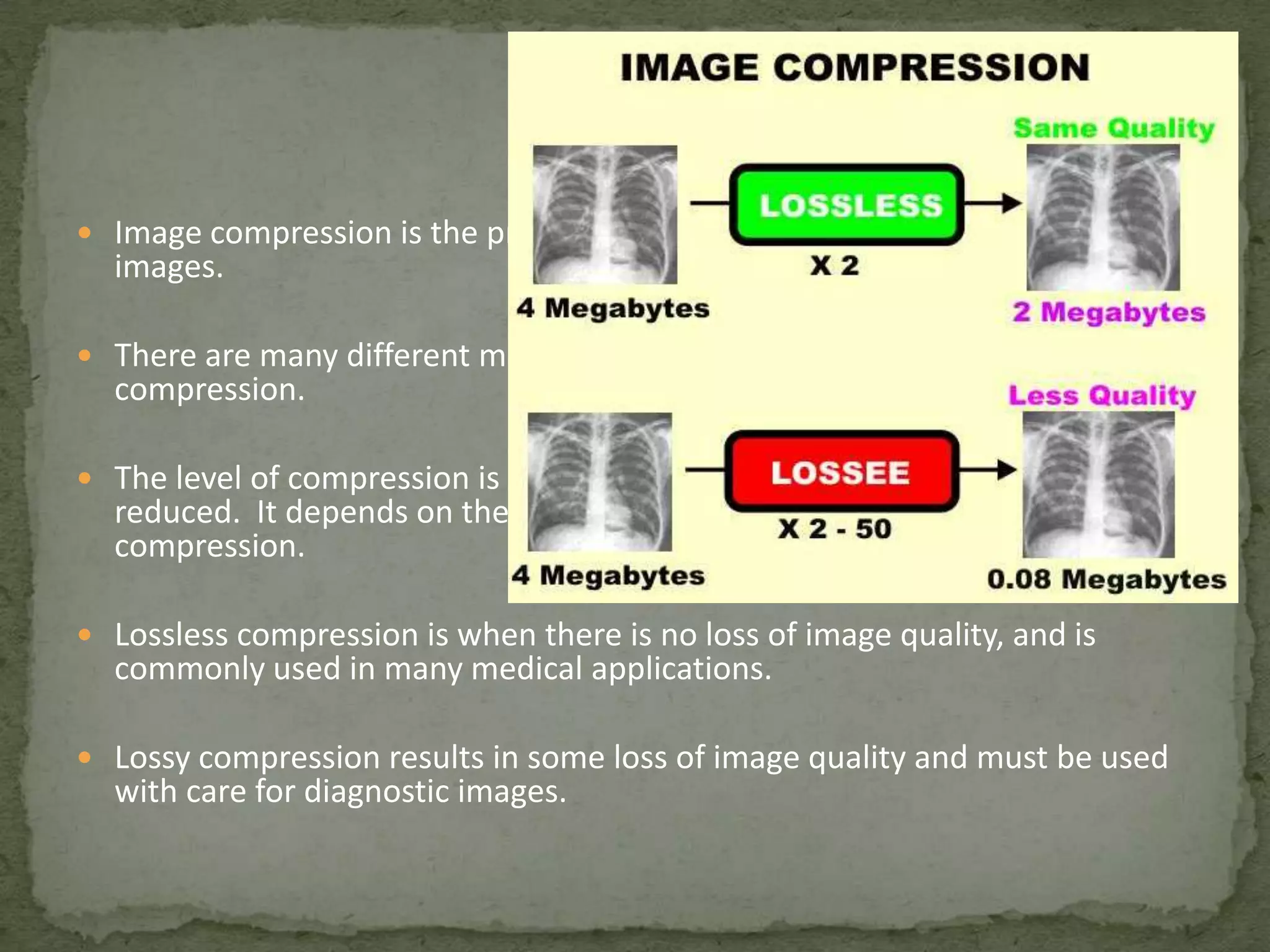
Different matrix sizes for imaging modalities; effects on image storage and compression techniques.


![[0] BASIC CONCEPT OF IMAGE.pdf](https://cdn.slidesharecdn.com/ss_thumbnails/0basicconceptofimage-230209023834-018ac04c-thumbnail.jpg?width=640&height=640&fit=bounds)


