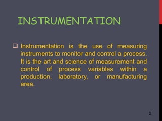
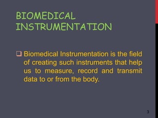
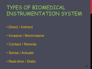
BIOMEDICAL
INSTRUMENTATION
PRESENTED BY: MD. SHAHEED AKHTER
BRANCH: EEE (3RD YEAR)
ROLL NO: 1200113019
PRESENTED TO: ER A. F. MINAI
ER F. A. KHAN
1
 Instrumentation is the use of measuring
instruments to monitor and control a process.
It is the art and science of measurement and
control of process variables within a
production, laboratory, or manufacturing
area.
INSTRUMENTATION
2
 Biomedical Instrumentation is the field
of creating such instruments that help
us to measure, record and transmit
data to or from the body.
BIOMEDICAL
INSTRUMENTATION
3
• Direct / Indirect
• Invasive / Noninvasive
• Contact / Remote
• Sense / Actuate
• Real-time / Static
TYPES OF BIOMEDICAL
INSTRUMENTATION SYSTEM
4
There are many instruments used in
biomedical such as:
 X-Rays
 Electrocardiography (ECG)
 MRI
 Ultrasound
 CT Scan
INSTRUMENTS USED
5
X-RAYS
 The frequency of x-rays as approximately
1020 Hz and its wave length is approximately
0.01 to 10 nanometer.
 It consist of high vacuum tube with a heater,
cathode and anode, vacuum tube, a large DC
voltage is used between cathode and anode of
x-rays tube.
6
HOW IT PRODUCED
 When heater is on and very high
anode to cathode voltage is applied
the electron emits from cathode and
travel toward the anode with very
high Velocity.
 This beam of electron strike the
metal anode such speed that new
rays are made from the slanting
surface of the anode.
 These rays are x-rays, seem to
bounce sideways out thought the
well of the tube.
7
BLOCK DIAGRAM
8
ELECTROCARDIOGRAPHY
 Electrocardiography is the recording of the
electrical activity of the heart.
 It picks up electrical impulses generated by
the polarization and depolarization of
cardiac tissue and translates into a
waveform.
9
 It detects and amplifies the tiny electrical
changes on the skin that are caused when
the heart muscle depolarizes during
each heartbeat.
 At rest, each heart muscle cell has a
negative charge, called the membrane
potential, across its cell membrane.
10
CONT…
ECG SCREEN
11
MAGNETIC RESONANCE
IMAGING
12
 Magnetic resonance imaging (MRI) makes use of the
magnetic properties of certain atomic nuclei.
 The hydrogen nuclei behave like compass needles
that are partially aligned by a strong magnetic field in
the scanner.
 MRI does not involve radioactivity or ionising radiation.
The frequencies used (typically 40-130 MHz) are in the
normal radiofrequency range, and there are no adverse
health effects.
13
Advantages:
MRI is particularly useful for the scanning and detection of
abnormalities in soft tissue structures in the body
 There is no involvement of any kind of radiations in the
MRI.
 MRI scan can provide information about the blood
circulation throughout the body and blood vessels.
14
Disadvantages:
 MRI scan is done in an enclosed space, i.e. fearful of
being in a closely enclosed surface, are facing
problems with MRI to be done.
 MRI scans involve really loud noises while
processing because they involve a really high amount
of electric current supply.
 MRI scanners are usually expensive.
15
ULTRASOUND
The frequencies of ultrasound required for medical
imaging are in the range 1 - 20 MHz.
Ultrasound is an oscillating sound pressure wave with
a frequency greater than the upper limit of the human
hearing range.
 Ultrasound can be used for medical imaging,
detection, measurement and cleaning.
16
17
ADVANTAGE
 Usually non-invasive, safe and relatively painless
 Uses no ionising radiation
 Does not usually require injection of a contrast
medium (dye)
DISADVANTAGES
 Quality and interpretation of the image highly depends on the skill
of the person doing the scan.
 Use of a special probe is required in some ultrasounds
 Special preparations may be required before a procedure (e.g.
fasting or a full bladder)
18
COMPUTERIZED TOMOGRAPHY
 A 'computerized tomography' (CT) uses a
computer that takes data from several X-
ray images of structures inside a human's
or animal's body and converts them into
pictures on a monitor.
19
WORKING
 A CT scanner emits a series of narrow beams
through the human body as it moves through an
arc.
 Inside the CT scanner there is an X-ray detector
which can see hundreds of different levels of
density. It can see tissues inside a solid organ. This
data is transmitted to a computer, which builds up a
3D cross-sectional picture of the part of the body
and displays it on the screen.
20
ADVANTAGES
Quick and painless
Can help diagnose and guide treatment for a wider
range of conditions than plain X-rays
Can detect or exclude the presence of more
serious problems
DISADVANTAGES
 Small increased risk of cancer in future from exposure
to ionising radiation.
 Uses higher doses of radiation, so the risks (while still
small) are in general greater than other imaging types 21
22
REFERENCE
Introduction to biomedical equipment
technology; J.J. Carr
Medical Instrumentation; Webster
Electronic devices; Boylestad
23
Biomedical instrumentation PPT

Biomedical instrumentation PPT

  • 1.
    BIOMEDICAL INSTRUMENTATION PRESENTED BY: MD.SHAHEED AKHTER BRANCH: EEE (3RD YEAR) ROLL NO: 1200113019 PRESENTED TO: ER A. F. MINAI ER F. A. KHAN 1
  • 2.
     Instrumentation isthe use of measuring instruments to monitor and control a process. It is the art and science of measurement and control of process variables within a production, laboratory, or manufacturing area. INSTRUMENTATION 2
  • 3.
     Biomedical Instrumentationis the field of creating such instruments that help us to measure, record and transmit data to or from the body. BIOMEDICAL INSTRUMENTATION 3
  • 4.
    • Direct /Indirect • Invasive / Noninvasive • Contact / Remote • Sense / Actuate • Real-time / Static TYPES OF BIOMEDICAL INSTRUMENTATION SYSTEM 4
  • 5.
    There are manyinstruments used in biomedical such as:  X-Rays  Electrocardiography (ECG)  MRI  Ultrasound  CT Scan INSTRUMENTS USED 5
  • 6.
    X-RAYS  The frequencyof x-rays as approximately 1020 Hz and its wave length is approximately 0.01 to 10 nanometer.  It consist of high vacuum tube with a heater, cathode and anode, vacuum tube, a large DC voltage is used between cathode and anode of x-rays tube. 6
  • 7.
    HOW IT PRODUCED When heater is on and very high anode to cathode voltage is applied the electron emits from cathode and travel toward the anode with very high Velocity.  This beam of electron strike the metal anode such speed that new rays are made from the slanting surface of the anode.  These rays are x-rays, seem to bounce sideways out thought the well of the tube. 7
  • 8.
  • 9.
    ELECTROCARDIOGRAPHY  Electrocardiography isthe recording of the electrical activity of the heart.  It picks up electrical impulses generated by the polarization and depolarization of cardiac tissue and translates into a waveform. 9
  • 10.
     It detectsand amplifies the tiny electrical changes on the skin that are caused when the heart muscle depolarizes during each heartbeat.  At rest, each heart muscle cell has a negative charge, called the membrane potential, across its cell membrane. 10 CONT…
  • 11.
  • 12.
  • 13.
     Magnetic resonanceimaging (MRI) makes use of the magnetic properties of certain atomic nuclei.  The hydrogen nuclei behave like compass needles that are partially aligned by a strong magnetic field in the scanner.  MRI does not involve radioactivity or ionising radiation. The frequencies used (typically 40-130 MHz) are in the normal radiofrequency range, and there are no adverse health effects. 13
  • 14.
    Advantages: MRI is particularlyuseful for the scanning and detection of abnormalities in soft tissue structures in the body  There is no involvement of any kind of radiations in the MRI.  MRI scan can provide information about the blood circulation throughout the body and blood vessels. 14
  • 15.
    Disadvantages:  MRI scanis done in an enclosed space, i.e. fearful of being in a closely enclosed surface, are facing problems with MRI to be done.  MRI scans involve really loud noises while processing because they involve a really high amount of electric current supply.  MRI scanners are usually expensive. 15
  • 16.
    ULTRASOUND The frequencies ofultrasound required for medical imaging are in the range 1 - 20 MHz. Ultrasound is an oscillating sound pressure wave with a frequency greater than the upper limit of the human hearing range.  Ultrasound can be used for medical imaging, detection, measurement and cleaning. 16
  • 17.
  • 18.
    ADVANTAGE  Usually non-invasive,safe and relatively painless  Uses no ionising radiation  Does not usually require injection of a contrast medium (dye) DISADVANTAGES  Quality and interpretation of the image highly depends on the skill of the person doing the scan.  Use of a special probe is required in some ultrasounds  Special preparations may be required before a procedure (e.g. fasting or a full bladder) 18
  • 19.
    COMPUTERIZED TOMOGRAPHY  A'computerized tomography' (CT) uses a computer that takes data from several X- ray images of structures inside a human's or animal's body and converts them into pictures on a monitor. 19
  • 20.
    WORKING  A CTscanner emits a series of narrow beams through the human body as it moves through an arc.  Inside the CT scanner there is an X-ray detector which can see hundreds of different levels of density. It can see tissues inside a solid organ. This data is transmitted to a computer, which builds up a 3D cross-sectional picture of the part of the body and displays it on the screen. 20
  • 21.
    ADVANTAGES Quick and painless Canhelp diagnose and guide treatment for a wider range of conditions than plain X-rays Can detect or exclude the presence of more serious problems DISADVANTAGES  Small increased risk of cancer in future from exposure to ionising radiation.  Uses higher doses of radiation, so the risks (while still small) are in general greater than other imaging types 21
  • 22.
  • 23.
    REFERENCE Introduction to biomedicalequipment technology; J.J. Carr Medical Instrumentation; Webster Electronic devices; Boylestad 23