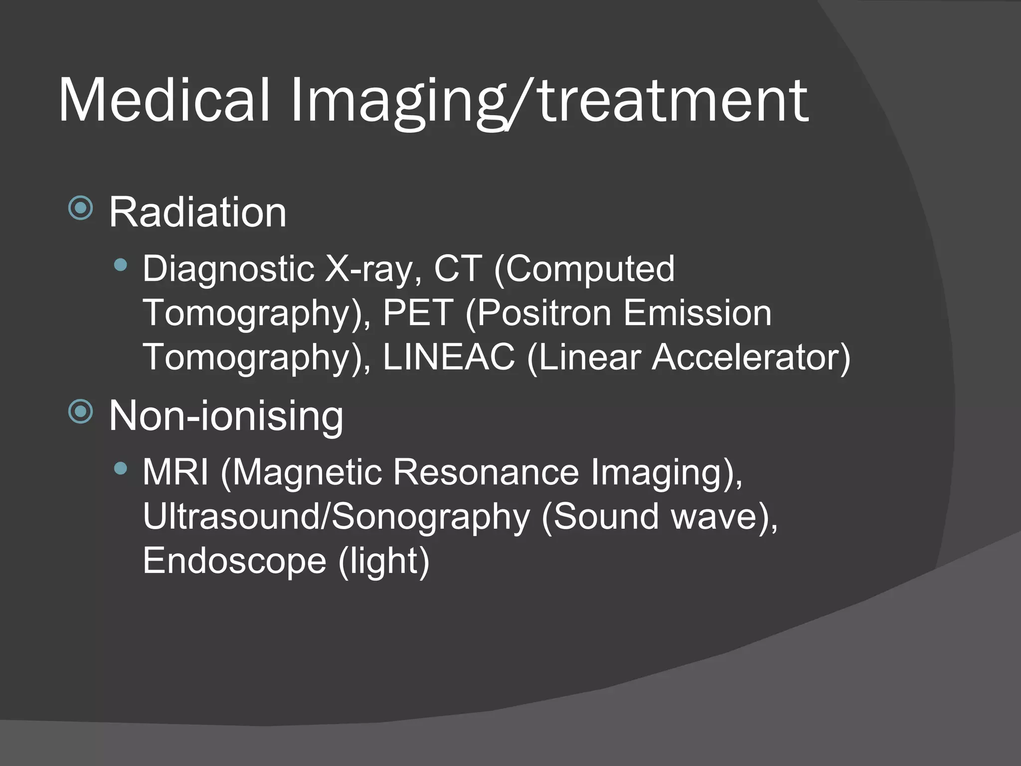
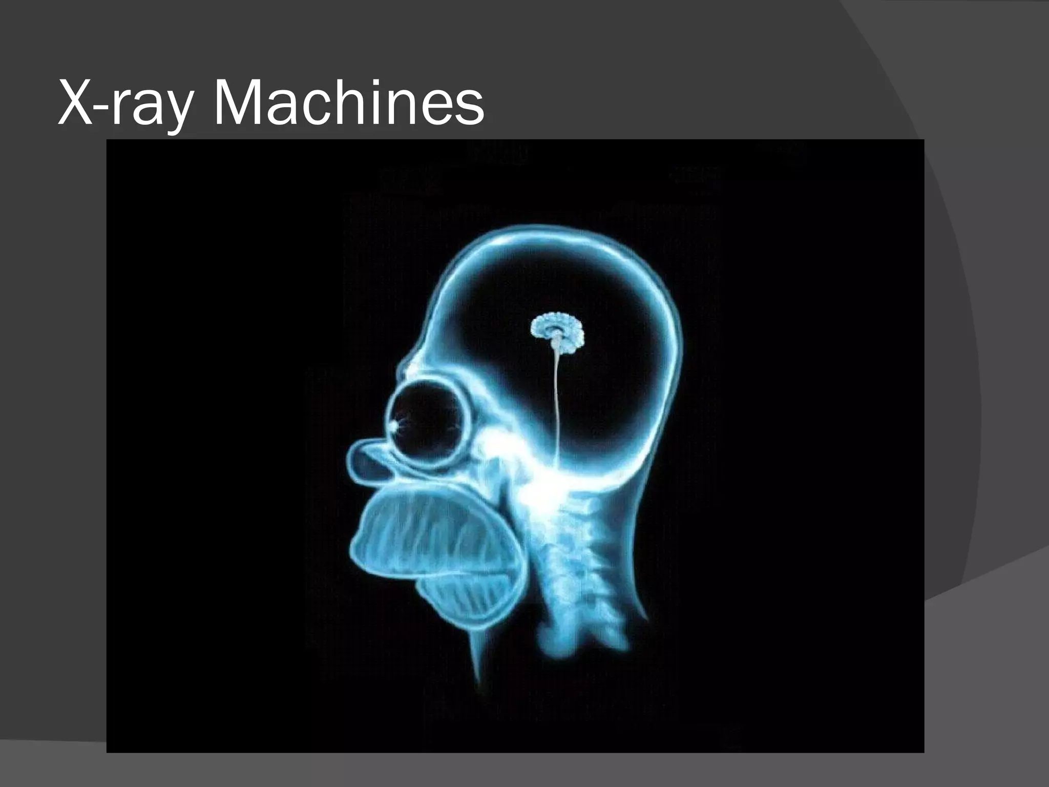
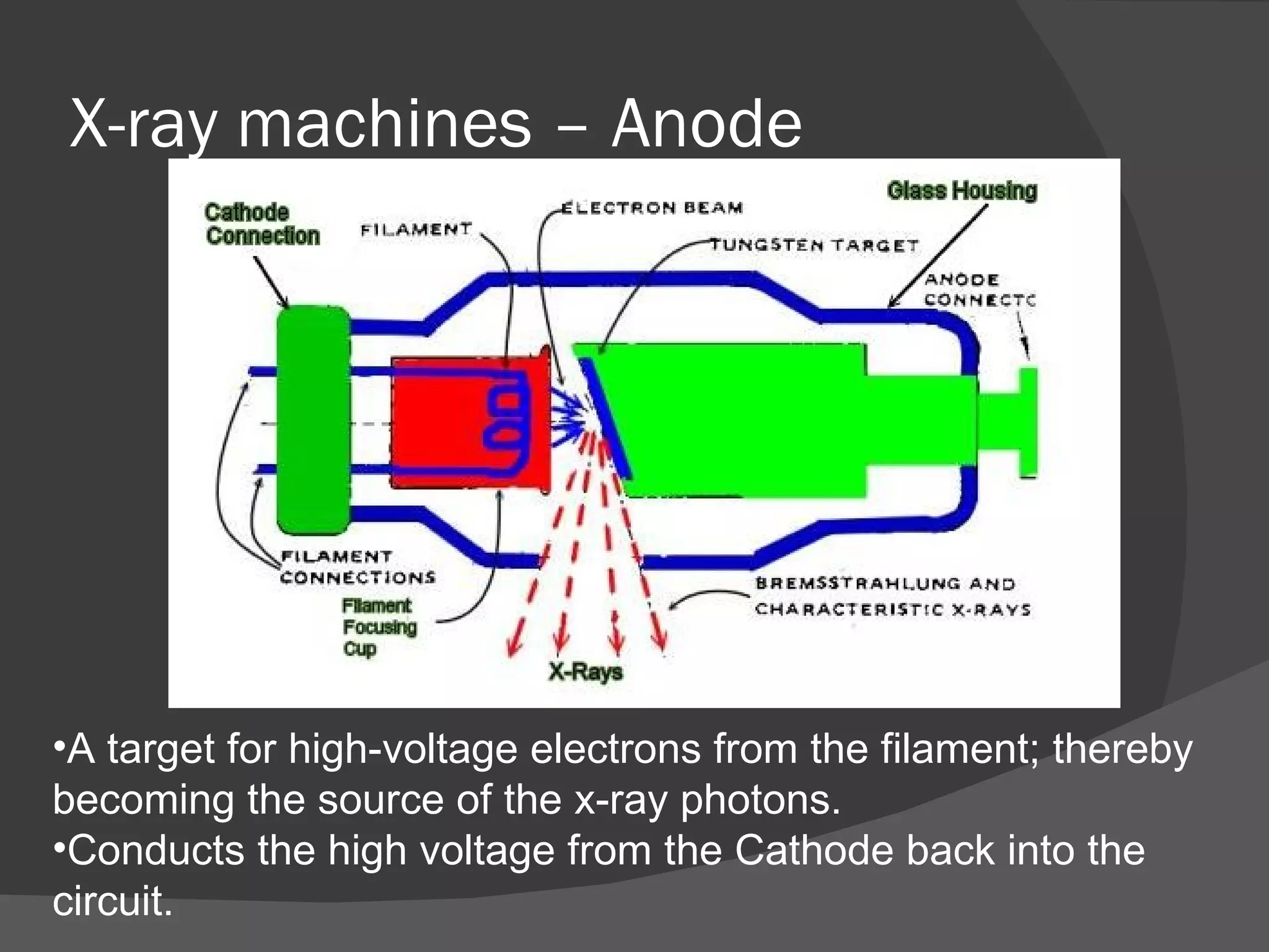
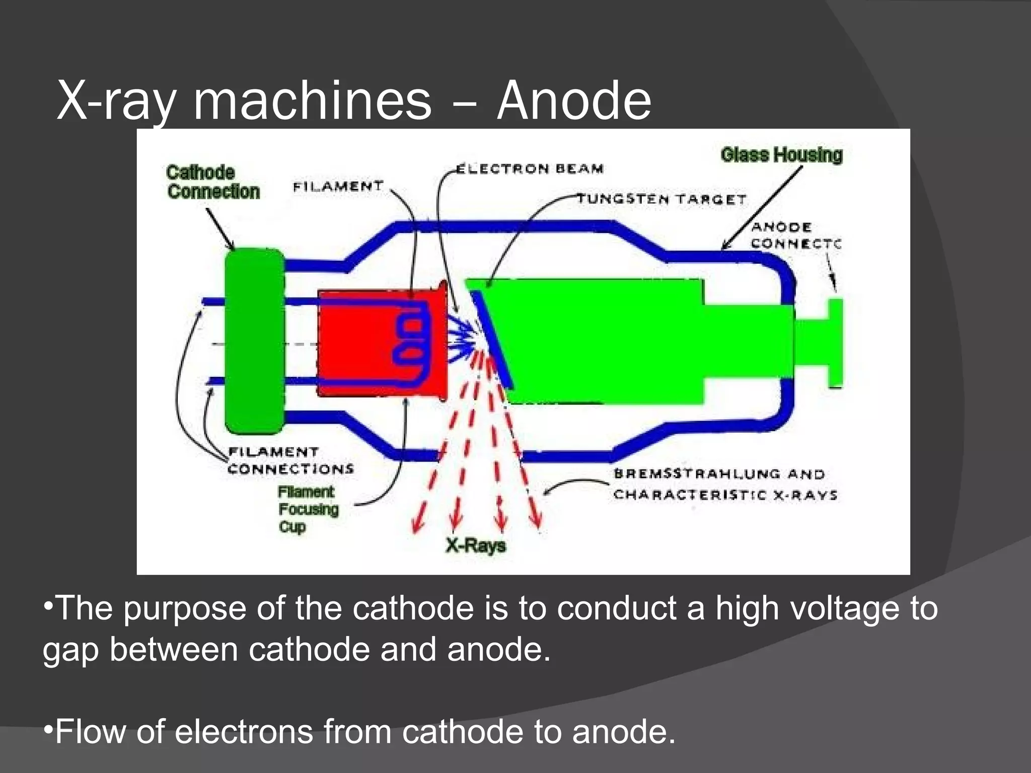
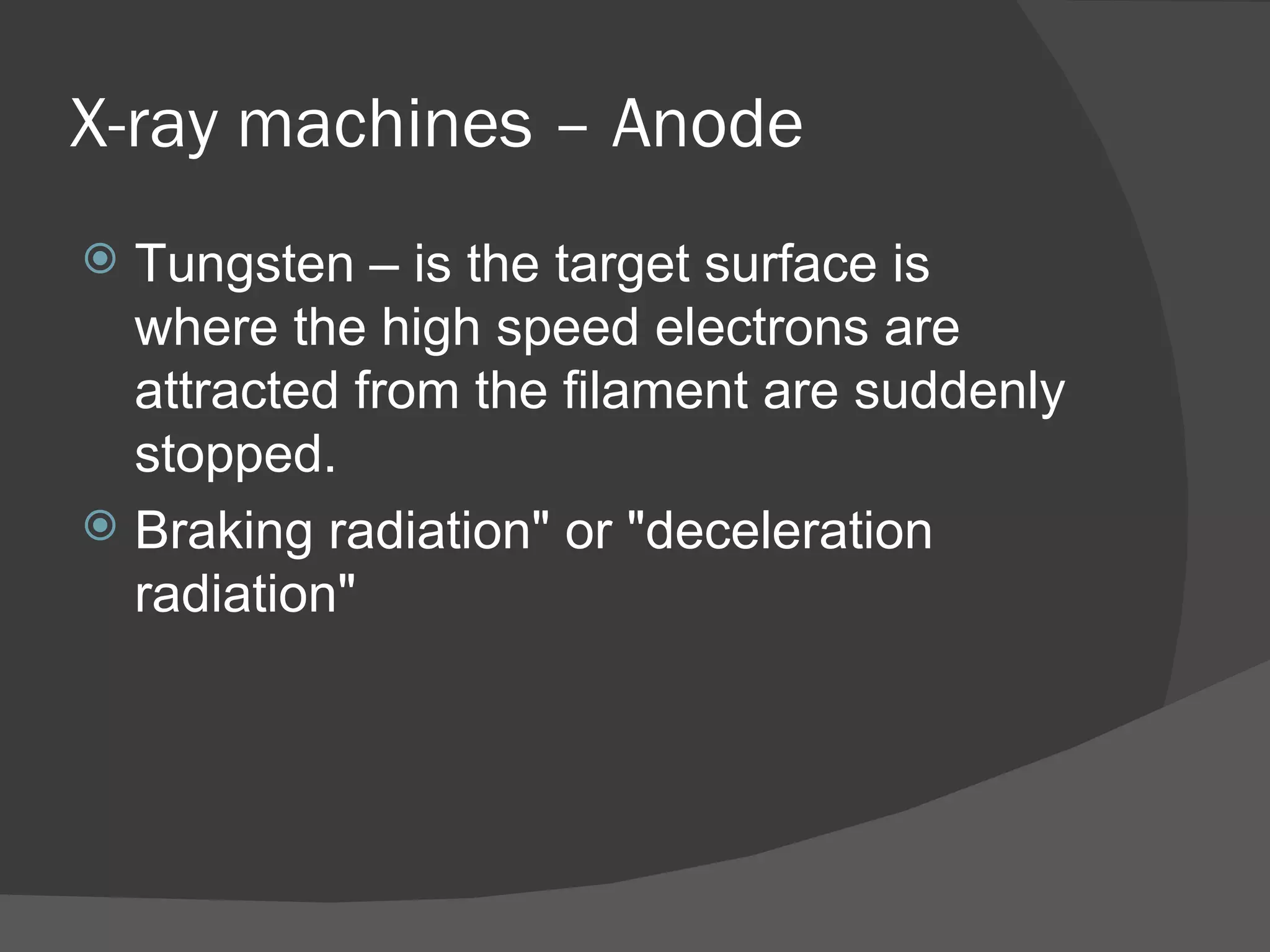
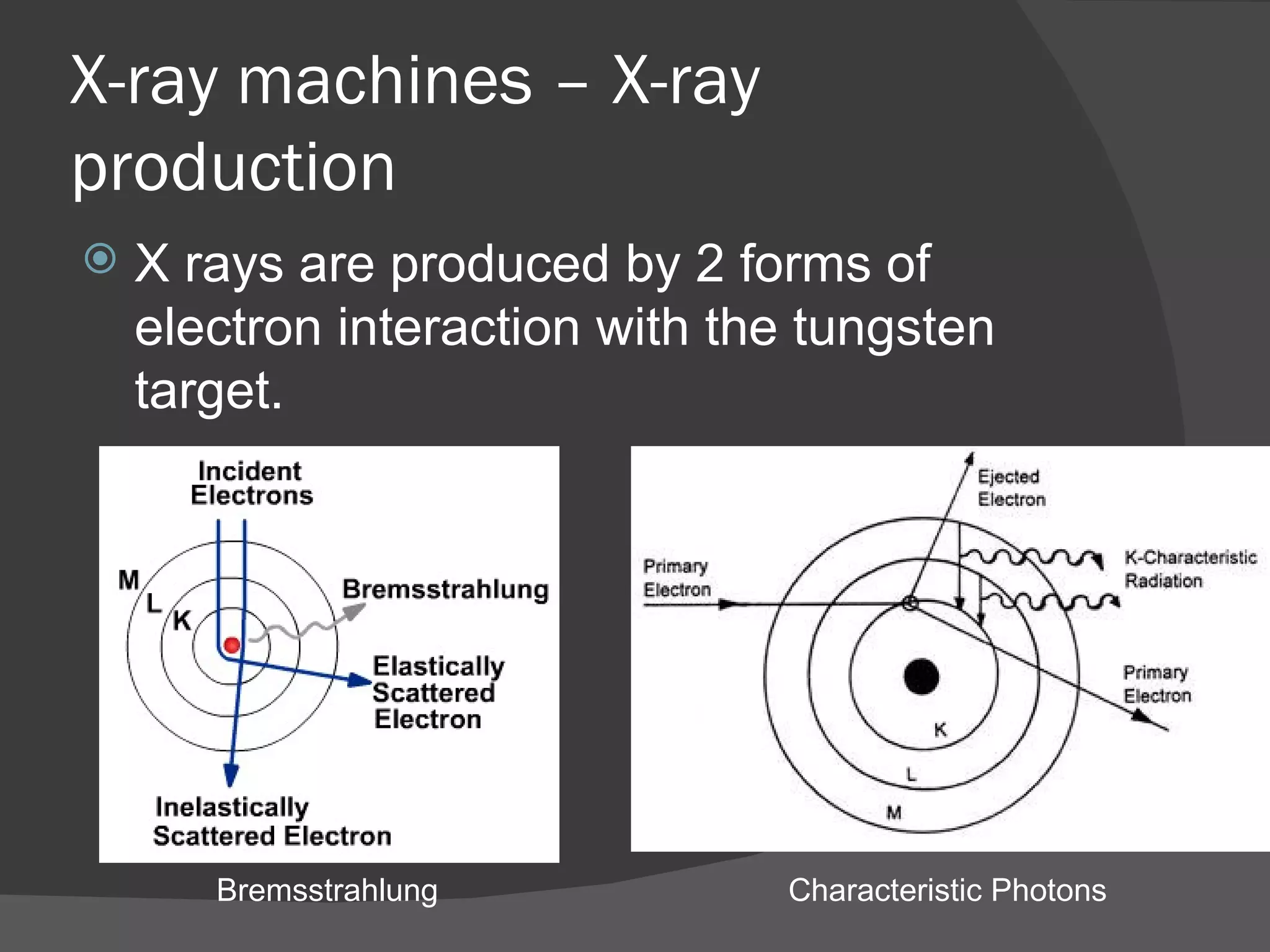
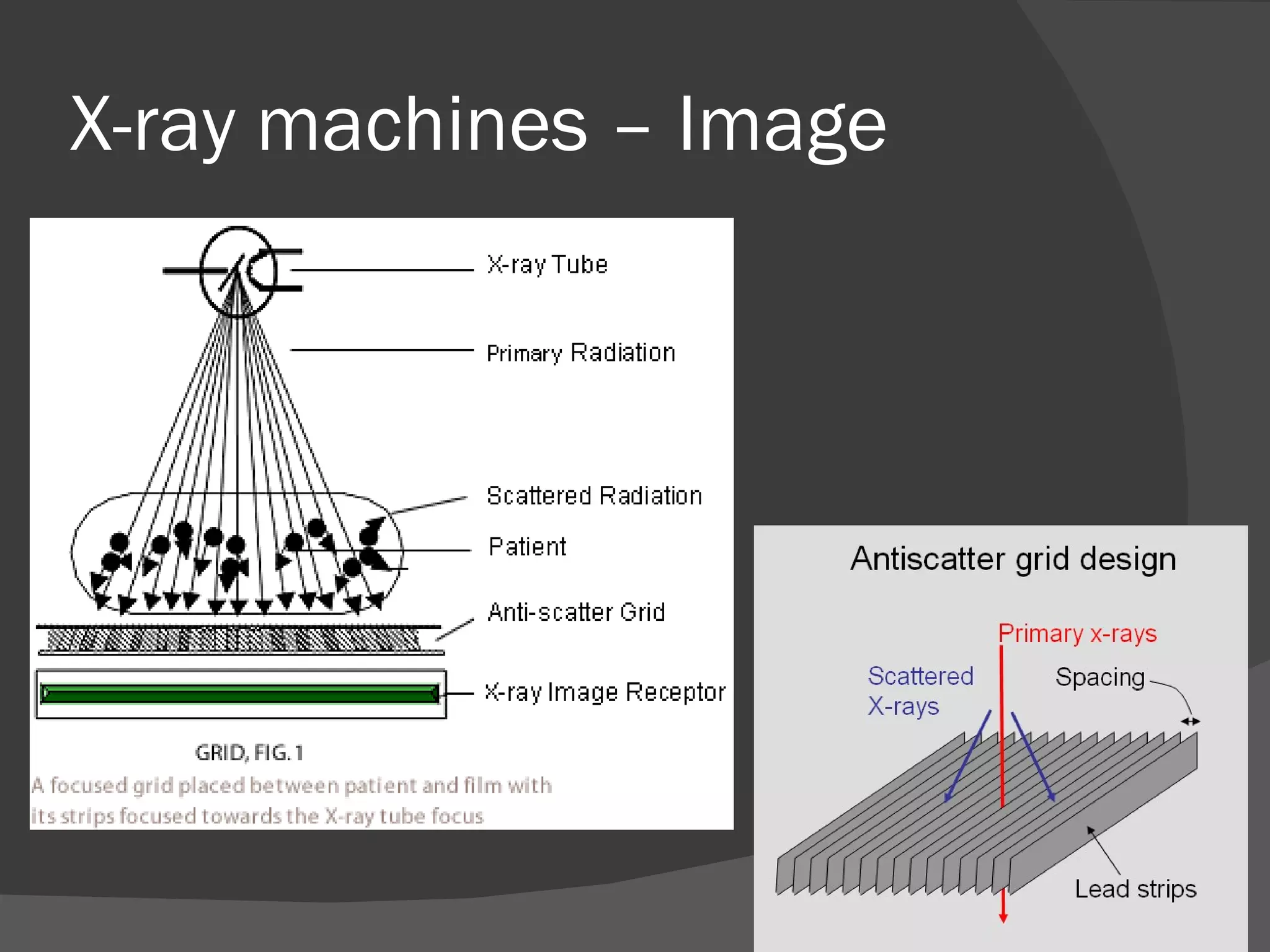
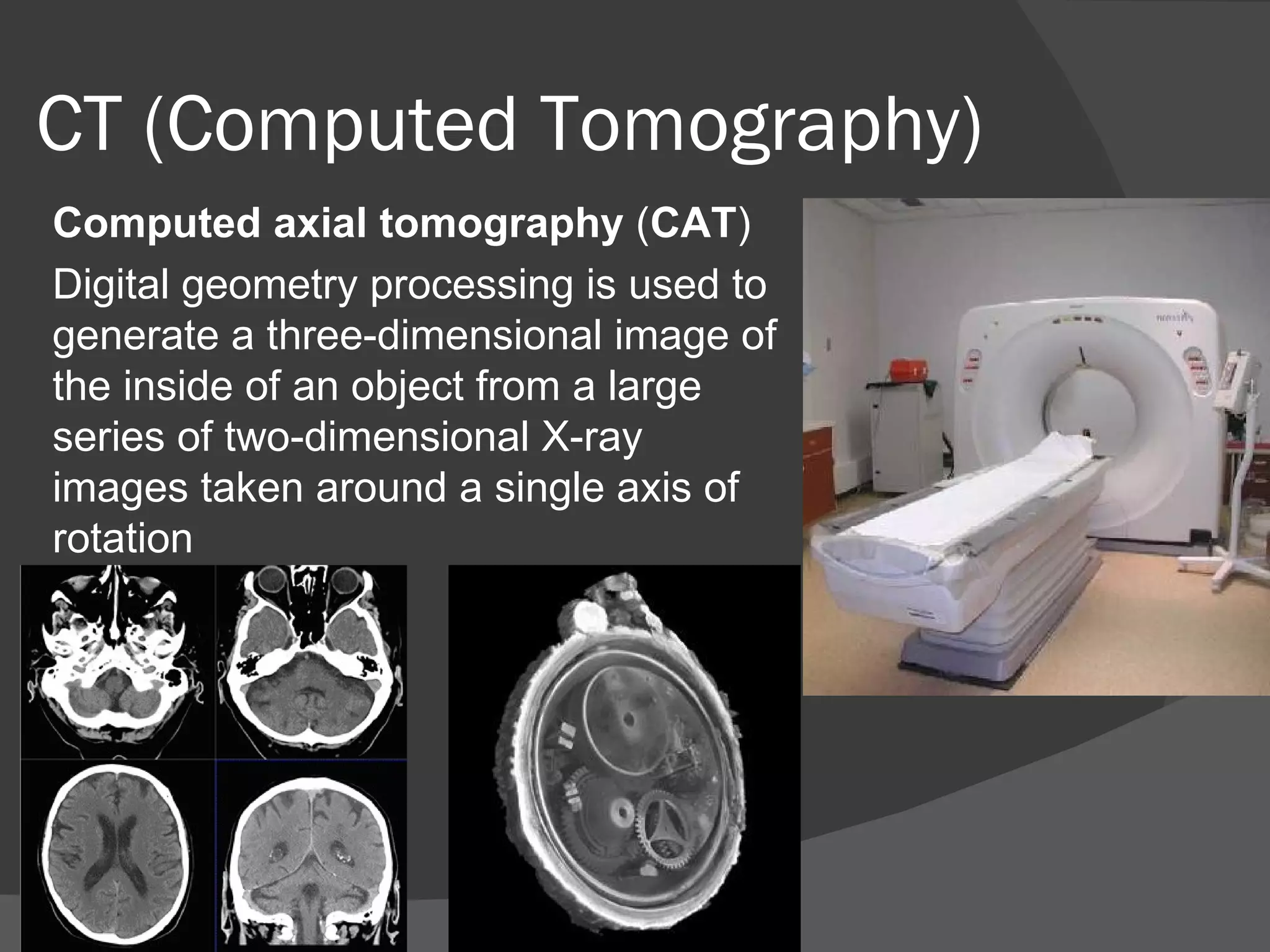
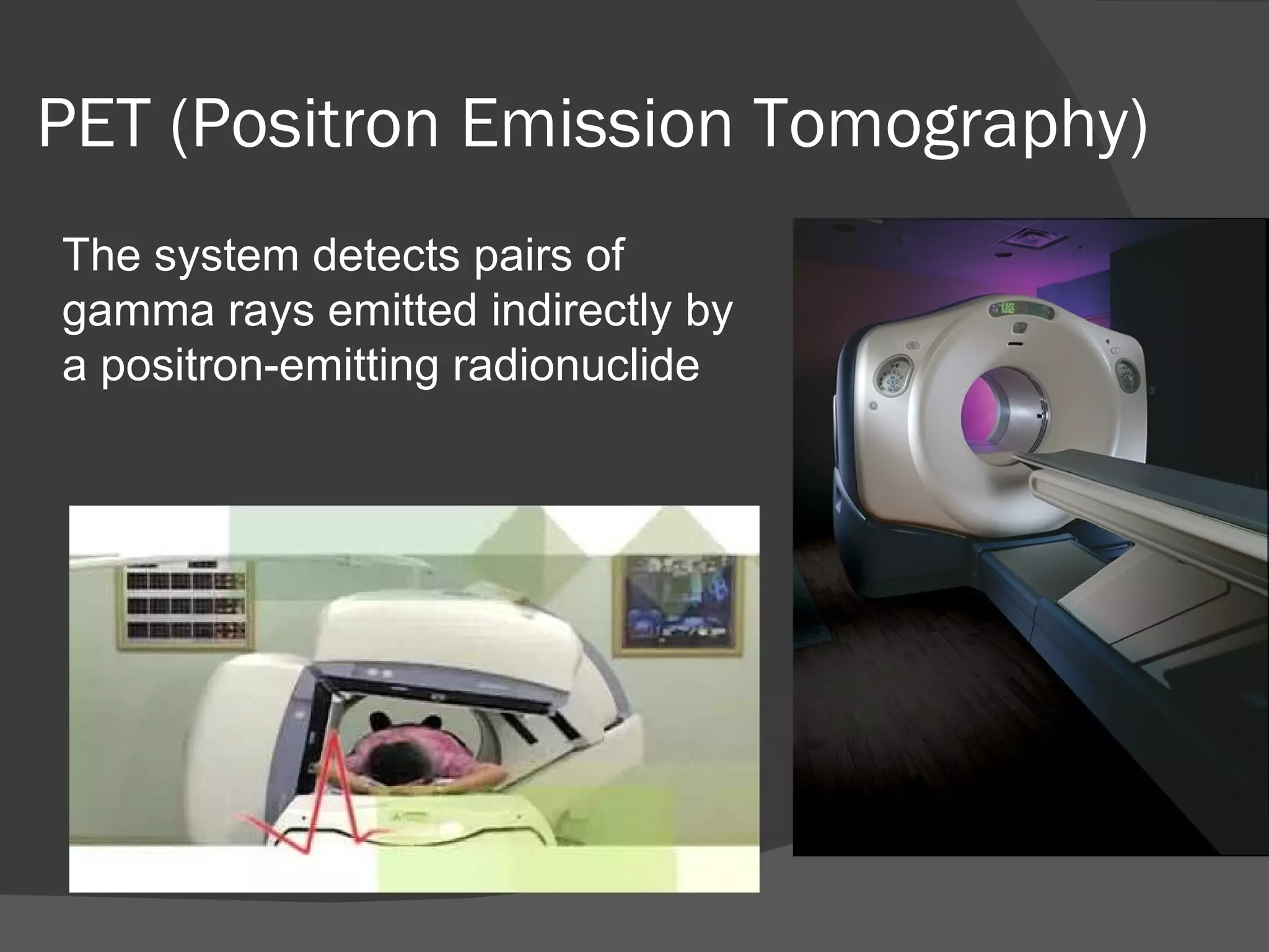
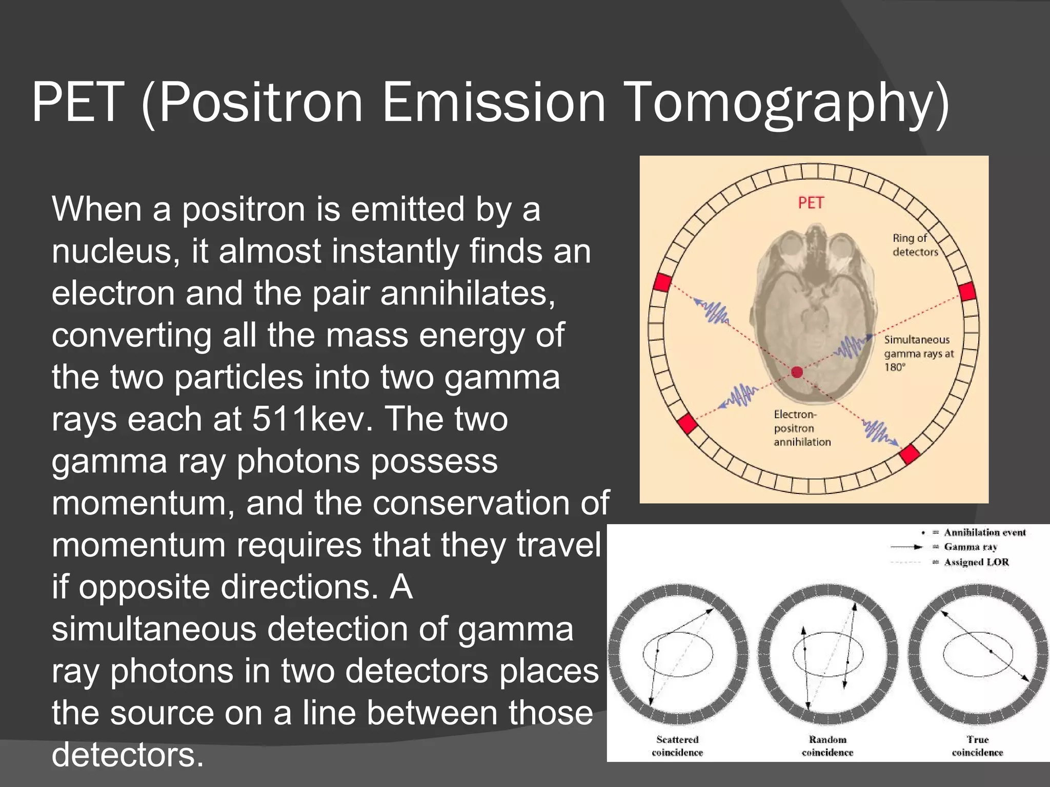
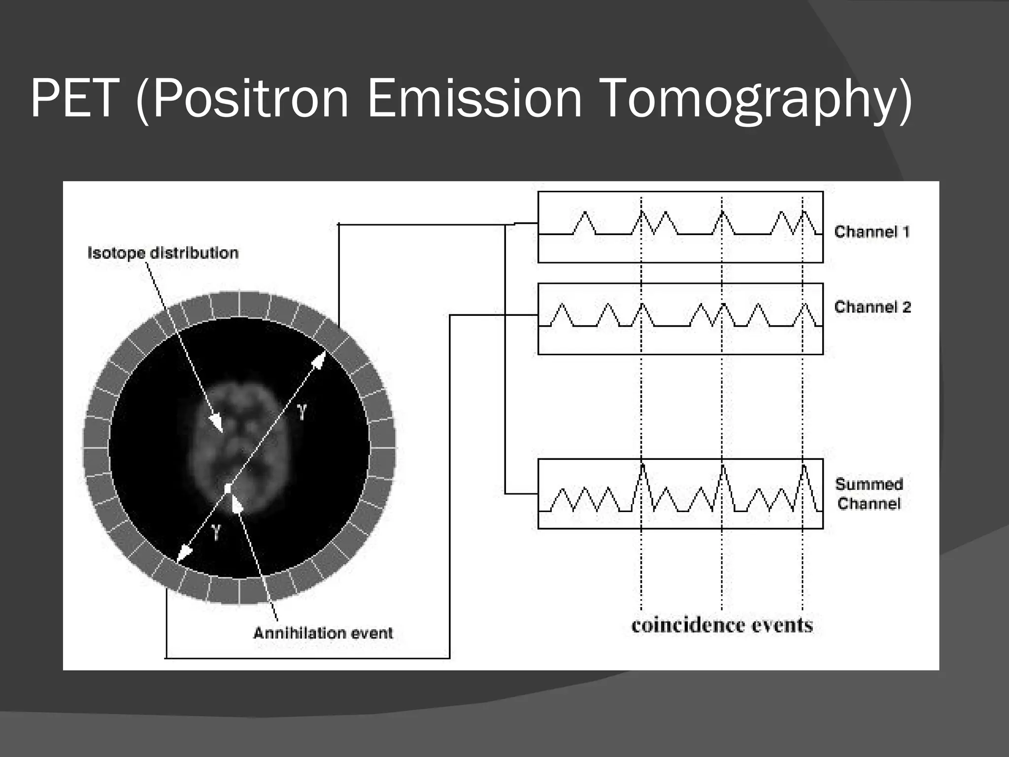
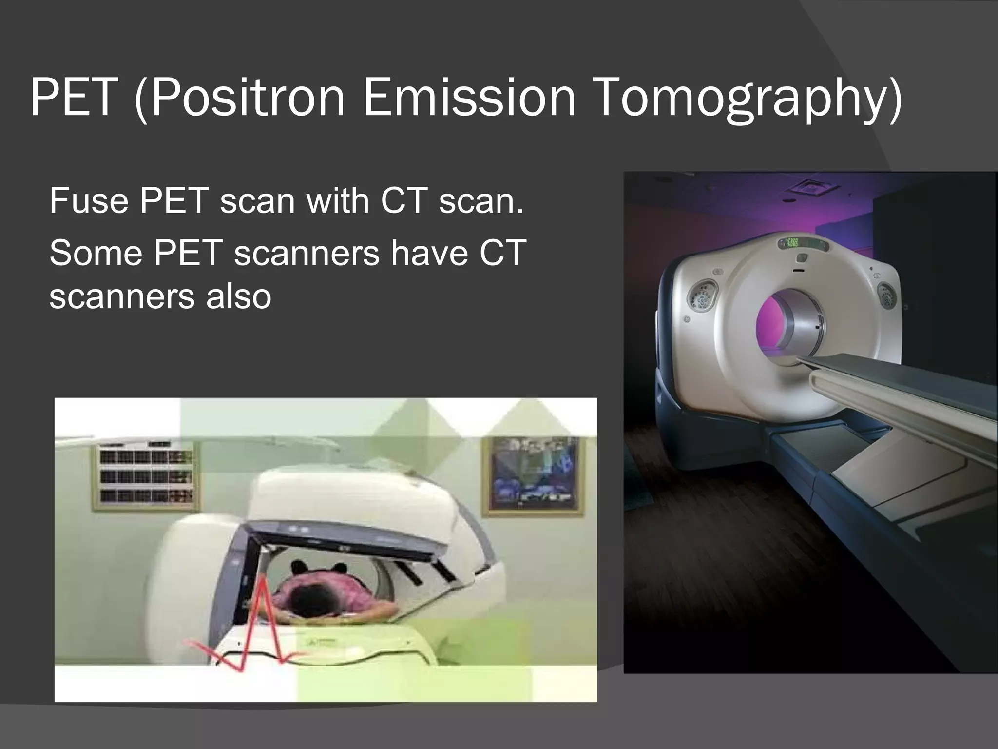
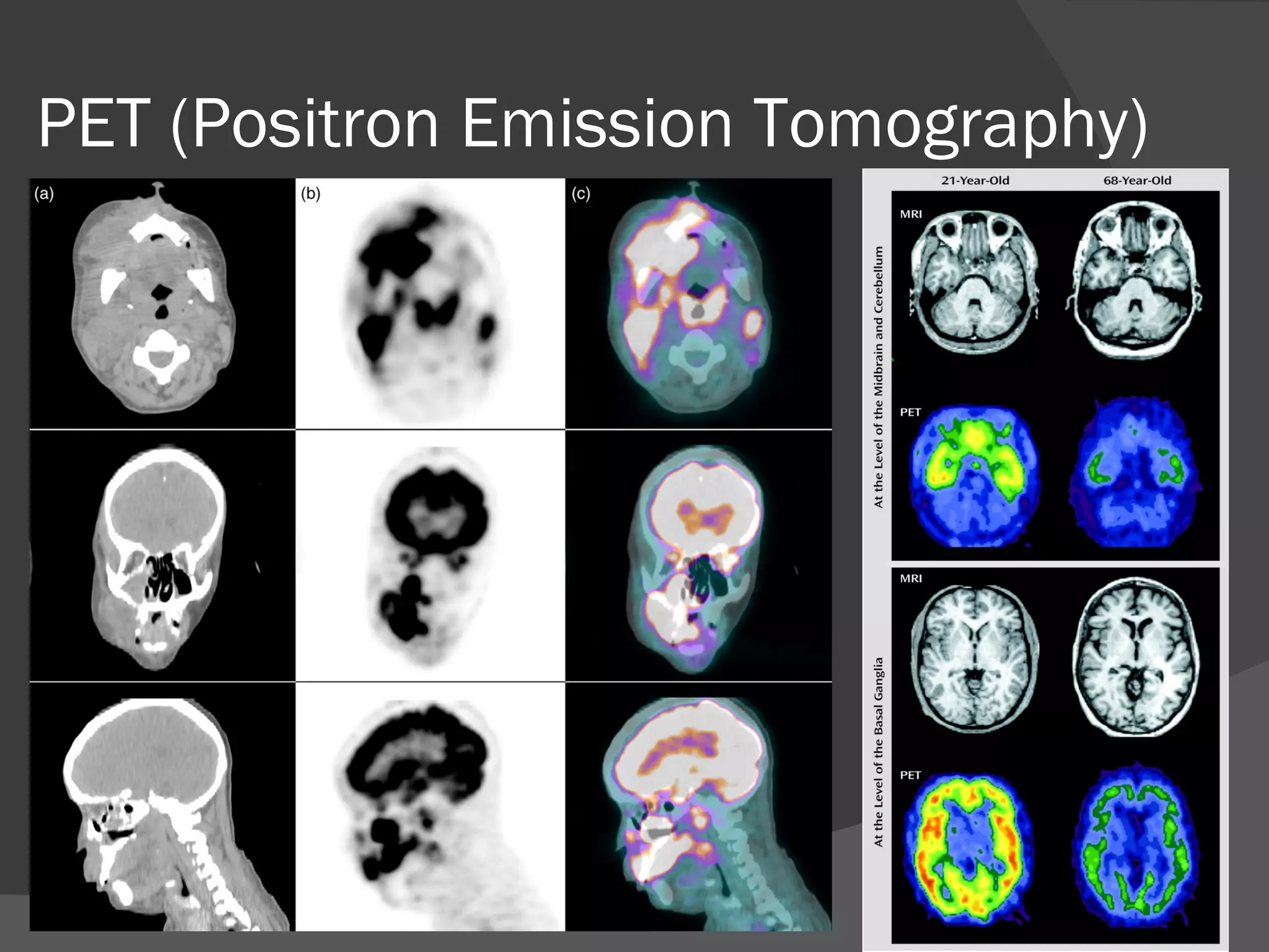
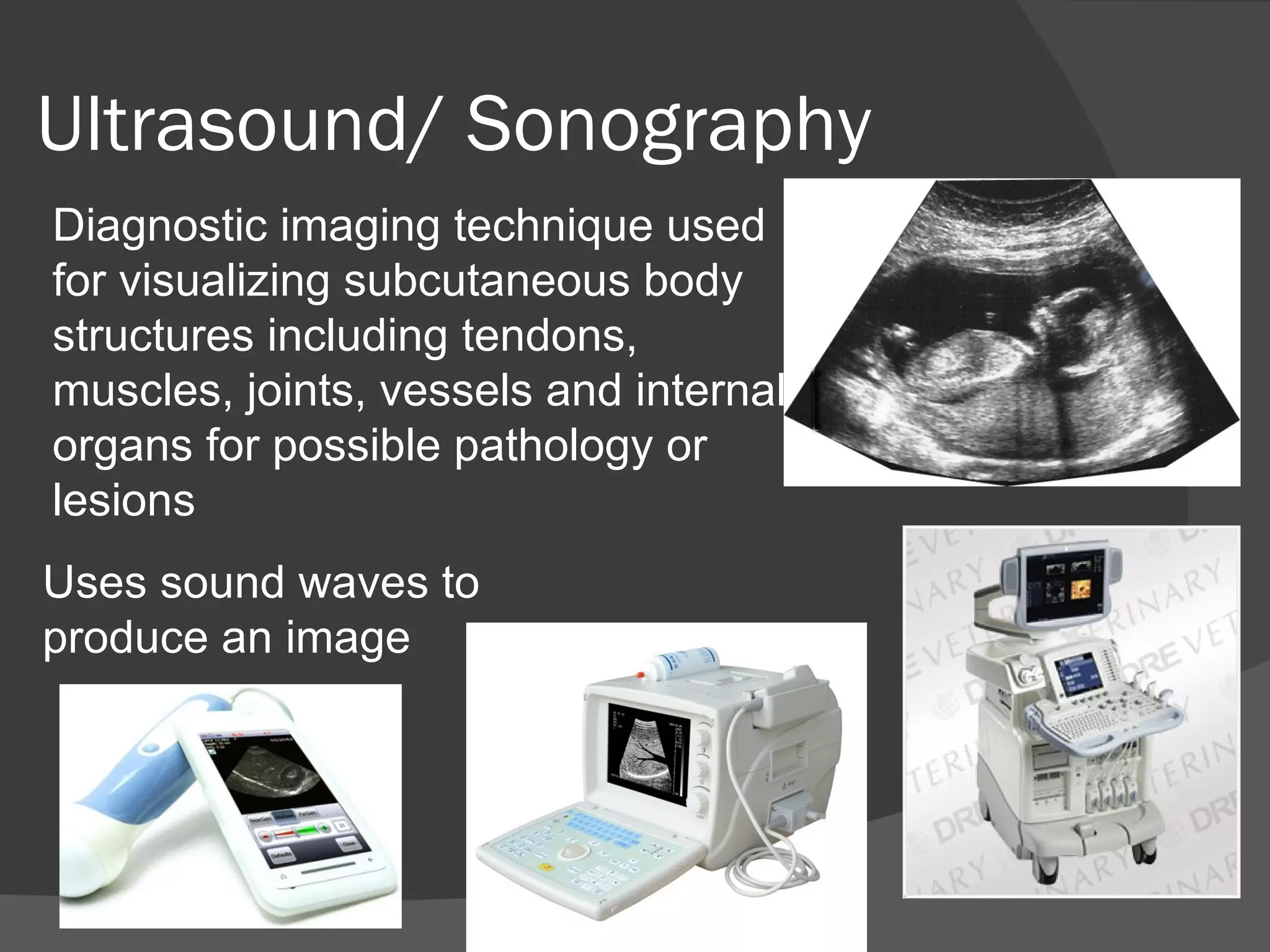
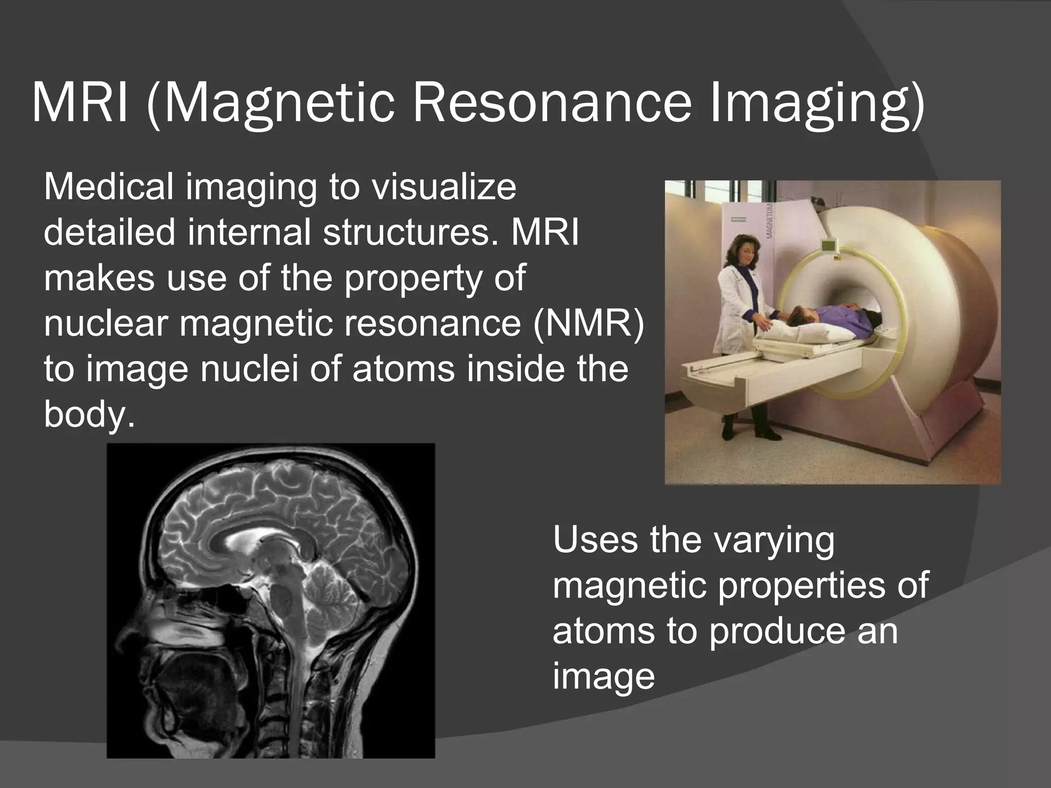
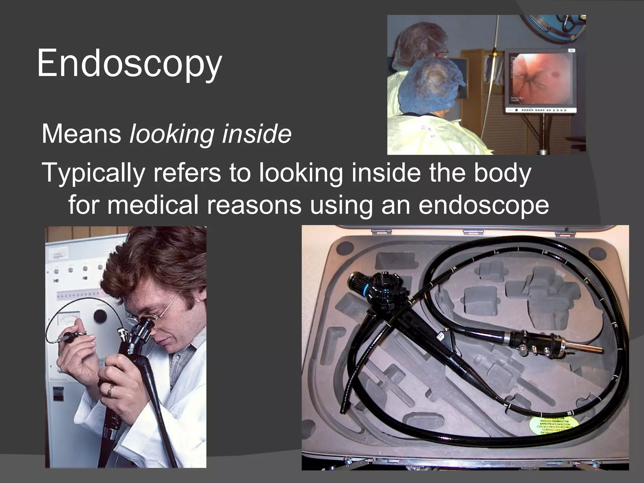
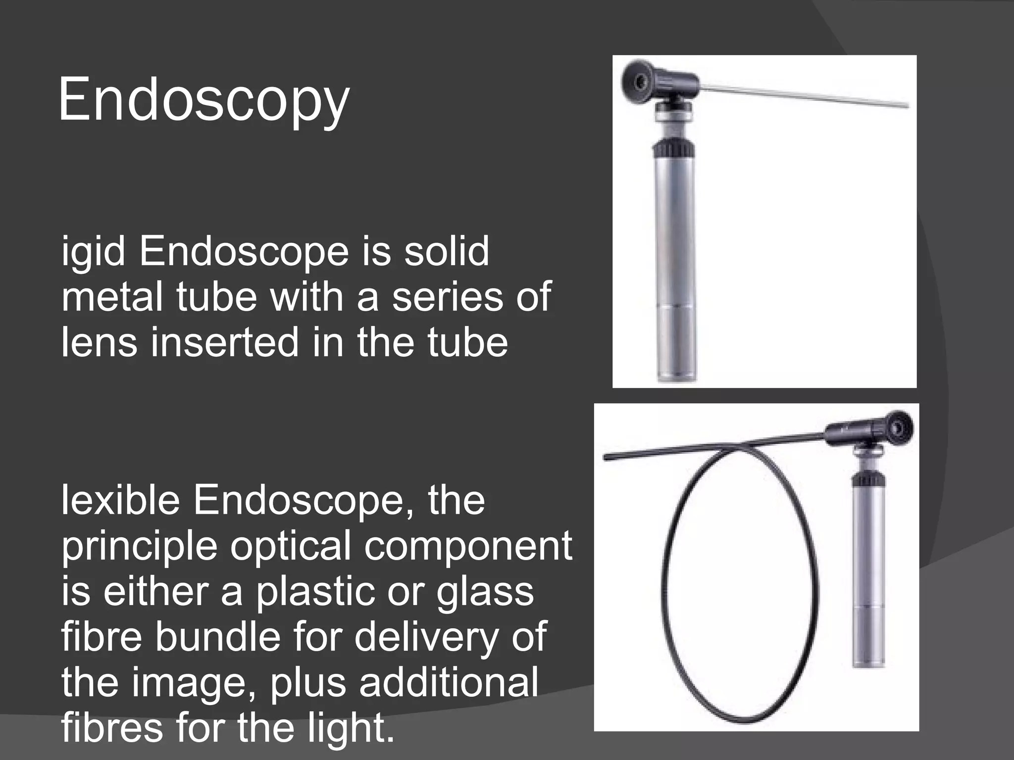
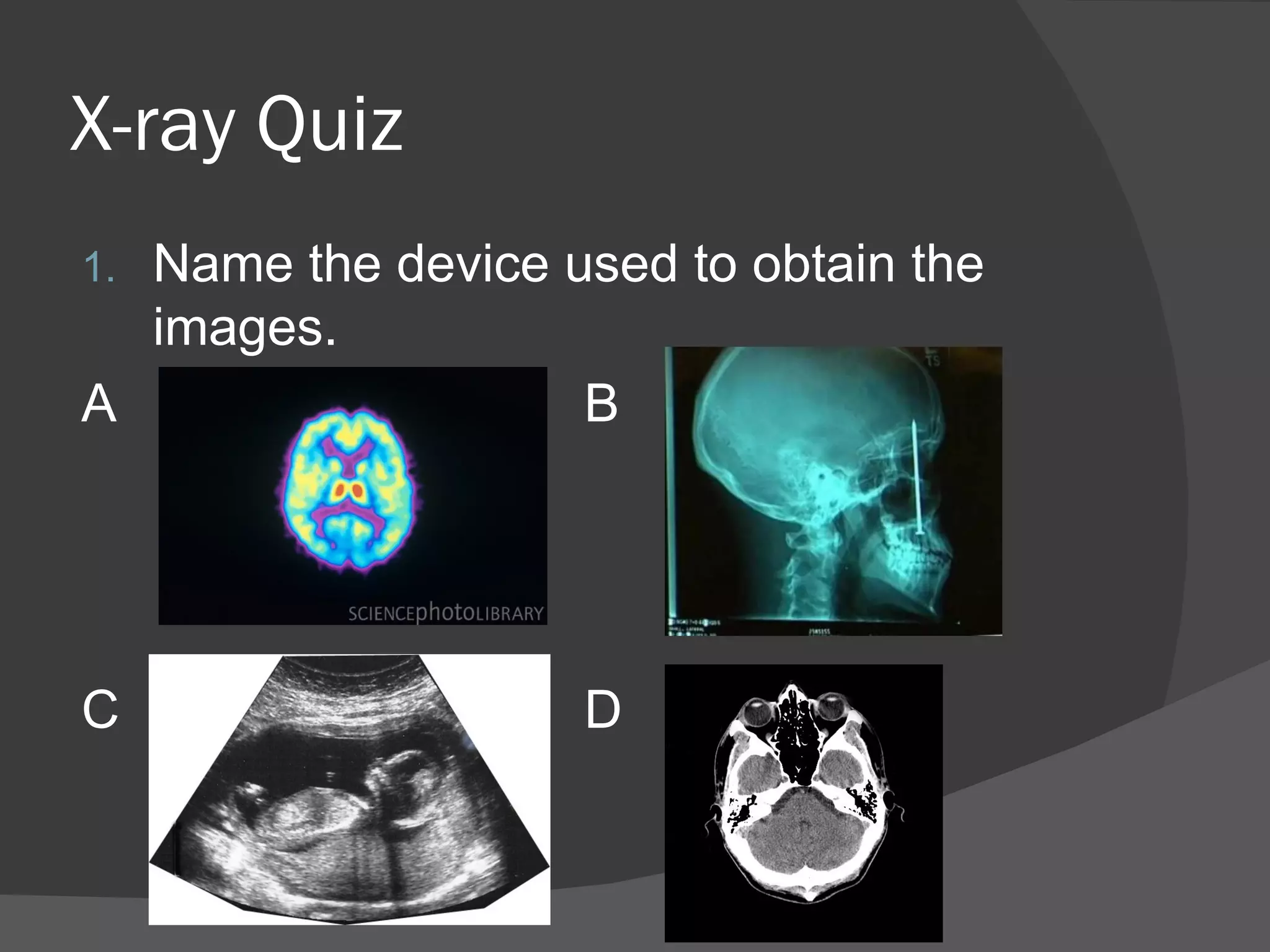
•Overview of various medical imaging/
treatment techniques
•Quiz
•Electricity
Why use medical imaging
Medical Imaging/treatment
   Radiation
     Diagnostic X-ray, CT (Computed
     Tomography), PET (Positron Emission
     Tomography), LINEAC (Linear Accelerator)
   Non-ionising
     MRI (Magnetic Resonance Imaging),
     Ultrasound/Sonography (Sound wave),
     Endoscope (light)
X-ray Machines
X-ray machines –
X-ray Photon Energy
X-ray machines – Anode


  Tungsten            High melting point
  Hi Atomic number    High-conducting
                        ability
X-ray machines – Anode


•A target for high-voltage electrons from the filament; thereby
becoming the source of the x-ray photons.
•Conducts the high voltage from the Cathode back into the
circuit.
X-ray machines – Anode


•The purpose of the cathode is to conduct a high voltage to
gap between cathode and anode.

•Flow of electrons from cathode to anode.
X-ray machines – Anode
 Tungsten – is the target surface is
  where the high speed electrons are
  attracted from the filament are suddenly
  stopped.
 Braking radiation" or "deceleration
  radiation"
X-ray machines – X-ray
production
   X rays are produced by 2 forms of
    electron interaction with the tungsten
    target.


       Bremsstrahlung           Characteristic Photons
X-ray machine - Filtration
 The bremsstrahlung photons generated within
  the target material are attenuated as they pass
  through typically 50 microns of target material.
  The beam is further attenuated by the
  aluminum or beryllium vacuum window.
 The results are an elimination of the low
  energy photons, 1 keV through l5 keV, and a
  significant reduction in the portion of the
  spectrum from 15 keV through 50 keV. The
  spectrum from an x-ray tube is further modified
  by the filtration caused by the selection of
  filters used in the setup.
X-ray machines – Image
CT (Computed Tomography)
Computed axial tomography (CAT)
Digital geometry processing is used to
generate a three-dimensional image of
the inside of an object from a large
series of two-dimensional X-ray
images taken around a single axis of
rotation
CT (Computed Tomography)
Tomography Digital geometry
processing is used to generate a
three-dimensional image of the inside
of an object from a large series of two-
dimensional X-ray images taken
around a single axis of rotation
Tomography Reconstruction
PET (Positron Emission Tomography)
The system detects pairs of
gamma rays emitted indirectly by
a positron-emitting radionuclide
PET (Positron Emission Tomography)
When a positron is emitted by a
nucleus, it almost instantly finds an
electron and the pair annihilates,
converting all the mass energy of
the two particles into two gamma
rays each at 511kev. The two
gamma ray photons possess
momentum, and the conservation of
momentum requires that they travel
if opposite directions. A
simultaneous detection of gamma
ray photons in two detectors places
the source on a line between those
detectors.
PET (Positron Emission Tomography)
Radionuclide, placed on a
chemical - glucose.
Patient fasts for 12 hours so body
is starved from energy.
When injected glucose will
concentrate to more active cells,
as patient lying still glucose will to
cancer cell. Cancer cells are more
active than normal cells as they
are rapidly dividing.
PET (Positron Emission Tomography)
Fuse PET scan with CT scan.
Some PET scanners have CT
scanners also
PET (Positron Emission Tomography)
Ultrasound/ Sonography
Diagnostic imaging technique used
for visualizing subcutaneous body
structures including tendons,
muscles, joints, vessels and internal
organs for possible pathology or
lesions
Uses sound waves to
produce an image
Doppler effect
The received frequency is higher (compared to
the emitted frequency) during the approach, it is
identical at the instant of passing by, and it is
lower during the recession.

Is the change in frequency of a wave for an
observer moving relative to the source of the
wave.
Doppler effect
MRI (Magnetic Resonance Imaging)
Medical imaging to visualize
detailed internal structures. MRI
makes use of the property of
nuclear magnetic resonance (NMR)
to image nuclei of atoms inside the
body.


                             Uses the varying
                             magnetic properties of
                             atoms to produce an
                             image
MRI (Magnetic Resonance Imaging)
Endoscopy
Means looking inside
Typically refers to looking inside the body
  for medical reasons using an endoscope
Endoscopy

igid Endoscope is solid
metal tube with a series of
lens inserted in the tube


lexible Endoscope, the
principle optical component
is either a plastic or glass
fibre bundle for delivery of
the image, plus additional
fibres for the light.
X-ray Quiz
1. Name 2 types of imaging techniques
   that use radiation and 2 that don't.
2. When a patient is due to undergo a
   PET scan they are injected with a
   radionuclide. What is emitted from the
   radionuclide? What reaction takes
   place after?
3. How does a CT scan obtain an image
   that is a slice?
X-ray Quiz
1.Name the device used to obtain the
  images.
A                  B


C                   D
X-ray Quiz
1.   Ultrasound work below human hearing
     frequency T/F?
2.   PET scan are a relatively quick procedure
     T/F?
3.   Ultrasound cannot travel through 2
     mediums in the body what are they?
4.   There are white area black areas on an X-
     ray image are showing variation in what?
5.   What is the main principal behind an MRI
     machine and the subject being
     investigated?
X-ray Quiz
   Organise these in terms of flow from
    electron source to X-ray. Start to finish.
       Key terminology
    Characteristic Photon, Patient, Anode,
    Tungsten Target, Film, Filament,
    Cathode, Bremsstrahlung, Scatter Filter,
    Casset.
X-ray Quiz
1. Cathode,
1. Filament,
2. Tungsten Target,
2. Anode,
3. Bremsstrahlung, Characteristic Photons,
4. Patient,
5. Scatter Filter,
6. Cassette,
7. Film.
Thank You

Medical imaging summary 1

  • 1.
    •Overview of variousmedical imaging/ treatment techniques •Quiz •Electricity
  • 2.
  • 3.
    Medical Imaging/treatment  Radiation  Diagnostic X-ray, CT (Computed Tomography), PET (Positron Emission Tomography), LINEAC (Linear Accelerator)  Non-ionising  MRI (Magnetic Resonance Imaging), Ultrasound/Sonography (Sound wave), Endoscope (light)
  • 4.
  • 5.
  • 6.
    X-ray machines –Anode  Tungsten  High melting point  Hi Atomic number  High-conducting ability
  • 7.
    X-ray machines –Anode •A target for high-voltage electrons from the filament; thereby becoming the source of the x-ray photons. •Conducts the high voltage from the Cathode back into the circuit.
  • 8.
    X-ray machines –Anode •The purpose of the cathode is to conduct a high voltage to gap between cathode and anode. •Flow of electrons from cathode to anode.
  • 9.
    X-ray machines –Anode  Tungsten – is the target surface is where the high speed electrons are attracted from the filament are suddenly stopped.  Braking radiation" or "deceleration radiation"
  • 10.
    X-ray machines –X-ray production  X rays are produced by 2 forms of electron interaction with the tungsten target. Bremsstrahlung Characteristic Photons
  • 11.
    X-ray machine -Filtration  The bremsstrahlung photons generated within the target material are attenuated as they pass through typically 50 microns of target material. The beam is further attenuated by the aluminum or beryllium vacuum window.  The results are an elimination of the low energy photons, 1 keV through l5 keV, and a significant reduction in the portion of the spectrum from 15 keV through 50 keV. The spectrum from an x-ray tube is further modified by the filtration caused by the selection of filters used in the setup.
  • 12.
  • 13.
  • 14.
  • 15.
    CT (Computed Tomography) Computedaxial tomography (CAT) Digital geometry processing is used to generate a three-dimensional image of the inside of an object from a large series of two-dimensional X-ray images taken around a single axis of rotation
  • 16.
    CT (Computed Tomography) TomographyDigital geometry processing is used to generate a three-dimensional image of the inside of an object from a large series of two- dimensional X-ray images taken around a single axis of rotation
  • 17.
  • 18.
    PET (Positron EmissionTomography) The system detects pairs of gamma rays emitted indirectly by a positron-emitting radionuclide
  • 19.
    PET (Positron EmissionTomography) When a positron is emitted by a nucleus, it almost instantly finds an electron and the pair annihilates, converting all the mass energy of the two particles into two gamma rays each at 511kev. The two gamma ray photons possess momentum, and the conservation of momentum requires that they travel if opposite directions. A simultaneous detection of gamma ray photons in two detectors places the source on a line between those detectors.
  • 20.
  • 21.
    PET (Positron EmissionTomography) Radionuclide, placed on a chemical - glucose. Patient fasts for 12 hours so body is starved from energy. When injected glucose will concentrate to more active cells, as patient lying still glucose will to cancer cell. Cancer cells are more active than normal cells as they are rapidly dividing.
  • 22.
    PET (Positron EmissionTomography) Fuse PET scan with CT scan. Some PET scanners have CT scanners also
  • 23.
  • 24.
    Ultrasound/ Sonography Diagnostic imagingtechnique used for visualizing subcutaneous body structures including tendons, muscles, joints, vessels and internal organs for possible pathology or lesions Uses sound waves to produce an image
  • 25.
    Doppler effect The receivedfrequency is higher (compared to the emitted frequency) during the approach, it is identical at the instant of passing by, and it is lower during the recession. Is the change in frequency of a wave for an observer moving relative to the source of the wave.
  • 26.
  • 27.
    MRI (Magnetic ResonanceImaging) Medical imaging to visualize detailed internal structures. MRI makes use of the property of nuclear magnetic resonance (NMR) to image nuclei of atoms inside the body. Uses the varying magnetic properties of atoms to produce an image
  • 28.
  • 29.
  • 30.
    Endoscopy Means looking inside Typicallyrefers to looking inside the body for medical reasons using an endoscope
  • 31.
    Endoscopy igid Endoscope issolid metal tube with a series of lens inserted in the tube lexible Endoscope, the principle optical component is either a plastic or glass fibre bundle for delivery of the image, plus additional fibres for the light.
  • 32.
    X-ray Quiz 1. Name2 types of imaging techniques that use radiation and 2 that don't. 2. When a patient is due to undergo a PET scan they are injected with a radionuclide. What is emitted from the radionuclide? What reaction takes place after? 3. How does a CT scan obtain an image that is a slice?
  • 33.
    X-ray Quiz 1.Name thedevice used to obtain the images. A B C D
  • 34.
    X-ray Quiz 1. Ultrasound work below human hearing frequency T/F? 2. PET scan are a relatively quick procedure T/F? 3. Ultrasound cannot travel through 2 mediums in the body what are they? 4. There are white area black areas on an X- ray image are showing variation in what? 5. What is the main principal behind an MRI machine and the subject being investigated?
  • 35.
    X-ray Quiz  Organise these in terms of flow from electron source to X-ray. Start to finish. Key terminology Characteristic Photon, Patient, Anode, Tungsten Target, Film, Filament, Cathode, Bremsstrahlung, Scatter Filter, Casset.
  • 36.
    X-ray Quiz 1. Cathode, 1.Filament, 2. Tungsten Target, 2. Anode, 3. Bremsstrahlung, Characteristic Photons, 4. Patient, 5. Scatter Filter, 6. Cassette, 7. Film.
  • 37.