This document discusses the potential role of point-of-care ultrasound (POCUS) in medical school. It begins with an overview of ultrasound fundamentals and image interpretation. It then examines how POCUS can efficiently address focused clinical questions at the bedside, such as detecting pneumothorax, pleural effusion, and pericardial effusion. Emerging evidence demonstrates POCUS has diagnostic accuracy comparable to other imaging modalities. The document argues POCUS could improve patient care, be a valuable clinical skill, and enhance career satisfaction if physicians receive proper training. It encourages readers to consider incorporating POCUS into their practice.
Brief discussion on ultrasonography of the chest: Benefits, Techniques and Instrumentation, Normal Anatomy, Diagnostic US of the chest, Limitations of Thoracic US, US based differential diagnosis, Take home points.
Brief discussion on ultrasonography of the chest: Benefits, Techniques and Instrumentation, Normal Anatomy, Diagnostic US of the chest, Limitations of Thoracic US, US based differential diagnosis, Take home points.
Ultrasound Physics Made easy - By Dr Chandni WadhwaniChandni Wadhwani
History of ultrasound, Principle of Ultrasound.
Ultrasound wave and its interactions
Ultrasound machine and its parts, Image display, Artifacts and their clinical importance
what is Doppler ultrasound, Elastography and Recent advances in field of ultrasound.
Safety issues in ultrasound.
Body mind balance through vibroacoustic therapyAviMan
what is bodymind balance? what is homeostasis, attunement, harmony ?
This presentation describes how to use Vibroacoustic therapy to balance mental and physical system to optimize vitality and usage of energies to live happy, healthy life.
Basic oral health surveys provide a sound basis for assessing the current oral health status of a population and its future needs for oral health care. The World Health Organization (WHO) has a long tradition of epidemiological survey methodology, which includes a description of the diagnostic criteria that can be readily understood and applied in public health programmes worldwide. The WHO manual Oral Health Surveys – Basic Methods has encouraged countries to conduct standardized oral health surveys that are comparable
internationally.
Ultra sonography indications in maxillofacial region /prosthodontic coursesIndian dental academy
The Indian Dental Academy is the Leader in continuing dental education , training dentists in all aspects of dentistry and
offering a wide range of dental certified courses in different formats.
This is a chapter from Grainger and Allison. I have Coolected all images from chapter 21 with caption in this presentation.
In my opinion it will be very benificial to have this in your android.
Using off-the-shelf ultrasound imagers, and transition to portable system-on-chip ultrasound imagers such as Butterfly IQ.
Embedded devices such as Butterfly IQ can be further improved by integrating deep learning / artificial intelligence at device level, and naturally at the post-processing and analysis levels
Alternative download link:
https://www.dropbox.com/s/rlwv7m29mh6y2w6/pupillometry_throughTheEyelids.pdf?dl=0
Synthetic Fiber Construction in lab .pptxPavel ( NSTU)
Synthetic fiber production is a fascinating and complex field that blends chemistry, engineering, and environmental science. By understanding these aspects, students can gain a comprehensive view of synthetic fiber production, its impact on society and the environment, and the potential for future innovations. Synthetic fibers play a crucial role in modern society, impacting various aspects of daily life, industry, and the environment. ynthetic fibers are integral to modern life, offering a range of benefits from cost-effectiveness and versatility to innovative applications and performance characteristics. While they pose environmental challenges, ongoing research and development aim to create more sustainable and eco-friendly alternatives. Understanding the importance of synthetic fibers helps in appreciating their role in the economy, industry, and daily life, while also emphasizing the need for sustainable practices and innovation.
2024.06.01 Introducing a competency framework for languag learning materials ...Sandy Millin
http://sandymillin.wordpress.com/iateflwebinar2024
Published classroom materials form the basis of syllabuses, drive teacher professional development, and have a potentially huge influence on learners, teachers and education systems. All teachers also create their own materials, whether a few sentences on a blackboard, a highly-structured fully-realised online course, or anything in between. Despite this, the knowledge and skills needed to create effective language learning materials are rarely part of teacher training, and are mostly learnt by trial and error.
Knowledge and skills frameworks, generally called competency frameworks, for ELT teachers, trainers and managers have existed for a few years now. However, until I created one for my MA dissertation, there wasn’t one drawing together what we need to know and do to be able to effectively produce language learning materials.
This webinar will introduce you to my framework, highlighting the key competencies I identified from my research. It will also show how anybody involved in language teaching (any language, not just English!), teacher training, managing schools or developing language learning materials can benefit from using the framework.
Read| The latest issue of The Challenger is here! We are thrilled to announce that our school paper has qualified for the NATIONAL SCHOOLS PRESS CONFERENCE (NSPC) 2024. Thank you for your unwavering support and trust. Dive into the stories that made us stand out!
Ethnobotany and Ethnopharmacology:
Ethnobotany in herbal drug evaluation,
Impact of Ethnobotany in traditional medicine,
New development in herbals,
Bio-prospecting tools for drug discovery,
Role of Ethnopharmacology in drug evaluation,
Reverse Pharmacology.
Welcome to TechSoup New Member Orientation and Q&A (May 2024).pdfTechSoup
In this webinar you will learn how your organization can access TechSoup's wide variety of product discount and donation programs. From hardware to software, we'll give you a tour of the tools available to help your nonprofit with productivity, collaboration, financial management, donor tracking, security, and more.
Instructions for Submissions thorugh G- Classroom.pptxJheel Barad
This presentation provides a briefing on how to upload submissions and documents in Google Classroom. It was prepared as part of an orientation for new Sainik School in-service teacher trainees. As a training officer, my goal is to ensure that you are comfortable and proficient with this essential tool for managing assignments and fostering student engagement.
Unit 8 - Information and Communication Technology (Paper I).pdfThiyagu K
This slides describes the basic concepts of ICT, basics of Email, Emerging Technology and Digital Initiatives in Education. This presentations aligns with the UGC Paper I syllabus.
How to Split Bills in the Odoo 17 POS ModuleCeline George
Bills have a main role in point of sale procedure. It will help to track sales, handling payments and giving receipts to customers. Bill splitting also has an important role in POS. For example, If some friends come together for dinner and if they want to divide the bill then it is possible by POS bill splitting. This slide will show how to split bills in odoo 17 POS.
Palestine last event orientationfvgnh .pptxRaedMohamed3
An EFL lesson about the current events in Palestine. It is intended to be for intermediate students who wish to increase their listening skills through a short lesson in power point.
Digital Tools and AI for Teaching Learning and Research
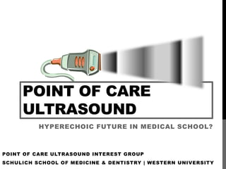
Point of Care Ultrasound - Hyperechoic Future in Medical School?
1. POINT OF CARE
ULTRASOUND
HYPERECHOIC FUTURE IN MEDICAL SCHOOL?
POINT OF CARE ULTRASOUND INTEREST GROUP
SCHULICH SCHOOL OF MEDICINE & DENTISTRY | WESTERN UNIVERSITY
4. ULTRASOUND FUNDAMENTALS
An understanding of ultrasound physics (groan) is a necessary
evil in the quest to applying and mastering ultrasound at the point
of care.
5. ULTRASOUND FUNDAMENTALS
Ultrasound machines measure the amplitude or strength of a
returning echo. The term echo is used to describe an ultrasound
beam returning to its source.
6. ULTRASOUND FUNDAMENTALS
Strong returning echoes appear as bright & white (formally,
hyperechoic) areas on the ultrasound screen. Weak returning
echoes appear as dark gray & black (formally, hypoechoic)
areas.
HYPERECHOIC HYPOECHOIC
8. ULTRASOUND FUNDAMENTALS
An ultrasound beam reflects back to its source when it encounters
an interface between different tissues or media.
LIVER KIDNEY
TISSUE
INTERFACE
9. ULTRASOUND FUNDAMENTALS
Reflection at an interface increases when the density difference
between two tissues at an interface increases.
LIVER LUNG
TISSUE
INTERFACE
16. ULTRASOUND FUNDAMENTALS
All of these factors contribute to the attenuation or weakening of
an ultrasound beam, which in turn impacts image acquisition and
quality.
REFRACTION SCATTER ABSORPTION
17. ULTRASOUND FUNDAMENTALS
An ultrasound beam is generated within the ultrasound probe by
the piezoelectric effect, which is the production of a pressure
wave when an applied voltage deforms a crystal element.
18. ULTRASOUND FUNDAMENTALS
The crystal element is also deformed by returning pressure
waves. This generates an electric current that the ultrasound
machine translates into a pixel.
PIXEL
GENERATED
ON SCREEN
19. ULTRASOUND FUNDAMENTALS
Many types of probes (also known as transducers) have been
developed. A few examples are shown below:
CONVEX PROBE
LINEAR PROBE
PHASED-ARRAY
PROBE
20. ULTRASOUND FUNDAMENTALS
A convex probe uses a lower frequency range, permitting deeper
tissue penetration. A linear probe uses a higher frequency range,
allowing higher image resolution.
CONVEX PROBE
LINEAR PROBE
23. ULTRASOUND FUNDAMENTALS
The convention when the screen marker is on the left of the
screen is that the probe marker should be directed to the patient’s
head or to the patient’s right side when scanning.
HEAD OR FEET OR
RIGHT SIDE LEFT SIDE
24. UNDERSTANDING THE IMAGE
I’m no meteorologist, but it
looks like London is getting
some rain today.
25. UNDERSTANDING THE IMAGE
There are a variety of scanning modes used in point of care
ultrasound. Here we will discuss B- or brightness mode, M-
mode or motion mode and D- or doppler mode.
B-MODE M-MODE DOPPLER
26. UNDERSTANDING THE IMAGE
B-mode (also called 2D mode) converts echo waveforms into a
256 shade grayscale image. The shade of gray depends on the
amplitude of the returning echo.
INTERNAL
JUGULAR VEIN
CAROTID
ARTERY
27. UNDERSTANDING THE IMAGE
M-mode plots the motion of a structure of interest. The probe’s
image plane is plotted on a vertical axis and time is plotted on a
horizontal axis.
IMAGE PLANE IMAGE
REPRESENTED ON PLANE
2D IMAGE
TIME
28. UNDERSTANDING THE IMAGE
Doppler mode can determine movement of reflected ultrasound
waves toward or away from the probe. This can be represented
by colour changes or graphical peaks.
BLUE REPRESENTS
MOTION AWAY FROM
TRANSDUCER
RED REPRESENTS
MOTION TOWARDS COLOUR DOPPLER SPECTRAL DOPPLER
TRANSDUCER
29. UNDERSTANDING THE IMAGE
Image artifacts are due to false assumptions made by the
ultrasound machine. They are an important concept! Some
artifacts aid image interpretation. Other artifacts interfere with
interpretation. A few examples (there are many more) …
30. UNDERSTANDING THE IMAGE
Acoustic shadowing occurs when an ultrasound beam
encounters structures much denser (such as bone) or much less
dense (such as air) than soft tissue.
SCATTER AND REFLECTION LEAD TO A LOSS OF SIGNAL DISTAL TO AIR OR BONE
31. UNDERSTANDING THE IMAGE
Reverberation occurs when ultrasound beams bounce between
two reflective interfaces. Below, equidistant lines on the
ultrasound screen represent reflections between the
transducer/skin interface and pleura.
TRANSDUCER/SKIN PLEURA
32. UNDERSTANDING THE IMAGE
Enhancement is artificial brightness deep to a hypoechoic
structure, commonly a cystic structure (such as the bladder) or
blood vessel.
BLADDER
ENHANCEMENT: THESE SOUND WAVES RETURN TO THE PROBE
WITH GREATER AMPLITUDE THAN THOSE FROM ADJACENT AREAS
33. POCUS IN MEDICAL SCHOOL?
Better sell my shares of
Ye Olde Stethoscopy,
Inc …
34. POCUS IN MEDICAL SCHOOL?
The goal today is not to teach you how to perform focused cardiac or
lung ultrasound exams.
Rather it is to get you to think about the role of point of care
ultrasound (pocus) in your future practice:
• Can I do this?
• Do I want to do this?
• Will it improve patient-centred care?
• Does it compliment and enhance existing skills and knowledge?
• Could it improve career satisfaction?
35. POCUS IN MEDICAL SCHOOL?
MAYBE SOMETHING TO
THINK ABOUT …
ULTRASOUND IS ALSO A COMPONENT OF
THE PHYSICAL EXAM—THE VISUAL
STETHOSCOPE OF THE 21ST CENTURY!
36. POCUS IN MEDICAL SCHOOL?
Skeptical? Let’s see if we can build a case for having this discussion …
37. POCUS IN MEDICAL SCHOOL?
There are many benefits of ultrasound:
• Has comparable or superior diagnostic capability to the status quo in
a growing number of scenarios
• Delivers no ionizing radiation
• Cost-effective imaging modality
• An effective educational tool
• Increases patient satisfaction
SOURCE: www.ultrasoundfirst.org (includes citations of peer-reviewed literature)
38. POCUS IN MEDICAL SCHOOL?
In the context of pocus:
• Provides new, immediate and real-time information at the bedside
that—like the stethoscope—helps address focused clinical questions
• Should be viewed as an extension of the physical exam, not a
replacement for definitive diagnostic tests
39. POCUS IN MEDICAL SCHOOL?
Pocus is used in many medical and surgical specialities.
Some current applications of pocus …
SOURCE: Point-of-Care Ultrasonography. Christopher L. Moore, M.D., and Joshua A. Copel, M.D.. N Engl J
Med 2011; 364:749-757.
40.
41. POCUS IN MEDICAL SCHOOL?
Recent advances in technology have transformed the once
cumbersome ultrasound machine into a handheld device that is
becoming increasingly practical and affordable for the physician to use
at the bedside.
42. POCUS IN MEDICAL SCHOOL?
It is important to remember that pocus is a user-dependent tool
requiring practice and expertise to develop appropriate technique and
skill (don’t forget that most aspects of the physical exam are also user-
dependent!). Like any skill in medicine, know your limits!
43. POCUS IN MEDICAL SCHOOL?
How to make a case for using pocus in your practice?
• Physician must be appropriately trained
• Efficient use of time
• Reassure the difficult patient requesting unnecessary investigations
(e.g., chest x-ray when clinical picture consistent with bronchitis)
• Detect pathology before onset of symptoms where earlier
intervention makes a patient-centred difference (e.g., global cardiac
systolic function in patient at risk of heart failure)
• Provide convincing evidence against life threatening pathology in the
symptomatic patient by answering focused clinical questions:
• Is there a pneumothorax?
• Is there a pleural effusion?
• Is there a pericardial effusion?
44. POCUS IN MEDICAL SCHOOL?
A patient presents to your office with undifferentiated shortness of
breath. Focused clinical question: Is there a pneumothorax?
SOURCE: SonoCloud
45. POCUS IN MEDICAL SCHOOL?
A patient presents to your office with undifferentiated shortness of
breath. Focused clinical question: Is there a pneumothorax?
SOURCE: SonoCloud
46. POCUS IN MEDICAL SCHOOL?
• Lung ultrasound (LUS) in the diagnosis of pneumothorax
Authors Patients Standard Sens Spec PPV NPV
Blaivas ’05 172 blunt CT, chest 98 99 98 99
trauma tube
patients
Rowan ’02 27 ED CT 100 94 92 100
trauma
getting CT
Dulchavsky 382 trauma CXR 94 100 95 99.4
’01 patients
Lichtenstein 115 ICU CXR, CT 100 96.5 89 100
’99 patients
Litchenstein 111 CXR, CT 95.3 91.1 87 100
‘95 hemithoraces
in ICU
SOURCE: Ultrasound Podcast, Episode 31 Lung Ultrasound with Vicki Noble
48. POCUS IN MEDICAL SCHOOL?
A patient presents to your office with undifferentiated shortness of
breath. Focused clinical question: Is there a pleural effusion?
SOURCE: SonoCloud
49. POCUS IN MEDICAL SCHOOL?
• Lung ultrasound (LUS) in the diagnosis of pleural effusion
Authors Patients Standard Sens Spec PPV NPV
Ma ’97 240 trauma CT/tube 96 100 100 99.5
patients thorocostomy
Sisley ‘98 360 trauma CXR 97.5 99 97.4 99.1
patients
Abboud ‘04 155 trauma CT 12.5 98.4 50 90
patients
Brooks ‘04 61 trauma CXR/tube 92 100 100 98
patients thorocostomy
SOURCE: Ultrasound Podcast, Episode 31 Lung Ultrasound with Vicki Noble
50. POCUS IN MEDICAL SCHOOL?
A patient presents to your office with undifferentiated shortness of
breath. Focused clinical question: Is there a pericardial effusion?
SOURCE: SonoCloud
51. POCUS IN MEDICAL SCHOOL?
A patient presents to your office with undifferentiated shortness of
breath. Focused clinical question: Is there a pericardial effusion?
SOURCE: SonoCloud
52. POCUS IN MEDICAL SCHOOL?
Emerging evidence …
CAP may be diagnosed and followed up by lung sonography (LUS), a technique that shows excellent
sensitivity and specificity that is at least comparable with that of chest X-ray in two planes. LUS may be
performed with any abdomen-sonography device. Therefore, LUS is a readily available diagnostic tool that
does not involve radiation exposure and has wide applications especially in situations where X-ray is not
available and/or not applicable. An X-ray or CT of the chest should be performed in cases of negative lung
sonography and if other differential diagnoses or complications are suspected.
53. POCUS IN MEDICAL SCHOOL?
So, as said on the Ultrasound Podcast …
Get out there, ultrasound some hearts, some lungs, some IVCs and let
others know how you feel about it!