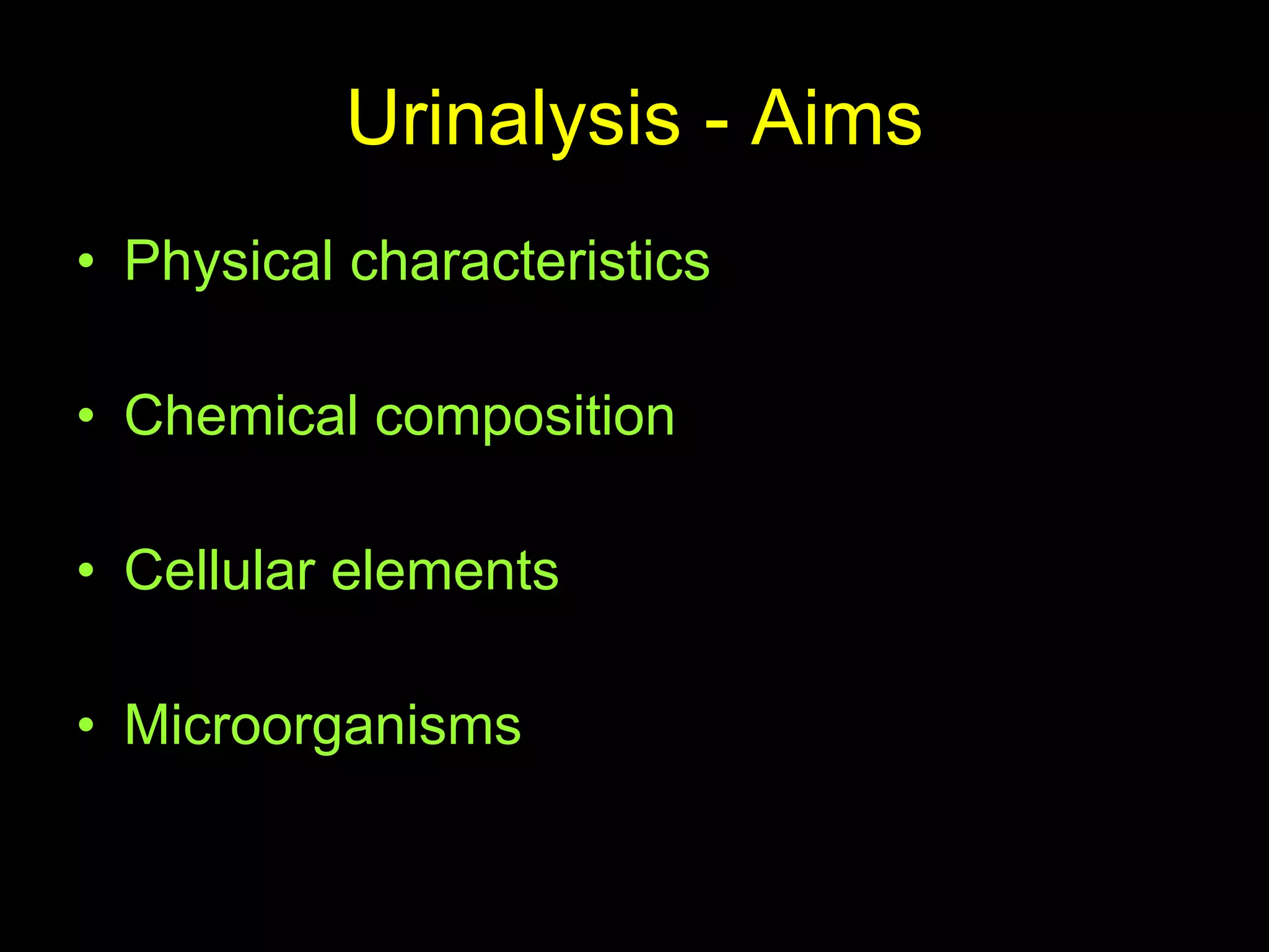
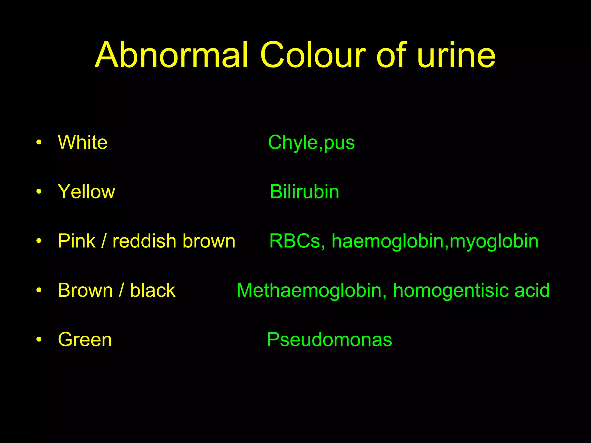
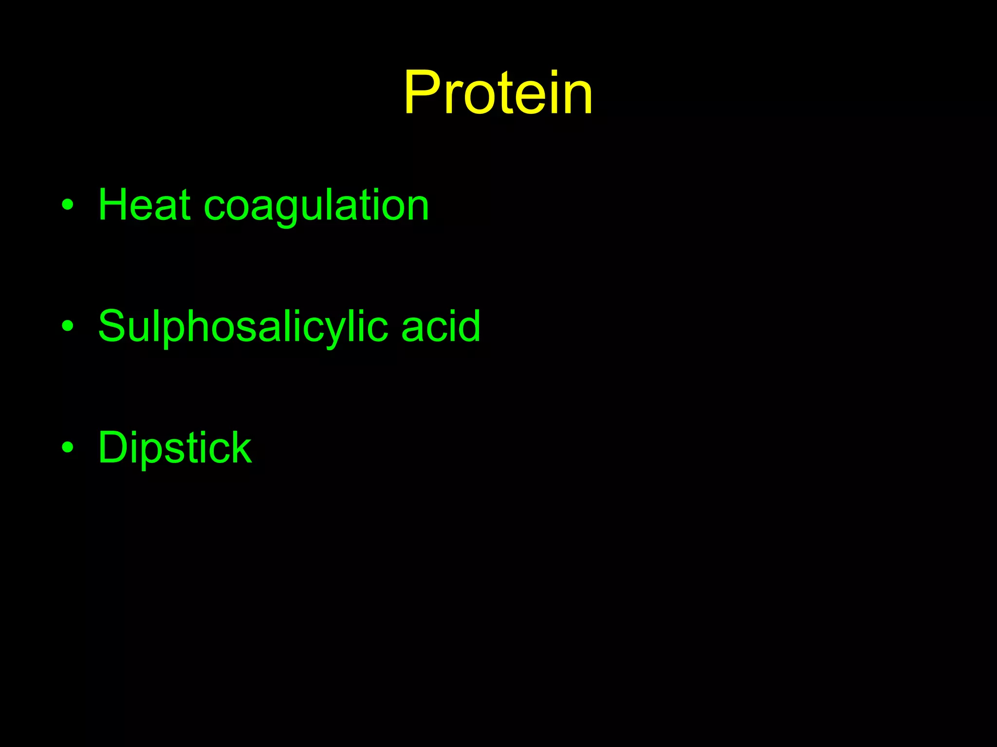
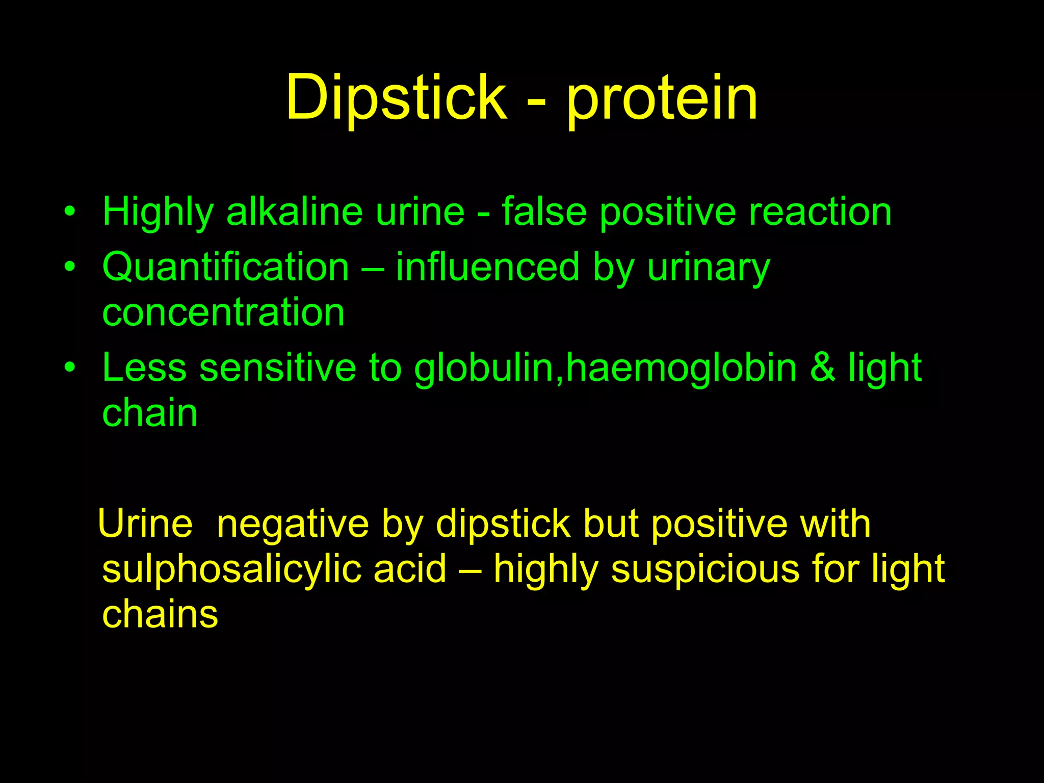
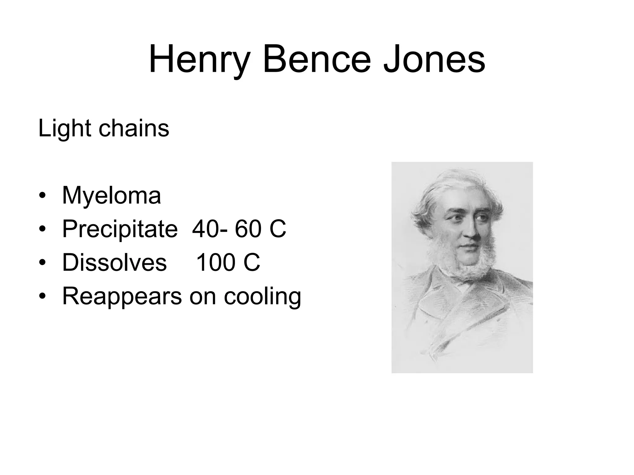
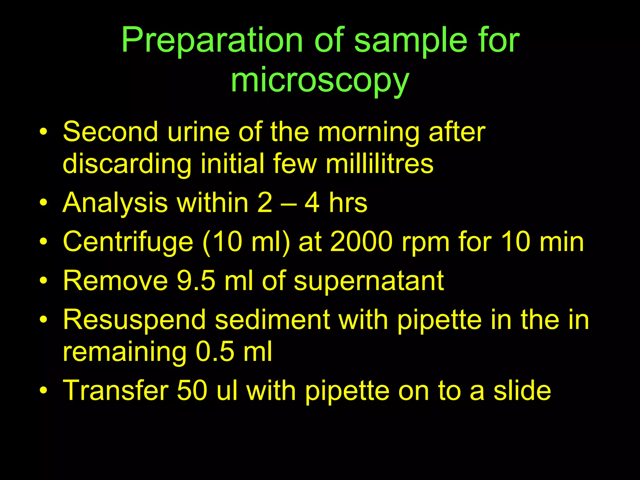
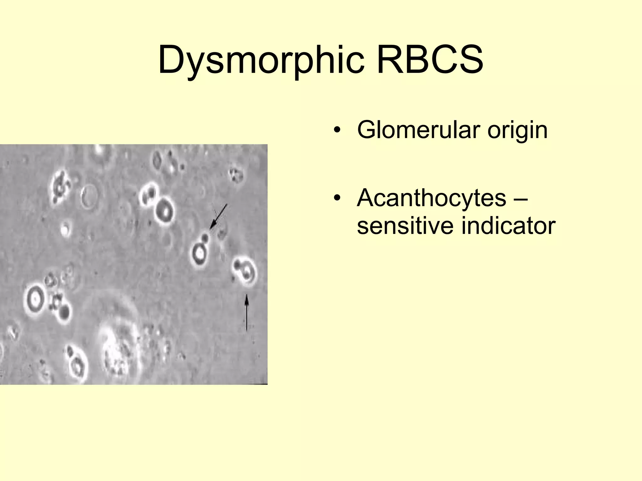
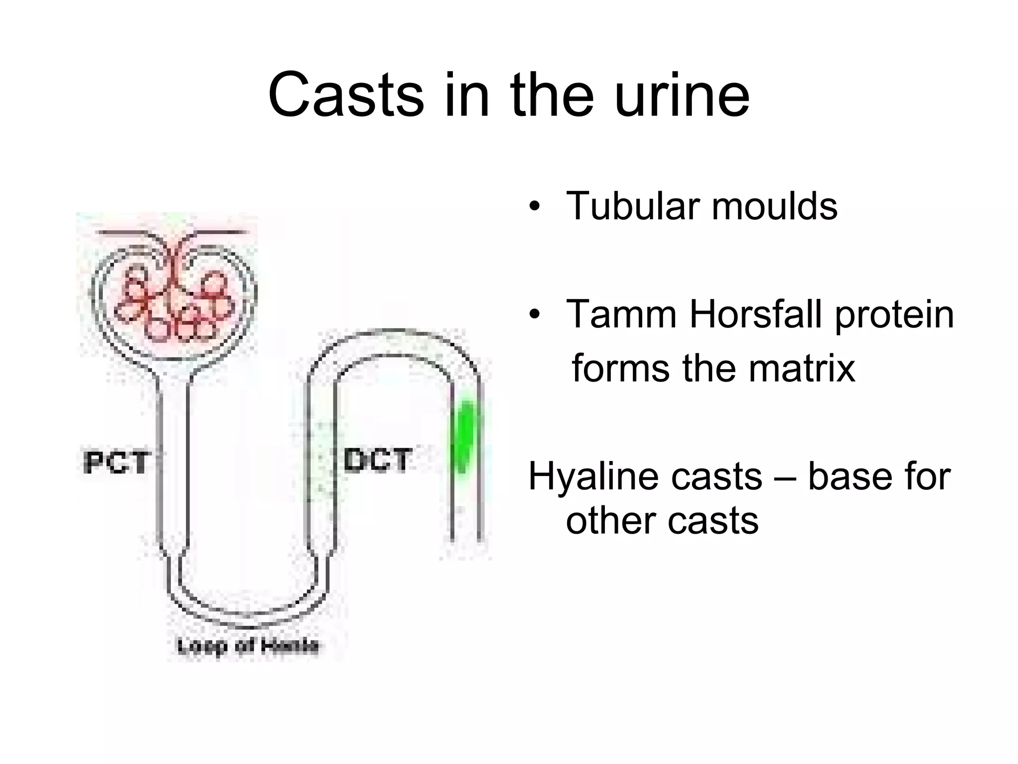
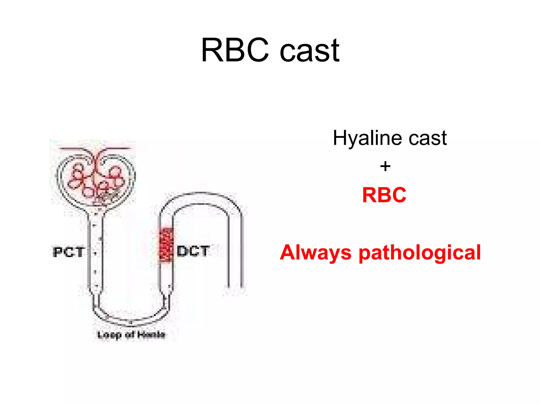
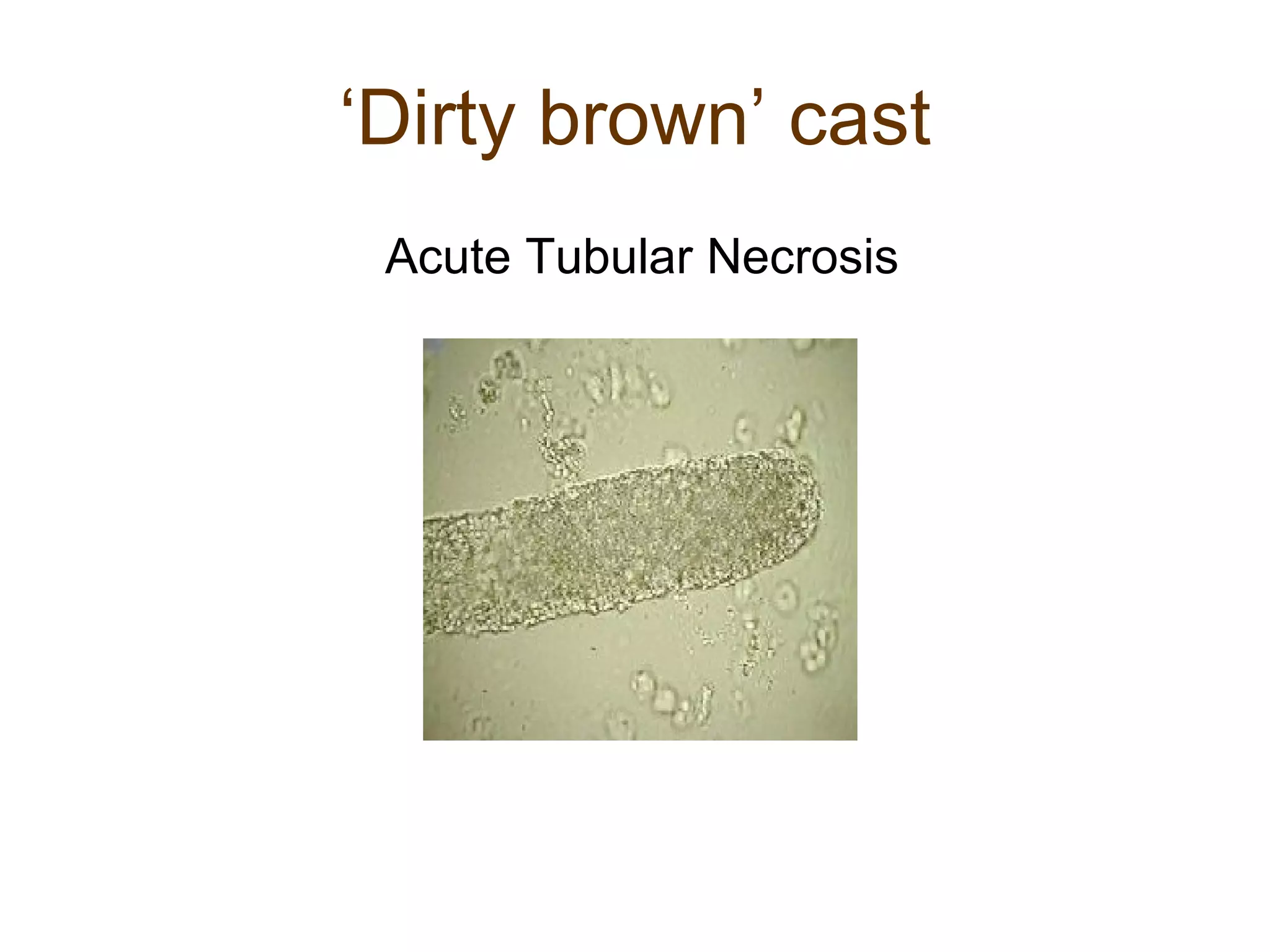
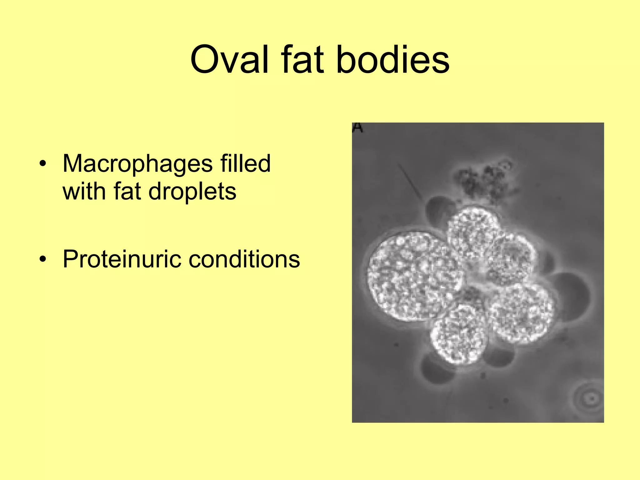
URINALYSIS – The ‘cornerstone’ of Clinical Nephrology N.Gopalakrishnan, MD, MRCP, DM
‘ Sick Lady and Doctor’ 16 th century painting by  Caspar Netscher (AJKD,2007)
Urinalysis - Aims Physical characteristics Chemical composition Cellular elements Microorganisms
Abnormal Colour of urine White  Chyle,pus Yellow  Bilirubin Pink / reddish brown  RBCs, haemoglobin,myoglobin Brown / black  Methaemoglobin, homogentisic acid Green  Pseudomonas
Dipstick  pH Protein Blood Specific gravity Glucose Leucocytes Nitrite Bilirubin Urobilinogen Ketones
pH pH Dipstick – pH 4.5 to 8.5;  less accurate when pH is < 5 or >8 For accurate estimation, pH meter with glass electrode is mandatory
Specific gravity Denotes weight of solutes Inaccurate surrogate of osmolality Methods:  Hydrometer Refractometer Dipstick Range: 1.001 – 1.035 Assesses concentrating ability Differentiation between pre renal azotemia & ATN
Protein Heat coagulation Sulphosalicylic acid Dipstick
Dipstick  - protein Protein indicator strips – tetrabromophenol blue or bromocresol blue Colour change according to protein concentration Trace to 4+ trace  5 – 20 mg/dl 1+  -  30,  2+  -  100, 3+  -  300,  4+  -  > 2000 mg/dl
Dipstick - protein Highly alkaline urine - false positive reaction  Quantification – influenced by urinary concentration Less sensitive to globulin,haemoglobin & light chain Urine  negative by dipstick but positive with sulphosalicylic acid – highly suspicious for light chains
Henry Bence Jones Light chains Myeloma Precipitate  40- 60 C Dissolves  100 C Reappears on cooling
Preparation of sample for microscopy Second urine of the morning after  discarding initial few millilitres Analysis within 2 – 4 hrs Centrifuge (10 ml) at 2000 rpm for 10 min Remove 9.5 ml of supernatant Resuspend sediment with pipette in the in remaining 0.5 ml Transfer 50 ul with pipette on to a slide
Microscopy Phase contrast microscope – ideal High sensitivity for hyaline casts & RBCs with low Hb content For RBC morphology Filters to polarize light – for lipids & unusual crystals
Erythrocytes > 2 -3 / HPF --- Haematuria Isomorphic haematuria – from collecting system Dysmorphic  haematuria – from renal parenchyma
Dysmorphic RBCS Glomerular origin Acanthocytes – sensitive indicator
Leucocytes Indicate urinary tract inflammation ‘ Glitter cells’ – swollen polymorphs with prominent granules UTI, interstitial nephritis, glomerulonephritis
Sterile pyuria Tuberculosis Partially treated UTI Steroids Calculi Prostatitis Bladder tumour Papillary necrosis
Eosinophiluria Acute allergic interstitial nephritis Atheroembolism Rapidly progressive glomerulonephritis Schistosomiasis Hansel’s stain
Renal tubular epithelial cells (RTECs) Larger than WBCs Few cells are normal Indicate tubular damage or inflammation ATN, interstitial nephritis
Casts in the urine Tubular moulds Tamm Horsfall protein forms the matrix  Hyaline casts – base for other casts
RBC cast Hyaline cast + RBC Always pathological
RBC cast Hallmark of Nephritic sediment
WBC cast Pyelonephritis Glomerulonephritis
‘ Dirty brown’ cast Acute Tubular Necrosis
Fine granular cast Derived from altered serum proteins Usually, pathological
Coarse granular cast Result from degeneration of embedded cells Usually pathological
Broad casts (Waxy cast) Form in dilated, atrophic tubules  More refractile than hyaline casts Usual in chronic renal insufficiency
Telescoped urine sediment Plethora of findings RBCs, RBC casts, WBCs, granular casts, broad casts,etc Classically seen in SLE
Crystals in urine Common crystals Pathological crystals Crystals due to drugs
Pathological crystals Cystine  -  Cystinuria Cholesterol –  massive proteinuria 2,8 dihydroxyadenine –  Adeninie phosphoribosyl transferase deficiency
Drugs causing crystalluria Drug overdose Dehydration Hypoalbuminemia Urine pH Acyclovir Indinavir Sulphadiazine Amoxycillin Vitamin C (oxalate)
Cystine crystals Always pathological Pathognomonic of cystinuria
Uric acid crystals Rhomboid  Hyperuricosuric conditions Acute uric acid nephropathy
Calcium Oxalate Monohydrate:’dumb bell’ Byhydrate:  ‘envelope’
Triple phosphate crystals ‘ Coffin lid’shaped Rectangular with beveled ends
Blood Depends on peroxidase activity of haemoglobin Blood, haemoglobin & myoglobin test positive Dipstick positive for blood, but no RBCs in microscopy – haemoglobin or myoglobin
Microorganisms Bacteria Yeasts Trichomonas
Nitrite screening test For detection of bacteriuria Bacteria convert urinary nitrate to nitrite which activates a chromogen False negative – Enterococcus
Oval fat bodies Macrophages filled with fat droplets Proteinuric conditions
Microalbumin dipsticks  For screening for microalbuminuria Colorimetric detection of albumin bound to gold – conjugated antibody
Let us uphold the ancient tradition! THANK  YOU

CME - Urinalysis

  • 1.
    URINALYSIS – The‘cornerstone’ of Clinical Nephrology N.Gopalakrishnan, MD, MRCP, DM
  • 2.
    ‘ Sick Ladyand Doctor’ 16 th century painting by Caspar Netscher (AJKD,2007)
  • 3.
    Urinalysis - AimsPhysical characteristics Chemical composition Cellular elements Microorganisms
  • 4.
    Abnormal Colour ofurine White Chyle,pus Yellow Bilirubin Pink / reddish brown RBCs, haemoglobin,myoglobin Brown / black Methaemoglobin, homogentisic acid Green Pseudomonas
  • 5.
    Dipstick pHProtein Blood Specific gravity Glucose Leucocytes Nitrite Bilirubin Urobilinogen Ketones
  • 6.
    pH pH Dipstick– pH 4.5 to 8.5; less accurate when pH is < 5 or >8 For accurate estimation, pH meter with glass electrode is mandatory
  • 7.
    Specific gravity Denotesweight of solutes Inaccurate surrogate of osmolality Methods: Hydrometer Refractometer Dipstick Range: 1.001 – 1.035 Assesses concentrating ability Differentiation between pre renal azotemia & ATN
  • 8.
    Protein Heat coagulationSulphosalicylic acid Dipstick
  • 9.
    Dipstick -protein Protein indicator strips – tetrabromophenol blue or bromocresol blue Colour change according to protein concentration Trace to 4+ trace 5 – 20 mg/dl 1+ - 30, 2+ - 100, 3+ - 300, 4+ - > 2000 mg/dl
  • 10.
    Dipstick - proteinHighly alkaline urine - false positive reaction Quantification – influenced by urinary concentration Less sensitive to globulin,haemoglobin & light chain Urine negative by dipstick but positive with sulphosalicylic acid – highly suspicious for light chains
  • 11.
    Henry Bence JonesLight chains Myeloma Precipitate 40- 60 C Dissolves 100 C Reappears on cooling
  • 12.
    Preparation of samplefor microscopy Second urine of the morning after discarding initial few millilitres Analysis within 2 – 4 hrs Centrifuge (10 ml) at 2000 rpm for 10 min Remove 9.5 ml of supernatant Resuspend sediment with pipette in the in remaining 0.5 ml Transfer 50 ul with pipette on to a slide
  • 13.
    Microscopy Phase contrastmicroscope – ideal High sensitivity for hyaline casts & RBCs with low Hb content For RBC morphology Filters to polarize light – for lipids & unusual crystals
  • 14.
    Erythrocytes > 2-3 / HPF --- Haematuria Isomorphic haematuria – from collecting system Dysmorphic haematuria – from renal parenchyma
  • 15.
    Dysmorphic RBCS Glomerularorigin Acanthocytes – sensitive indicator
  • 16.
    Leucocytes Indicate urinarytract inflammation ‘ Glitter cells’ – swollen polymorphs with prominent granules UTI, interstitial nephritis, glomerulonephritis
  • 17.
    Sterile pyuria TuberculosisPartially treated UTI Steroids Calculi Prostatitis Bladder tumour Papillary necrosis
  • 18.
    Eosinophiluria Acute allergicinterstitial nephritis Atheroembolism Rapidly progressive glomerulonephritis Schistosomiasis Hansel’s stain
  • 19.
    Renal tubular epithelialcells (RTECs) Larger than WBCs Few cells are normal Indicate tubular damage or inflammation ATN, interstitial nephritis
  • 20.
    Casts in theurine Tubular moulds Tamm Horsfall protein forms the matrix Hyaline casts – base for other casts
  • 21.
    RBC cast Hyalinecast + RBC Always pathological
  • 22.
    RBC cast Hallmarkof Nephritic sediment
  • 23.
    WBC cast PyelonephritisGlomerulonephritis
  • 24.
    ‘ Dirty brown’cast Acute Tubular Necrosis
  • 25.
    Fine granular castDerived from altered serum proteins Usually, pathological
  • 26.
    Coarse granular castResult from degeneration of embedded cells Usually pathological
  • 27.
    Broad casts (Waxycast) Form in dilated, atrophic tubules More refractile than hyaline casts Usual in chronic renal insufficiency
  • 28.
    Telescoped urine sedimentPlethora of findings RBCs, RBC casts, WBCs, granular casts, broad casts,etc Classically seen in SLE
  • 29.
    Crystals in urineCommon crystals Pathological crystals Crystals due to drugs
  • 30.
    Pathological crystals Cystine - Cystinuria Cholesterol – massive proteinuria 2,8 dihydroxyadenine – Adeninie phosphoribosyl transferase deficiency
  • 31.
    Drugs causing crystalluriaDrug overdose Dehydration Hypoalbuminemia Urine pH Acyclovir Indinavir Sulphadiazine Amoxycillin Vitamin C (oxalate)
  • 32.
    Cystine crystals Alwayspathological Pathognomonic of cystinuria
  • 33.
    Uric acid crystalsRhomboid Hyperuricosuric conditions Acute uric acid nephropathy
  • 34.
    Calcium Oxalate Monohydrate:’dumbbell’ Byhydrate: ‘envelope’
  • 35.
    Triple phosphate crystals‘ Coffin lid’shaped Rectangular with beveled ends
  • 36.
    Blood Depends onperoxidase activity of haemoglobin Blood, haemoglobin & myoglobin test positive Dipstick positive for blood, but no RBCs in microscopy – haemoglobin or myoglobin
  • 37.
  • 38.
    Nitrite screening testFor detection of bacteriuria Bacteria convert urinary nitrate to nitrite which activates a chromogen False negative – Enterococcus
  • 39.
    Oval fat bodiesMacrophages filled with fat droplets Proteinuric conditions
  • 40.
    Microalbumin dipsticks For screening for microalbuminuria Colorimetric detection of albumin bound to gold – conjugated antibody
  • 41.
    Let us upholdthe ancient tradition! THANK YOU

Editor's Notes