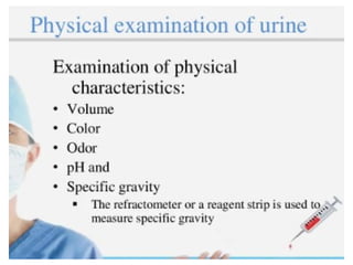
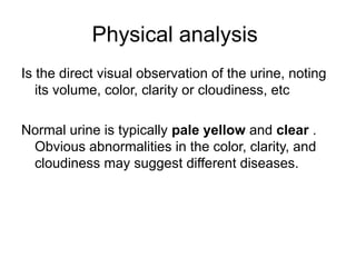
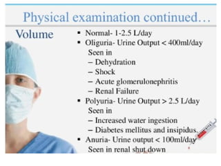
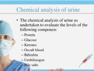
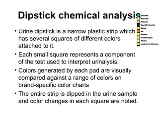
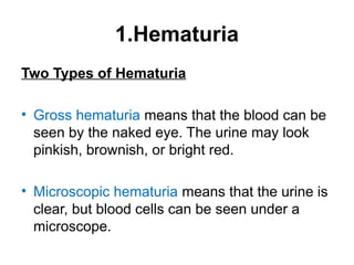
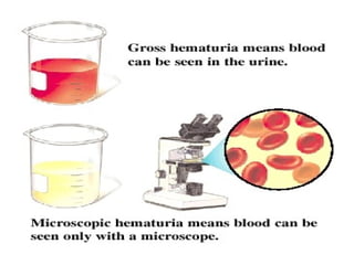
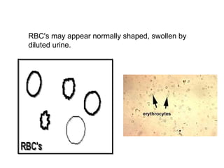
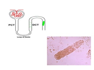
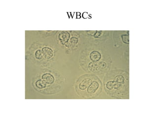
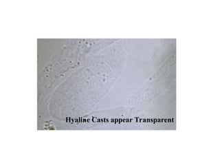
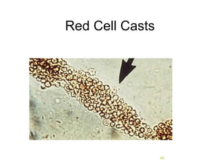
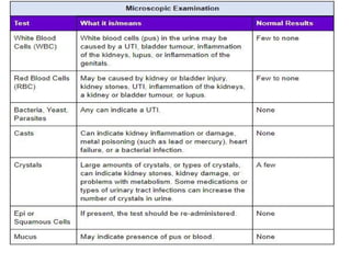
Urine analysis
What is a Urinalysis?
• A test that determines the content of the
urine.
– Because urine removes toxins and excess liquids from the body, its
contents can provide vital health information.
– Urinalysis can be used to detect some types of disease, particularly
in the case of kidney disease.
– Urinalysis can also be used to uncover evidence of drug abuse.
Kidney Function
• The role of the kidneys is to maintain
homeostasis by controlling the chemical
composition of the blood. The kidneys do
this by:
– Removing waste products from the blood
– Leaving nutrients such as proteins and glucose in the
blood
– Regulating water and electrolyte balance
Proper Kidney Function
• Therefore, if the kidneys are functioning properly
and person is in good over-all health, urine will
be normal.
• If urine shows abnormalities, this can indicate
problem with kidneys. The following conditions
can cause kidney problems:
– Heart failure
– Injury to glomerulus or tubules from drugs, heavy
metals, and viral infections.
– Diabetes, hypertension, and kidney stones
Collection of urine specimens
• Improper collection---- may invalidate the results
• Containers for collection of urine should be wide mouthed, clean and dry.
• Analysed within 2 hours of collection else requires refrigeration.
Urinalysis Basics
• Urinalysis is carried out by following steps:
– Physical Examination
– Chemical Analysis
– Microscopic Examination
Physical analysis
Is the direct visual observation of the urine, noting
its volume, color, clarity or cloudiness, etc
Normal urine is typically pale yellow and clear .
Obvious abnormalities in the color, clarity, and
cloudiness may suggest different diseases.
Normal Urine Abnormal Urine
Dipstick chemical analysis
• Urine dipstick is a narrow plastic strip which
has several squares of different colors
attached to it.
• Each small square represents a component
of the test used to interpret urinalysis.
• Colors generated by each pad are visually
compared against a range of colors on
brand-specific color charts
• The entire strip is dipped in the urine sample
and color changes in each square are noted.
The squares on the dipstick represent the
following components in the urine
Nitrite (suggestive of bacteria in urine)
Bilirubin ( possible liver disease or red blood cell break down)
Urobilinogen ( possible liver disease)
Parts of Chemical Analysis
• pH
– Test measures if urine is acidic, basic or neutral
– Normal urine ranges from 4.6 to 8.0
• Specific Gravity:
– Test measures the concentration of particles in the urine and evaluates
the body’s water balance.
– The more concentrated the urine, the higher the urine specific gravity.
– The most common increase in urine specific gravity is the result of
dehydration.
– Normal urine ranges between 1.002 to 1.028
Parts of Chemical Analysis
• Ketones:
– Test measures the presence or absence of ketones or excessive
fat breakdown, in the urine.
– Normal urine does not contain ketones
• Protein:
– Normal urine levels of proteins (called albumin) are very small
Parts of Chemical Analysis
• Glucose:
– The test measures the amount of sugar in a
urine sample.
– Normal urine does not contain glucose.
Microscopic Examination
• A variety of normal and abnormal cellular
elements may be seen in urine when
looked at under a microscope, including:
– Red blood cells
– White blood cells
– Epithelial cells
– Crystals
– Bacteria
Microscopic Examination
• Red blood cells are not found in normal urine.
• White blood cells and bacteria, signs of infections, are not found in
normal urine.
• Epithelial cells are found in urine as they are the cells that line the
urinary tract.
• Common crystals seen even in healthy patients include calcium
oxalate, triple phosphate crystals and amorphous phosphates.
– A large number of crystals, or certain types of crystals, may mean
kidney stones are present or there is a problem with how the body
is using food.
MICROSCOPIC URINALYSIS
Microscopic examination used to view elements that are not visible
without microscope. e.g cells
Red Blood Cells:
Hematuria is the presence of abnormal numbers of
red cells in urine due to:
a. Glomerular damage
b. Tumors
c. Urinary tract stones
d. Upper and lower urinary tract infections
1.Hematuria
Two Types of Hematuria
• Gross hematuria means that the blood can be
seen by the naked eye. The urine may look
pinkish, brownish, or bright red.
• Microscopic hematuria means that the urine is
clear, but blood cells can be seen under a
microscope.
RBC's may appear normally shaped, swollen by
diluted urine.
2.White Blood Cells
Pyuria refers to the presence of abnormal
numbers of leukocytes that may appear
with infection in either the upper or lower
urinary tract or with acute
glomerulonephritis.
Usually, the WBC's are granulocytes
WBCs - ≤2-5 WBCs/hpf
3. Epithelial Cells
• Renal tubular epithelial cells, contain a
large round or oval nucleus and normally
slough into the urine in small numbers.
However, with nephrotic syndrome the
number sloughed is increased.
4. Casts
• Urinary casts are cylindrical structures produced by the
kidney and present in the urine in certain disease states.
• They are formed in the distal convoluted tubule (DCT)
and collecting ducts of nephrons, then dislodge and pass
into the urine, where they can detected by microscopy.
-Urinary casts may be made up of cells (such as white
blood cells, red blood cells, kidney cells) or substances
such as protein.
Red blood cells may stick together and form
red blood cell casts. Such casts are indicative
of glomerulonephritis, with leakage of RBC's
from glomeruli
White blood cell casts may also be present
with glomerulonephritis. Their presence
indicates inflammation of the kidney,
because such casts will not form except in
the kidney.
Crenated RBC
RBCs
WBCs
Hyaline Casts appear Transparent
40
Red Cell Casts
41
White Cell Casts
Bence Jones proteins are small proteins found in the
urine. Testing for these proteins is done to diagnose and
monitor multiple myeloma and other similar diseases.
Bence Jones proteins are considered the first tumor
marker.
A tumor marker is a substance, made by the body, that
is linked to a certain cancer, or malignancy. Bence
Jones proteins are made by plasma cells, a type of white
blood cell. The presence of these proteins in a person's
urine is associated with a malignancy of plasma cells.
Bence Jones proteins
lecture 02. urine examination analysis.ppt

lecture 02. urine examination analysis.ppt

  • 1.
  • 2.
    What is aUrinalysis? • A test that determines the content of the urine. – Because urine removes toxins and excess liquids from the body, its contents can provide vital health information. – Urinalysis can be used to detect some types of disease, particularly in the case of kidney disease. – Urinalysis can also be used to uncover evidence of drug abuse.
  • 3.
    Kidney Function • Therole of the kidneys is to maintain homeostasis by controlling the chemical composition of the blood. The kidneys do this by: – Removing waste products from the blood – Leaving nutrients such as proteins and glucose in the blood – Regulating water and electrolyte balance
  • 4.
    Proper Kidney Function •Therefore, if the kidneys are functioning properly and person is in good over-all health, urine will be normal. • If urine shows abnormalities, this can indicate problem with kidneys. The following conditions can cause kidney problems: – Heart failure – Injury to glomerulus or tubules from drugs, heavy metals, and viral infections. – Diabetes, hypertension, and kidney stones
  • 6.
    Collection of urinespecimens • Improper collection---- may invalidate the results • Containers for collection of urine should be wide mouthed, clean and dry. • Analysed within 2 hours of collection else requires refrigeration.
  • 7.
    Urinalysis Basics • Urinalysisis carried out by following steps: – Physical Examination – Chemical Analysis – Microscopic Examination
  • 10.
    Physical analysis Is thedirect visual observation of the urine, noting its volume, color, clarity or cloudiness, etc Normal urine is typically pale yellow and clear . Obvious abnormalities in the color, clarity, and cloudiness may suggest different diseases.
  • 11.
  • 16.
    Dipstick chemical analysis •Urine dipstick is a narrow plastic strip which has several squares of different colors attached to it. • Each small square represents a component of the test used to interpret urinalysis. • Colors generated by each pad are visually compared against a range of colors on brand-specific color charts • The entire strip is dipped in the urine sample and color changes in each square are noted.
  • 17.
    The squares onthe dipstick represent the following components in the urine Nitrite (suggestive of bacteria in urine) Bilirubin ( possible liver disease or red blood cell break down) Urobilinogen ( possible liver disease)
  • 19.
    Parts of ChemicalAnalysis • pH – Test measures if urine is acidic, basic or neutral – Normal urine ranges from 4.6 to 8.0 • Specific Gravity: – Test measures the concentration of particles in the urine and evaluates the body’s water balance. – The more concentrated the urine, the higher the urine specific gravity. – The most common increase in urine specific gravity is the result of dehydration. – Normal urine ranges between 1.002 to 1.028
  • 20.
    Parts of ChemicalAnalysis • Ketones: – Test measures the presence or absence of ketones or excessive fat breakdown, in the urine. – Normal urine does not contain ketones • Protein: – Normal urine levels of proteins (called albumin) are very small
  • 22.
    Parts of ChemicalAnalysis • Glucose: – The test measures the amount of sugar in a urine sample. – Normal urine does not contain glucose.
  • 23.
    Microscopic Examination • Avariety of normal and abnormal cellular elements may be seen in urine when looked at under a microscope, including: – Red blood cells – White blood cells – Epithelial cells – Crystals – Bacteria
  • 24.
    Microscopic Examination • Redblood cells are not found in normal urine. • White blood cells and bacteria, signs of infections, are not found in normal urine. • Epithelial cells are found in urine as they are the cells that line the urinary tract. • Common crystals seen even in healthy patients include calcium oxalate, triple phosphate crystals and amorphous phosphates. – A large number of crystals, or certain types of crystals, may mean kidney stones are present or there is a problem with how the body is using food.
  • 25.
    MICROSCOPIC URINALYSIS Microscopic examinationused to view elements that are not visible without microscope. e.g cells Red Blood Cells: Hematuria is the presence of abnormal numbers of red cells in urine due to: a. Glomerular damage b. Tumors c. Urinary tract stones d. Upper and lower urinary tract infections
  • 26.
    1.Hematuria Two Types ofHematuria • Gross hematuria means that the blood can be seen by the naked eye. The urine may look pinkish, brownish, or bright red. • Microscopic hematuria means that the urine is clear, but blood cells can be seen under a microscope.
  • 28.
    RBC's may appearnormally shaped, swollen by diluted urine.
  • 29.
    2.White Blood Cells Pyuriarefers to the presence of abnormal numbers of leukocytes that may appear with infection in either the upper or lower urinary tract or with acute glomerulonephritis. Usually, the WBC's are granulocytes WBCs - ≤2-5 WBCs/hpf
  • 31.
    3. Epithelial Cells •Renal tubular epithelial cells, contain a large round or oval nucleus and normally slough into the urine in small numbers. However, with nephrotic syndrome the number sloughed is increased.
  • 33.
    4. Casts • Urinarycasts are cylindrical structures produced by the kidney and present in the urine in certain disease states. • They are formed in the distal convoluted tubule (DCT) and collecting ducts of nephrons, then dislodge and pass into the urine, where they can detected by microscopy. -Urinary casts may be made up of cells (such as white blood cells, red blood cells, kidney cells) or substances such as protein.
  • 35.
    Red blood cellsmay stick together and form red blood cell casts. Such casts are indicative of glomerulonephritis, with leakage of RBC's from glomeruli White blood cell casts may also be present with glomerulonephritis. Their presence indicates inflammation of the kidney, because such casts will not form except in the kidney.
  • 37.
  • 38.
  • 39.
  • 40.
  • 41.
  • 42.
    Bence Jones proteinsare small proteins found in the urine. Testing for these proteins is done to diagnose and monitor multiple myeloma and other similar diseases. Bence Jones proteins are considered the first tumor marker. A tumor marker is a substance, made by the body, that is linked to a certain cancer, or malignancy. Bence Jones proteins are made by plasma cells, a type of white blood cell. The presence of these proteins in a person's urine is associated with a malignancy of plasma cells. Bence Jones proteins