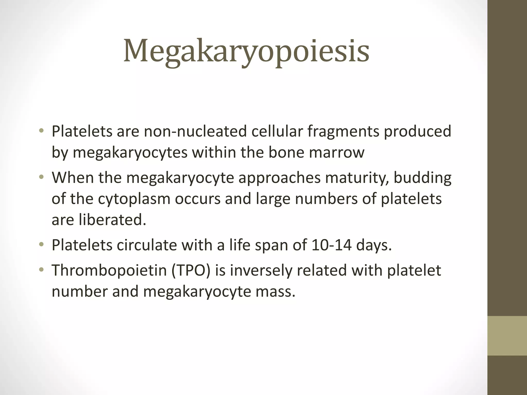
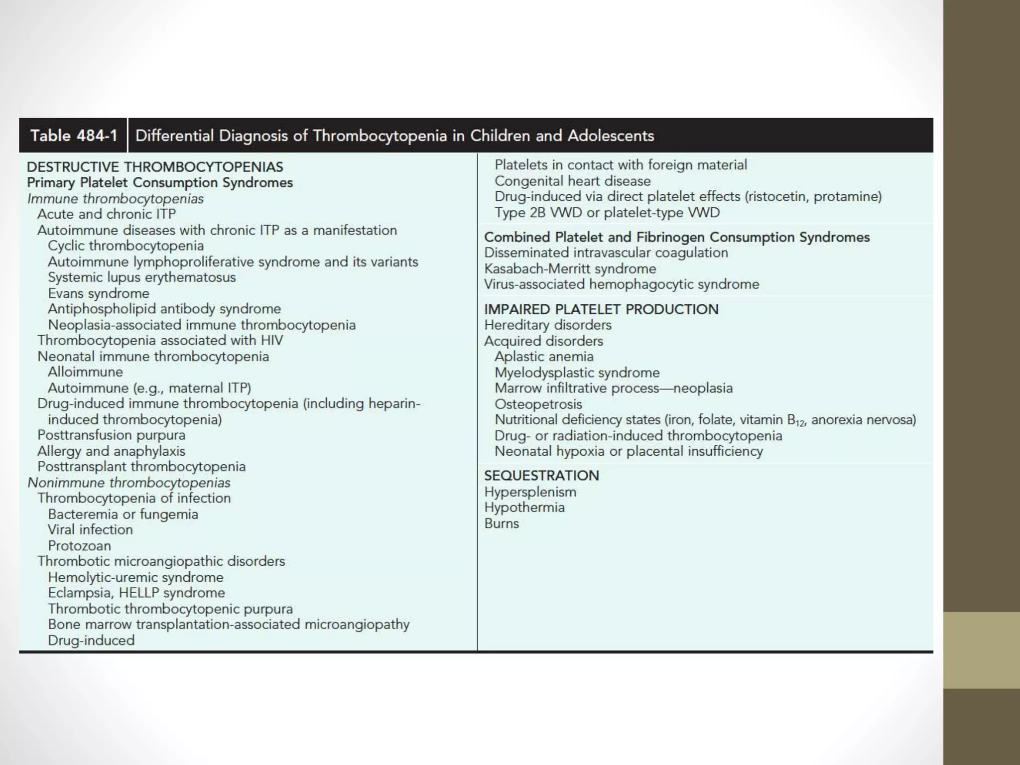
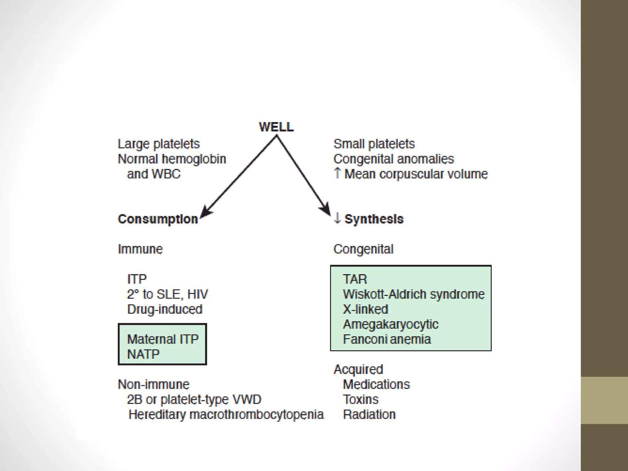
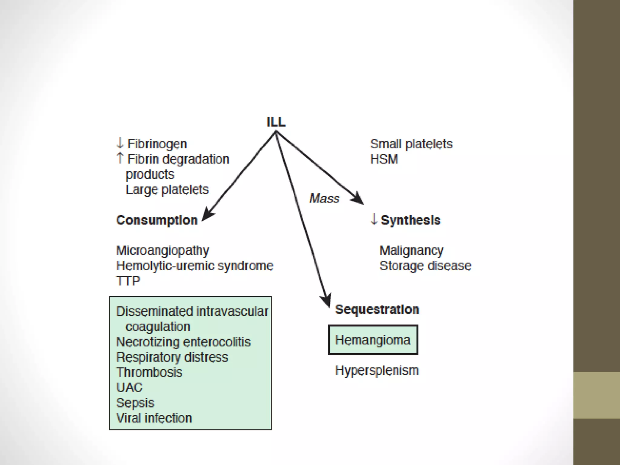
Platelet disorders
By
P.Padma Priyanka
Megakaryopoiesis
• Platelets are non-nucleated cellular fragments produced
by megakaryocytes within the bone marrow
• When the megakaryocyte approaches maturity, budding
of the cytoplasm occurs and large numbers of platelets
are liberated.
• Platelets circulate with a life span of 10-14 days.
• Thrombopoietin (TPO) is inversely related with platelet
number and megakaryocyte mass.
Thrombocytopenia
• Reduction in platelet count to <1.5lakhs
• Causes
• Decreased production-congential /acquired
• Sequestration
• Increased destruction-immune/nonimmune
Idiopathic thrombocytopenic
purpura
• Most common cause
• 1 in 20,000
• Peak age is 1-4yr
• M=F
• Late winter and spring
• 1-4week after exposure to a common viral infection(50-65%)
an autoantibody directed against the platelet surface
• The antibody binds to the platelet surface , circulating
antibody-coated platelets are recognized by the Fc receptor on
splenic macrophages, ingested, and destroyed
• Viruses associated with ITP – EBV and HIV
• Clinical manifestations
• previously healthy 1-4 yr old child with sudden onset of
generalized petechiae and purpura
• Bleeding from gums and mucous membranes(<1lakh)
• Preceding viral infection
• Rare-splenomegaly,lymphadenopathy,bone pain,pallor
• Outcome
• Spontaneous resolution occurs within 6months(70-80%)
• <1% of patients develop ICH
• 20% with acute ITP progress to chronic ITP
• Outcome may be related to age
• Laboratory findings
• Severe thrombocytopenia
• Platelet size –normal/increased
• Hb,TC,DC-normal
• Bone marrow-normal/increased megakaryocytes.Some-
immature
• Indications of bone marrow aspiration/biopsy
• Abnormal TC/DC
• unexplained anemia with findings suggestive of a bone marrow
failure syndrome or malignancy
• Other tests
• HIV
• Platelet antibody testing
• DCT
• Treatment
• Antiplatelet antibodies bind to transfused platelets so it is
contraindicated
• Education and counseling
• Single dose of IVIG(0.8-1g/kg) for 1-2days
• Prednisolone 1-4mg/kg/day
• IV anti-D therapy(50-75microg/kg)
• IVH-platelet transfusion,IVIG,high-dose corticosteroids
• Splenectomy-
• ≥4yr,severe ITP,>1yr
• Not easily controlled with therapy
• ICH
Secondary to SLE/HIV
• When the onset is insidious, especially in an adolescent, the
possibility of a systemic illness, such as systemic lupus
erythematosus (SLE), is more likely.
• HIV-associated ITP is usually chronic
Drug-induced
thrombocytopenia
• Result of either an immune process/megakaryocyte injury
• Valproic acid, phenytoin, carbamazepine, sulfonamides,
vancomycin, and trimethoprim-sulfamethoxazole
• Heparin-induced thrombocytopenia occurs after exposure to
heparin, the patient has an antibody directed against the
heparin–platelet factor 4 complex.
Maternal ITP
• Have lower risk of serious hemorrhage
• Treatment
• prenatal administration of corticosteroids to the mother
• administration of IVIG
• sometimes corticosteroids to the infant after delivery
• Usually resolves within 2-4months after delivery
Neonatal alloimmune
thrombocytopenic purpura
• Development of maternal antibodies against antigens present
on fetal platelets that are shared with the father
• 1 in 4000-5000 live births
• C/F-apparently well child -generalised petechiae and purpura
within 1st few days
• 30% may have ICH
• Diagnosis-presence of maternal alloantibodies directed
against the father’s platelets
• Treatment -administration of IVIG prenatally to the mother
from 2nd trimester throughout pregnancy
Type 2B VWD
• Results from mutations that increase the ability of VWF to
bind platelets.
• This leads to increased clearance of both VWF and platelets
from circulation and results in decreased VWF activity
• Diagnosis-direct measurement of the increased platelet
binding/increased response to low-dose ristocetin on platelet
aggregation testing.
• Thrombocytopenia is not always present and may be more
prominent during times of stress such as surgery or
pregnancy.
Platelet-type VWD
• Mutation in platelet GPIb causes spontaneous binding to VWF
• Presents with decreased VWF activity, loss of high-molecular-
weight multimers, and thrombocytopenia similar to type 2B
VWD.
• Treatment-platelet transfusion
Thrombocytopenia-absent
radius syndrome
• Thrombocytopenia (absence or hypoplasia of megakaryocytes)
that presents in early infancy
• Bilateral radial anomalies of variable severity, ranging from
mild changes to marked limb shortening
Wiskott-aldrich syndrome
• Characterized by eczema, and recurrent infection as a
consequence of immune deficiency.
• It is inherited as an X-linked disorder.
• Splenectomy often corrects the thrombocytopenia
X-linked thrombocytopenia
• point mutation within the WAS gene
Congenital amegakaryocytic
thrombocytopenia
• Manifests within the 1st few days to week of life
• Child presents with petechiae and purpura caused by profound
thrombocytopenia.
• A rare defect in hematopoiesis as a result of a mutation in the stem
cell TPO receptor.
• Bone marrow shows an absence of megakaryocytes.
• Often progress to marrow failure (aplasia) over time.
• Hematopoietic stem cell transplantation is curative
Microangiopathy
• DIC,HUS,TTP
• RBC destruction and consumptive thrombocytopenia due to
platelet and fibrin deposition in the microvasculature
• Presence of RBC fragments including helmet
cells,schistocytes,spherocytes and burr cells
Hemolytic uremic syndrome
• It is a common cause of community acquired AKI
• Triad-microangiopathic hemolytic anemia,
thrombocytopenia, and renal insufficiency
• Most common - toxin-producing Escherichia coli that
causes prodromal acute enteritis (diarrhea-associated
HUS)
• Asia,southern Africa-Shigella dysenteriae type 1
• Western countries-verotoxin or Shiga-like toxin producing
E. coli (STEC)
• Genetic –deficiencies of vWF cleaving
protease/complement factor H, I, or B, and defects in
vitamin B12 metabolism(absence of preceding diarrhea
prodrome)
• Pathogenesis
• Microvascular injury with endothelial cell damage
• Endogenous IgM recognises T antigen causing microvascular
angiopathy
• Clinical manifestations
• E.coli
• Fever,vomiting,abdominal pain and diarrhea
• Pallor,irritability,weakness and lethargy
• oliguria
• Pneumococci
• Pneumonia,empyema,bacteremia
• Majority –irritability,lethargy,encephalopathy,seizures
• Treatment
• prompt correction of volume deficit
• control of hypertension
• early institution of dialysis
Thrombotic thrombocytopenic
purpura
• Pentad of
• Fever
• Microangiopathic hemolytic anemia
• Thrombocytopenia
• Abnormal renal function
• Central nervous system changes
• Microvascular thrombi within the central nervous system
cause subtle neurologic signs
• Initial manifestations are often nonspecific
• Diagnosis-microangiopathic hemolytic anemia characterized
by morphologically abnormal RBCs, with schistocytes,
spherocytes, helmet cells, elevated reticulocyte count
• Treatment-plasmapheresis (effective in 80-95% of cases)
based on thrombocytopenia and microangiopathic hemolytic
anemia
• Rituximab, corticosteroids, and splenectomy are reserved for
refractory cases.
• The majority of cases of TTP are caused by an autoantibody–
mediated deficiency of a metalloproteinase (ADAMTS-13) that
is responsible for cleaving the high-molecular-weight
multimers of VWF
• ADAMTS- 13 deficiency can be treated by repeated infusions
of fresh-frozen plasma.
Sequestration
• Thrombocytopenia develops in individuals with massive
splenomegaly because the spleen acts as a sponge for
platelets and sequesters large numbers.
• Most such patients also have mild leukopenia and anemia on
CBC.
• Individuals who have thrombocytopenia caused by splenic
sequestration should be diagnosed for etiology of
splenomegaly, including infections, inflammatory, infiltrative,
neoplastic, obstructive, and hemolytic causes.
Kasabach –Merritt syndrome
• The association of a giant hemangioma with localized
intravascular coagulation causing thrombocytopenia and
hypofibrinogenemia
• In most patients, the site of the hemangioma is obvious, but
retroperitoneal and intraabdominal hemangiomas may
require body imaging for detection.
• Inside the hemangioma there is platelet trapping and
activation of coagulation, with fibrinogen consumption and
generation of fibrinogen degradation products.
Thrombocytopenia from
acquired disorders
• Infiltrative disorders like malignancies(ALL),histiocytosis,
lymphomas, and storage disease
• Aplastic processes may present as isolated thrombocytopenia
or along with leukopenia, neutropenia, anemia, or
macrocytosis
• Bone marrow examination should be performed when
thrombocytopenia is associated with abnormalities found on
physical examination or on examination of the other blood cell
lines.
Thrombocytosis
• Increased platelet count above normal >4 lakhs
• Primary
• Myeloproliferative disorders
• Essential thrombocythemia,PCV,CML,idiopathic myelofibrosis
• Leucocytosis,immature white cells,nucleated red cells,defective
platelet function,splenomegaly
• Secondary
• Hemorrhage ,trauma ,infections ,iron deficiency ,malignancy ,
splenectomy ,chronic inflammatory disease
• Features of underlying causative disorder are evident
Thank You
Fanconi anemia
• Radial anomalies
• Short stature
• Microcephaly
• Hyperpigmentation

Quantitative platelet disorders

  • 1.
  • 2.
    Megakaryopoiesis • Platelets arenon-nucleated cellular fragments produced by megakaryocytes within the bone marrow • When the megakaryocyte approaches maturity, budding of the cytoplasm occurs and large numbers of platelets are liberated. • Platelets circulate with a life span of 10-14 days. • Thrombopoietin (TPO) is inversely related with platelet number and megakaryocyte mass.
  • 4.
    Thrombocytopenia • Reduction inplatelet count to <1.5lakhs • Causes • Decreased production-congential /acquired • Sequestration • Increased destruction-immune/nonimmune
  • 7.
    Idiopathic thrombocytopenic purpura • Mostcommon cause • 1 in 20,000 • Peak age is 1-4yr • M=F • Late winter and spring • 1-4week after exposure to a common viral infection(50-65%) an autoantibody directed against the platelet surface • The antibody binds to the platelet surface , circulating antibody-coated platelets are recognized by the Fc receptor on splenic macrophages, ingested, and destroyed • Viruses associated with ITP – EBV and HIV
  • 8.
    • Clinical manifestations •previously healthy 1-4 yr old child with sudden onset of generalized petechiae and purpura • Bleeding from gums and mucous membranes(<1lakh) • Preceding viral infection • Rare-splenomegaly,lymphadenopathy,bone pain,pallor
  • 9.
    • Outcome • Spontaneousresolution occurs within 6months(70-80%) • <1% of patients develop ICH • 20% with acute ITP progress to chronic ITP • Outcome may be related to age • Laboratory findings • Severe thrombocytopenia • Platelet size –normal/increased • Hb,TC,DC-normal • Bone marrow-normal/increased megakaryocytes.Some- immature
  • 10.
    • Indications ofbone marrow aspiration/biopsy • Abnormal TC/DC • unexplained anemia with findings suggestive of a bone marrow failure syndrome or malignancy • Other tests • HIV • Platelet antibody testing • DCT
  • 11.
    • Treatment • Antiplateletantibodies bind to transfused platelets so it is contraindicated • Education and counseling • Single dose of IVIG(0.8-1g/kg) for 1-2days • Prednisolone 1-4mg/kg/day • IV anti-D therapy(50-75microg/kg) • IVH-platelet transfusion,IVIG,high-dose corticosteroids • Splenectomy- • ≥4yr,severe ITP,>1yr • Not easily controlled with therapy • ICH
  • 12.
    Secondary to SLE/HIV •When the onset is insidious, especially in an adolescent, the possibility of a systemic illness, such as systemic lupus erythematosus (SLE), is more likely. • HIV-associated ITP is usually chronic
  • 13.
    Drug-induced thrombocytopenia • Result ofeither an immune process/megakaryocyte injury • Valproic acid, phenytoin, carbamazepine, sulfonamides, vancomycin, and trimethoprim-sulfamethoxazole • Heparin-induced thrombocytopenia occurs after exposure to heparin, the patient has an antibody directed against the heparin–platelet factor 4 complex.
  • 14.
    Maternal ITP • Havelower risk of serious hemorrhage • Treatment • prenatal administration of corticosteroids to the mother • administration of IVIG • sometimes corticosteroids to the infant after delivery • Usually resolves within 2-4months after delivery
  • 15.
    Neonatal alloimmune thrombocytopenic purpura •Development of maternal antibodies against antigens present on fetal platelets that are shared with the father • 1 in 4000-5000 live births • C/F-apparently well child -generalised petechiae and purpura within 1st few days • 30% may have ICH • Diagnosis-presence of maternal alloantibodies directed against the father’s platelets • Treatment -administration of IVIG prenatally to the mother from 2nd trimester throughout pregnancy
  • 16.
    Type 2B VWD •Results from mutations that increase the ability of VWF to bind platelets. • This leads to increased clearance of both VWF and platelets from circulation and results in decreased VWF activity • Diagnosis-direct measurement of the increased platelet binding/increased response to low-dose ristocetin on platelet aggregation testing. • Thrombocytopenia is not always present and may be more prominent during times of stress such as surgery or pregnancy.
  • 17.
    Platelet-type VWD • Mutationin platelet GPIb causes spontaneous binding to VWF • Presents with decreased VWF activity, loss of high-molecular- weight multimers, and thrombocytopenia similar to type 2B VWD. • Treatment-platelet transfusion
  • 19.
    Thrombocytopenia-absent radius syndrome • Thrombocytopenia(absence or hypoplasia of megakaryocytes) that presents in early infancy • Bilateral radial anomalies of variable severity, ranging from mild changes to marked limb shortening
  • 21.
    Wiskott-aldrich syndrome • Characterizedby eczema, and recurrent infection as a consequence of immune deficiency. • It is inherited as an X-linked disorder. • Splenectomy often corrects the thrombocytopenia X-linked thrombocytopenia • point mutation within the WAS gene
  • 22.
    Congenital amegakaryocytic thrombocytopenia • Manifestswithin the 1st few days to week of life • Child presents with petechiae and purpura caused by profound thrombocytopenia. • A rare defect in hematopoiesis as a result of a mutation in the stem cell TPO receptor. • Bone marrow shows an absence of megakaryocytes. • Often progress to marrow failure (aplasia) over time. • Hematopoietic stem cell transplantation is curative
  • 24.
    Microangiopathy • DIC,HUS,TTP • RBCdestruction and consumptive thrombocytopenia due to platelet and fibrin deposition in the microvasculature • Presence of RBC fragments including helmet cells,schistocytes,spherocytes and burr cells
  • 25.
    Hemolytic uremic syndrome •It is a common cause of community acquired AKI • Triad-microangiopathic hemolytic anemia, thrombocytopenia, and renal insufficiency • Most common - toxin-producing Escherichia coli that causes prodromal acute enteritis (diarrhea-associated HUS) • Asia,southern Africa-Shigella dysenteriae type 1 • Western countries-verotoxin or Shiga-like toxin producing E. coli (STEC) • Genetic –deficiencies of vWF cleaving protease/complement factor H, I, or B, and defects in vitamin B12 metabolism(absence of preceding diarrhea prodrome)
  • 26.
    • Pathogenesis • Microvascularinjury with endothelial cell damage • Endogenous IgM recognises T antigen causing microvascular angiopathy • Clinical manifestations • E.coli • Fever,vomiting,abdominal pain and diarrhea • Pallor,irritability,weakness and lethargy • oliguria • Pneumococci • Pneumonia,empyema,bacteremia • Majority –irritability,lethargy,encephalopathy,seizures
  • 27.
    • Treatment • promptcorrection of volume deficit • control of hypertension • early institution of dialysis
  • 28.
    Thrombotic thrombocytopenic purpura • Pentadof • Fever • Microangiopathic hemolytic anemia • Thrombocytopenia • Abnormal renal function • Central nervous system changes • Microvascular thrombi within the central nervous system cause subtle neurologic signs • Initial manifestations are often nonspecific • Diagnosis-microangiopathic hemolytic anemia characterized by morphologically abnormal RBCs, with schistocytes, spherocytes, helmet cells, elevated reticulocyte count
  • 29.
    • Treatment-plasmapheresis (effectivein 80-95% of cases) based on thrombocytopenia and microangiopathic hemolytic anemia • Rituximab, corticosteroids, and splenectomy are reserved for refractory cases. • The majority of cases of TTP are caused by an autoantibody– mediated deficiency of a metalloproteinase (ADAMTS-13) that is responsible for cleaving the high-molecular-weight multimers of VWF • ADAMTS- 13 deficiency can be treated by repeated infusions of fresh-frozen plasma.
  • 31.
    Sequestration • Thrombocytopenia developsin individuals with massive splenomegaly because the spleen acts as a sponge for platelets and sequesters large numbers. • Most such patients also have mild leukopenia and anemia on CBC. • Individuals who have thrombocytopenia caused by splenic sequestration should be diagnosed for etiology of splenomegaly, including infections, inflammatory, infiltrative, neoplastic, obstructive, and hemolytic causes.
  • 32.
    Kasabach –Merritt syndrome •The association of a giant hemangioma with localized intravascular coagulation causing thrombocytopenia and hypofibrinogenemia • In most patients, the site of the hemangioma is obvious, but retroperitoneal and intraabdominal hemangiomas may require body imaging for detection. • Inside the hemangioma there is platelet trapping and activation of coagulation, with fibrinogen consumption and generation of fibrinogen degradation products.
  • 34.
    Thrombocytopenia from acquired disorders •Infiltrative disorders like malignancies(ALL),histiocytosis, lymphomas, and storage disease • Aplastic processes may present as isolated thrombocytopenia or along with leukopenia, neutropenia, anemia, or macrocytosis • Bone marrow examination should be performed when thrombocytopenia is associated with abnormalities found on physical examination or on examination of the other blood cell lines.
  • 35.
    Thrombocytosis • Increased plateletcount above normal >4 lakhs • Primary • Myeloproliferative disorders • Essential thrombocythemia,PCV,CML,idiopathic myelofibrosis • Leucocytosis,immature white cells,nucleated red cells,defective platelet function,splenomegaly • Secondary • Hemorrhage ,trauma ,infections ,iron deficiency ,malignancy , splenectomy ,chronic inflammatory disease • Features of underlying causative disorder are evident
  • 36.
  • 37.
    Fanconi anemia • Radialanomalies • Short stature • Microcephaly • Hyperpigmentation