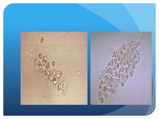
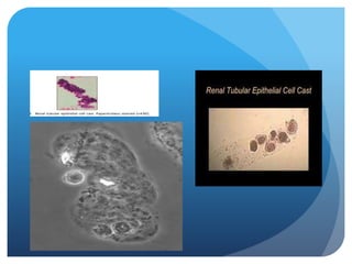
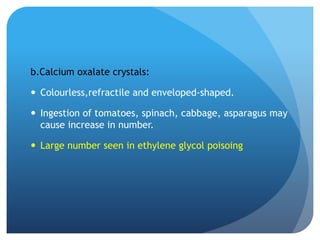
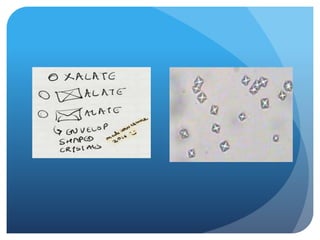
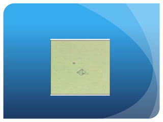
This document provides details on microscopic examination of urine sediment. Key points include:
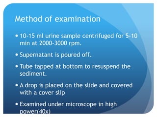
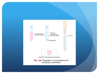
- Urine sample collection and preparation for examination under microscope by centrifuging and examining the sediment.
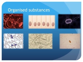
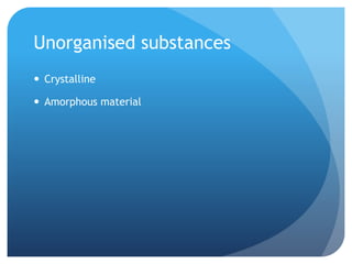
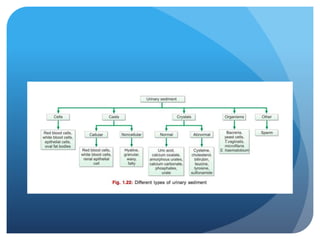
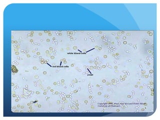
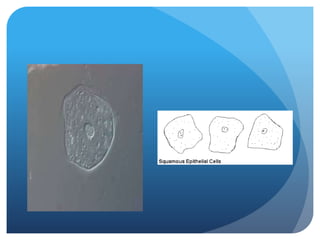
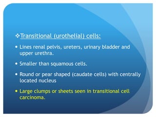
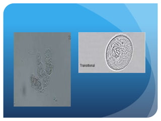
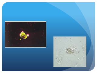
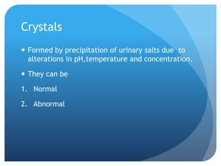
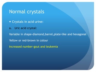
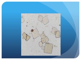
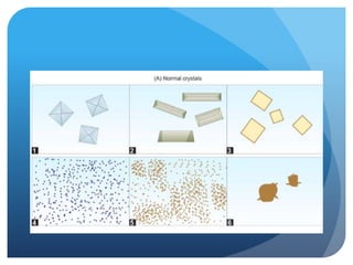
- Classification of findings as organised or unorganised substances, and types of cells, casts, crystals and other formed elements that may be observed.
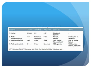
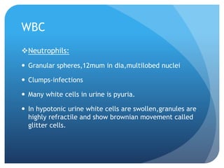
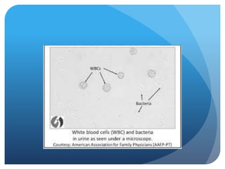
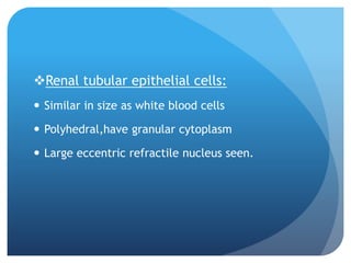
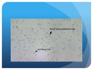
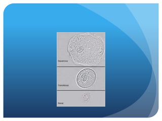
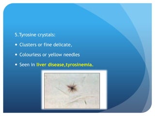
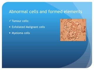
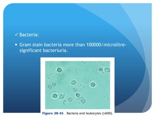
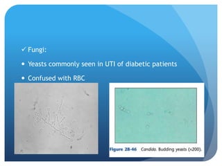
- Significance of various normal and abnormal findings in identifying renal and other diseases. Detailed morphology of different cell types, casts, crystals and other structures are described.