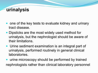
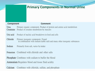
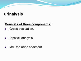
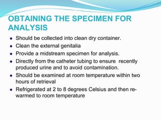
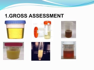
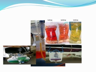
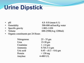
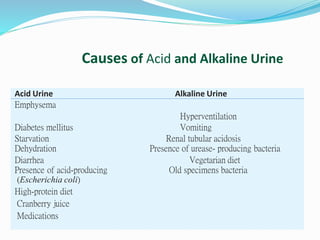
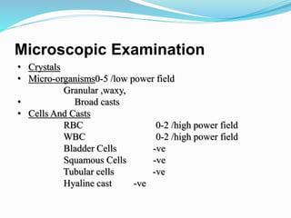
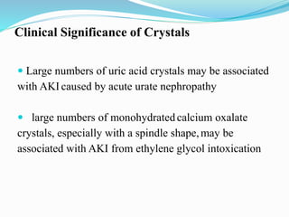
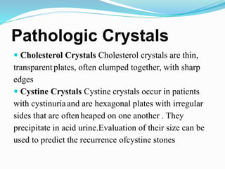
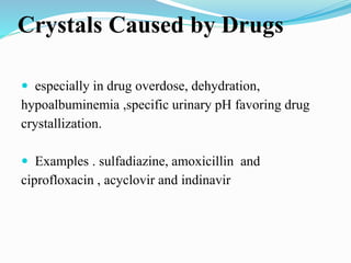
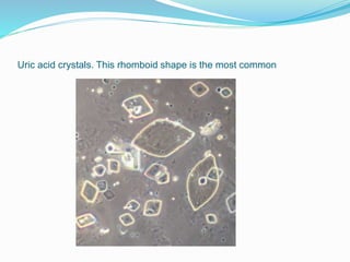
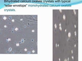
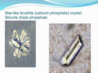
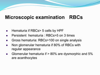
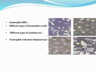
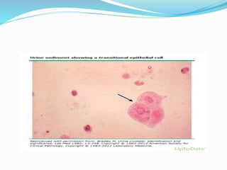
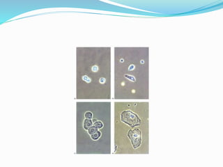
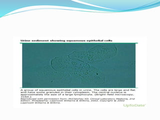
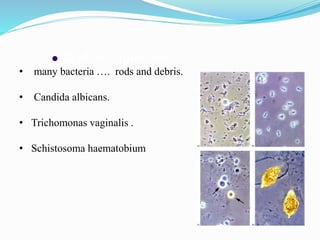
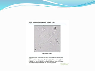
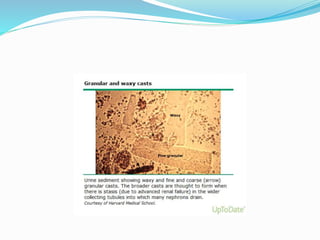
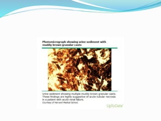
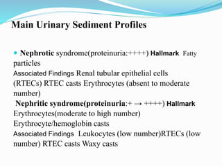
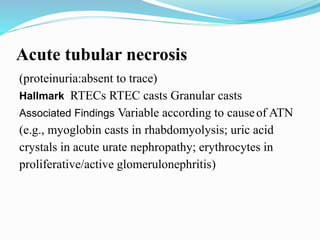
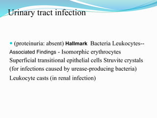
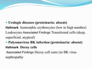
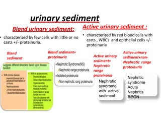
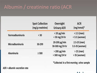
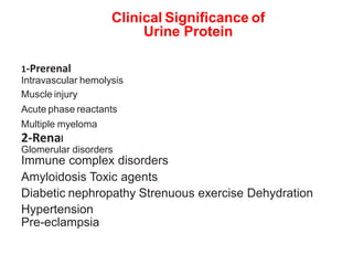
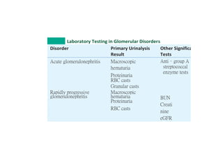
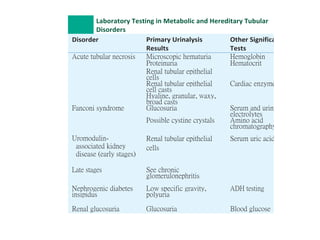
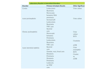
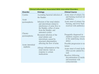
‫الجديد‬ ‫العام‬ ‫المنصوره‬ ‫مستشفى‬(‫الدولى‬)
urinalysis
Is a Poor man renal biopsy
urinalysis
 one of the key tests to evaluate kidney and urinary
tract disease.
 Dipsticks are the most widely used method for
urinalysis, but the nephrologist should be aware of
their limitations.
 Urine sediment examination is an integral part of
urinalysis, performed routinely in general clinical
laboratories.
 urine microscopy should be performed by trained
nephrologists rather than clinical laboratory personnel
urine Composition
urine consists of urea and other organic and inorganic
chemicals dissolved in water.
Urine is normally 95% water and 5% solutes, although
considerable variations in the concentrations of these
solutes can occur owing to the influence of factors such as
dietary intake, physical activity, body metabolism, and
endocrine functions
Primary Components in Normal Urine
Component Comment
Urea Primary organic component. Product of protein and amino acid metabolism
Creatinine Product of creatine metabolism by muscles
Uric acid Product of nucleic acid breakdown in food and cells
Chloride Primary inorganic component. Found
in combination with sodium (table salt) and many other inorganic substances
Sodium Primarily from salt, varies by intake
Potassium Combined with chloride and other salts
Phosphate Combines with sodium to buffer the blood
Ammonium Regulates blood and tissue fluid acidity
Calcium Combines with chloride, sulfate, and phosphate
urinalysis
Consists of three components:
 Gross evaluation.
 Dipstick analysis.
 M/E the urine sediment
OBTAINING THE SPECIMEN FOR
ANALYSIS
 Should be collected into clean dry container.
 Clean the external genitalia
 Provide a midstream specimen for analysis.
 Directly from the catheter tubing to ensure recently
produced urine and to avoid contamination.
 Should be examined at room temperature within two
hours of retrieval
 Refrigerated at 2 to 8 degrees Celsius and then re-
warmed to room temperature
Sample 24-Hour (Timed) Specimen
Collection Procedure
 Provide the patient with written instructions, and explain
the collection procedure.
Provide the patient with the proper collection container
and preservative.
Day 1: 7 a.m.: patient voids and discards specimen;
collects all urine for the next 24 hours.
Day 2: 7 a.m.: patient voids and adds this urine to
previously collected urine.
On arrival at laboratory, the entire 24-hour specimen is
thoroughly mixed, and the volume is measured and
recorded.
Clean-Catch Specimen Collection: Female
Cleansing Procedure
1. Wash hands.
2. Remove the lid from the container without touching the
inside of the container or lid.
3. Separate the skin folds (labia).
4. Cleanse from front to back on either side of the urinary
opening with an antiseptic towelette, using a clean one for
each side.
5. Hold the skin folds apart and begin to void into the toilet.
6. Bring the urine container into the stream of urine and collect
an adequate amount of urine. Do not touch the inside of the
container or allow the container to touch the genital area.
7. Finish voiding into the toilet.
8. Cover the specimen with the lid. Touch only the outside of
the lid andcontainer.
9. Label the container with the name and time of collection
and place in the specified area or follow institutional policy.
Clean-Catch Specimen Collection: Male
Cleansing Procedure
1. Wash hands.
2. Remove the lid from the sterile container without touching
the inside of the container or lid.
3. Cleanse the tip of the penis with antiseptic towelette and
let dry. Retract the foreskin if uncircumcised.
4. Void into the toilet. Hold back foreskin if necessary.
5. Bring the sterile urine container into the stream of urine and
collect an adequate amount of urine. Do not touch the
inside of the container or allow the container to touch the
genital area.
6. Finish voiding into the toilet.
7. Cover the specimen with the lid. Touch only the outside of
the lid andcontainer.
8. Label the container with the name and time of collection
and place in the specified area or follow institutional policy.
Urine Clarity
Clarity Term
Clear No visible particulates, transparent
Hazy Few particulates, print easily seen through urine
Cloudy Many particulates, print blurred through urine
Turbid Print cannot be seen through urine
Milky May precipitate or be clotted
1.GROSS ASSESSMENT
Colors
Urine Dipstick
• pH 4.8 -8.0 (mean 6.1)
• Osmolality 500-800 mOsm/Kg water
• Specific gravity 1.003-1.030
• Volume 600-2500(Avg 1200ml)
• Organic constituents per 24 Hours
Nitrogenous 25 - 35 gm
Urea 15-30 gm
Creatinine 1 -1.6 gm
Ammonia 0.7(0.3-1) gm
Uric acid 0.45 – (0.3 – 0.6) gm
Protein < 150 mg
Amylase 4-400 U/L
Table5–1
Causes of Acid and Alkaline Urine
Acid Urine Alkaline Urine
Emphysema
Hyperventilation
Diabetes mellitus Vomiting
Starvation Renal tubular acidosis
Dehydration Presence of urease- producing bacteria
Diarrhea Vegetarian diet
Presence of acid-producing Old specimens bacteria
(Escherichia coli)
High-protein diet
Cranberry juice
Medications
Microscopic Examination
• Crystals
• Micro-organisms0-5 /low power field
Granular ,waxy,
• Broad casts
• Cells And Casts
RBC 0-2 /high power field
WBC 0-2 /high power field
Bladder Cells -ve
Squamous Cells -ve
Tubular cells -ve
Hyaline cast -ve
Clinical Significance of Crystals
 Large numbers of uric acid crystals may be associated
with AKI caused by acute urate nephropathy
 large numbers of monohydratedcalcium oxalate
crystals, especially with a spindle shape,may be
associated with AKI from ethylene glycol intoxication
Pathologic Crystals
 Cholesterol Crystals Cholesterol crystals are thin,
transparentplates, often clumped together, with sharp
edges
 Cystine Crystals Cystine crystals occur in patients
with cystinuriaand are hexagonal plates with irregular
sides that are oftenheaped on one another . They
precipitate in acid urine.Evaluation of their size can be
used to predict the recurrence ofcystine stones
Crystals Caused by Drugs
 especially in drug overdose, dehydration,
hypoalbuminemia ,specific urinary pH favoring drug
crystallization.
 Examples . sulfadiazine, amoxicillin and
ciprofloxacin , acyclovir and indinavir
Uric acid crystals. This rhomboid shape is the most common
Bihydrated calcium oxalate crystals with typical
“letter envelope” monohydrated calcium oxalate
crystals.
Star-like brushite (calcium phosphate) crystal-
Struvite (triple phosphate
Cholesterol crystal- Cystine
crystals
Dihydroxyadenine crystal
Amoxicillin crystal
Microscopic examination : RBCs
 Hematuria if RBCs> 5 cells by HPF
 Persistent hematuria : RBCs>5 on 3 times
 Gross hematuria: RBCs>100 on single analysis
 Non glomerular hematuria if 80% of RBCs with
regular appearance
 Glomerular hematuria if > 80% are dysmorphic and 5%
are acanthocytes
• Isomorphic RBCs
• Different types of dysmorphic erythrocytes.
• Different types of acanthocytes .
• Neutrophils with their lobulated nucleus and granular cytoplasm
 3. Micro-organisms
• many bacteria …. rods and debris.
• Candida albicans.
• Trichomonas vaginalis .
• Schistosoma haematobium
Casts
• Elongated elements with a basic cylindrical
shape that has some possible variation due to
bending, wrinkling, and irregular edges.
• They form within the distal tubules and the
collecting ducts .
Casts
Clinical Significance of
Urinary Casts
 Hyaline Normal individual; renal disease
 Hyaline-granular Normal individual; renal disease
 Granular Renal disease; acute tubular necrosis
 Waxy Renal disease with function impairment
 Fatty Proteinuria; nephrotic syndrome
 Erythrocyte Glomerular hematuria; proliferative/necrotizing GN
 Leukocyte Acute interstitial nephritis; acute pyelonephritis;
proliferative GN
 Renal tubular epithelial cell(so-called epithelial casts)Acute tubular
necrosis; acute interstitial nephritis; proliferative GN; nephrotic
Syndrome
 Hemoglobin Same as for erythrocyte cast; hemoglobinuria
caused by intravascular hemolysis
 Myoglobin Rhabdomyolysis
 Bilirubin Jaundice caused by increased direct bilirubin
 Bacterial, fungal Bacterial or fungal infection in the
kidney
 Containing crystals; renalstonedisease;Normal
individual;crystalluricAKI
Main Urinary Sediment Profiles
 Nephrotic syndrome(proteinuria:++++) Hallmark Fatty
particles
Associated Findings Renal tubular epithelial cells
(RTECs) RTEC casts Erythrocytes (absent to moderate
number)
Nephritic syndrome(proteinuria:+ → ++++) Hallmark
Erythrocytes(moderate to high number)
Erythrocyte/hemoglobin casts
Associated Findings Leukocytes (low number)RTECs (low
number) RTEC casts Waxy casts
Acute tubular necrosis
(proteinuria:absent to trace)
Hallmark RTECs RTEC casts Granular casts
Associated Findings Variable according to causeof ATN
(e.g., myoglobin casts in rhabdomyolysis; uric acid
crystals in acute urate nephropathy; erythrocytes in
proliferative/active glomerulonephritis)
Urinary tract infection
 (proteinuria: absent) Hallmark Bacteria Leukocytes--
Associated Findings - Isomorphic erythrocytes
Superficial transitional epithelial cells Struvite crystals
(for infections caused by urease-producing bacteria)
Leukocyte casts (in renal infection)
 Urologic diseases (proteinuria: absent)
Hallmark Isomorphic erythrocytes (low to high number)
Leukocytes Associated Findings Transitional cells (deep,
superficial, atypical)
 Polyomavirus BK infection (proteinuria: absent)
Hallmark Decoy cells
Associated Findings Decoy cell casts (in BK virus
nephropathy
Urinary
indices
ATN Prerenal
(intact tubules)
Urinary Na+ >40 meq/L <20 meq/L
Fraction
excreation of
Na+ (FE Na+)
>2% <1%
urinary sediment
Bland urinary sediment:
• characterized by few cells with little or no
casts +/- proteinuria.
Active urinary sediment :
• characterized by red blood cells with
casts , WBCs and epithelial cells +/-
proteinuria
Bland sediment+
proteinuria
Bland
sediment
Nephrotic
syndrome
with active
sediment
Nephritic
syndrome
Acute
Nephritis
RPGN
Active urinary
sediment+
Nephrotic
range
proteinuria
Active urinary
sediment+non-
Nephrotic range
proteinuria
Albumin / creatinine ratio (ACR
Clinical Significance of
Urine Protein
1-Prerenal
Intravascular hemolysis
Muscle injury
Acute phase reactants
Multiple myeloma
2-Renal
Glomerular disorders
Immune complex disorders
Amyloidosis Toxic agents
Diabetic nephropathy Strenuous exercise Dehydration
Hypertension
Pre-eclampsia
3-Tubular Disorders
Fanconi syndrome
Toxic agents/heavy metals
Severe viral infections
4-Post renal
Lower urinary tract infections/ inflammation
Injury/trauma
Menstrual contamination
Prostatic fluid/spermatozoa
Vaginal secretions
Laboratory Testing in Glomerular Disorders
Disorder Primary Urinalysis
Result
Other Significant
Tests
Acute glomerulonephritis Macroscopic
hematuria
Anti–group A
streptococcal
enzyme tests
BUN
Creati
nine
eGFR
Proteinuria
RBC casts
Granular casts
Rapidly progressive
glomerulonephritis
Macroscopic
hematuria
Proteinuria
RBC casts
Laboratory Testing in Metabolic and Hereditary Tubular
Disorders
Disorder Primary Urinalysis
Results
Other Significant
Tests
Acute tubular necrosis Microscopic hematuria Hemoglobin
Proteinuria Hematocrit
Renal tubular epithelial
cells
Renal tubular epithelial
cell casts
Cardiac enzymes
Hyaline, granular, waxy,
broad casts
Fanconi syndrome Glucosuria Serum and urine
electrolytes
Possible cystine crystals Amino acid
chromatography
Uromodulin-
associated kidney
disease (early stages)
Renal tubular epithelial
cells
Serum uric acid
Late stages See chronic
glomerulonephritis
Nephrogenic diabetes
insipidus
Low specific gravity,
polyuria
ADH testing
Renal glucosuria Glucosuria Blood glucose
Laboratory Results in Interstitial Disorders
Disorder Primary Urinalysis Results Other Significant Tests
Cystitis Leukocyturia
Bacteriuria
Microscopic
hematuria Mild
proteinuria
Increased pH
Leukocyturia
Bacteriuria
WBC casts
Bacterial
casts
Microscopic
hematuria
Proteinuria
Leukocyturia
Bacteriuria
WBC casts
Bacterial
casts
Granular, waxy, broad casts
Hematuria
Proteinuria
Hematuria
Proteinuria
Leukocytu
ria WBC
casts
Urine culture
Acute pyelonephritis Urine culture
Chronic pyelonephritis Urine
culture
BUN
Creatinine
Acute interstitial nephritis
eGFR
Urine
eosinophils
BUN
Creatini
ne
eGFR
Clinical Information Associated With Interstitial Disorders
Disorder Etiology Clinical Course
Cystitis
Acute
pyelonephritis
Chronic
pyelonephritis
Acute interstitial
nephritis
Ascending bacterial infection of
the bladder
Infection of the renal tubules
and interstitium related to
interference of urine flow to
the bladder, reflux of urine
from the bladder, and
untreated cystitis
Recurrent infection of the
renal tubules and
interstitium caused by
structural abnormalities
affecting the flow of urine
Allergic inflammation of the
renal intersti- tium in
response to certain
medications
Acute onset of urinary frequency
and burning resolved with
antibiotics
Acute onset of urinary frequency,
burning, and lower back pain
resolved with antibiotics
Frequently diagnosed in
children; requires correction
of the underlying structural
defect
Possible progression to renal
failure
Acute onset of renal dysfunction
often accom- panied by a skin
rash
Resolves following
discontinuation of medica- tion
and treatment with
corticosteroids
References
• Comprehensive clinical nephrology 6th edtion
• Professor doctor/ iman sarahan Ain shams
university
• ESNT VIRTUAL ACADEMY
THANK YOU
Urine dr.hamada elsadaway

Urine dr.hamada elsadaway

  • 1.
    ‫الجديد‬ ‫العام‬ ‫المنصوره‬‫مستشفى‬(‫الدولى‬)
  • 2.
    urinalysis Is a Poorman renal biopsy
  • 3.
    urinalysis  one ofthe key tests to evaluate kidney and urinary tract disease.  Dipsticks are the most widely used method for urinalysis, but the nephrologist should be aware of their limitations.  Urine sediment examination is an integral part of urinalysis, performed routinely in general clinical laboratories.  urine microscopy should be performed by trained nephrologists rather than clinical laboratory personnel
  • 4.
    urine Composition urine consistsof urea and other organic and inorganic chemicals dissolved in water. Urine is normally 95% water and 5% solutes, although considerable variations in the concentrations of these solutes can occur owing to the influence of factors such as dietary intake, physical activity, body metabolism, and endocrine functions
  • 5.
    Primary Components inNormal Urine Component Comment Urea Primary organic component. Product of protein and amino acid metabolism Creatinine Product of creatine metabolism by muscles Uric acid Product of nucleic acid breakdown in food and cells Chloride Primary inorganic component. Found in combination with sodium (table salt) and many other inorganic substances Sodium Primarily from salt, varies by intake Potassium Combined with chloride and other salts Phosphate Combines with sodium to buffer the blood Ammonium Regulates blood and tissue fluid acidity Calcium Combines with chloride, sulfate, and phosphate
  • 6.
    urinalysis Consists of threecomponents:  Gross evaluation.  Dipstick analysis.  M/E the urine sediment
  • 7.
    OBTAINING THE SPECIMENFOR ANALYSIS  Should be collected into clean dry container.  Clean the external genitalia  Provide a midstream specimen for analysis.  Directly from the catheter tubing to ensure recently produced urine and to avoid contamination.  Should be examined at room temperature within two hours of retrieval  Refrigerated at 2 to 8 degrees Celsius and then re- warmed to room temperature
  • 8.
    Sample 24-Hour (Timed)Specimen Collection Procedure  Provide the patient with written instructions, and explain the collection procedure. Provide the patient with the proper collection container and preservative. Day 1: 7 a.m.: patient voids and discards specimen; collects all urine for the next 24 hours. Day 2: 7 a.m.: patient voids and adds this urine to previously collected urine. On arrival at laboratory, the entire 24-hour specimen is thoroughly mixed, and the volume is measured and recorded.
  • 9.
    Clean-Catch Specimen Collection:Female Cleansing Procedure 1. Wash hands. 2. Remove the lid from the container without touching the inside of the container or lid. 3. Separate the skin folds (labia). 4. Cleanse from front to back on either side of the urinary opening with an antiseptic towelette, using a clean one for each side. 5. Hold the skin folds apart and begin to void into the toilet. 6. Bring the urine container into the stream of urine and collect an adequate amount of urine. Do not touch the inside of the container or allow the container to touch the genital area. 7. Finish voiding into the toilet. 8. Cover the specimen with the lid. Touch only the outside of the lid andcontainer. 9. Label the container with the name and time of collection and place in the specified area or follow institutional policy.
  • 10.
    Clean-Catch Specimen Collection:Male Cleansing Procedure 1. Wash hands. 2. Remove the lid from the sterile container without touching the inside of the container or lid. 3. Cleanse the tip of the penis with antiseptic towelette and let dry. Retract the foreskin if uncircumcised. 4. Void into the toilet. Hold back foreskin if necessary. 5. Bring the sterile urine container into the stream of urine and collect an adequate amount of urine. Do not touch the inside of the container or allow the container to touch the genital area. 6. Finish voiding into the toilet. 7. Cover the specimen with the lid. Touch only the outside of the lid andcontainer. 8. Label the container with the name and time of collection and place in the specified area or follow institutional policy.
  • 11.
    Urine Clarity Clarity Term ClearNo visible particulates, transparent Hazy Few particulates, print easily seen through urine Cloudy Many particulates, print blurred through urine Turbid Print cannot be seen through urine Milky May precipitate or be clotted
  • 12.
  • 14.
    Urine Dipstick • pH4.8 -8.0 (mean 6.1) • Osmolality 500-800 mOsm/Kg water • Specific gravity 1.003-1.030 • Volume 600-2500(Avg 1200ml) • Organic constituents per 24 Hours Nitrogenous 25 - 35 gm Urea 15-30 gm Creatinine 1 -1.6 gm Ammonia 0.7(0.3-1) gm Uric acid 0.45 – (0.3 – 0.6) gm Protein < 150 mg Amylase 4-400 U/L
  • 15.
    Table5–1 Causes of Acidand Alkaline Urine Acid Urine Alkaline Urine Emphysema Hyperventilation Diabetes mellitus Vomiting Starvation Renal tubular acidosis Dehydration Presence of urease- producing bacteria Diarrhea Vegetarian diet Presence of acid-producing Old specimens bacteria (Escherichia coli) High-protein diet Cranberry juice Medications
  • 16.
    Microscopic Examination • Crystals •Micro-organisms0-5 /low power field Granular ,waxy, • Broad casts • Cells And Casts RBC 0-2 /high power field WBC 0-2 /high power field Bladder Cells -ve Squamous Cells -ve Tubular cells -ve Hyaline cast -ve
  • 17.
    Clinical Significance ofCrystals  Large numbers of uric acid crystals may be associated with AKI caused by acute urate nephropathy  large numbers of monohydratedcalcium oxalate crystals, especially with a spindle shape,may be associated with AKI from ethylene glycol intoxication
  • 18.
    Pathologic Crystals  CholesterolCrystals Cholesterol crystals are thin, transparentplates, often clumped together, with sharp edges  Cystine Crystals Cystine crystals occur in patients with cystinuriaand are hexagonal plates with irregular sides that are oftenheaped on one another . They precipitate in acid urine.Evaluation of their size can be used to predict the recurrence ofcystine stones
  • 19.
    Crystals Caused byDrugs  especially in drug overdose, dehydration, hypoalbuminemia ,specific urinary pH favoring drug crystallization.  Examples . sulfadiazine, amoxicillin and ciprofloxacin , acyclovir and indinavir
  • 21.
    Uric acid crystals.This rhomboid shape is the most common
  • 22.
    Bihydrated calcium oxalatecrystals with typical “letter envelope” monohydrated calcium oxalate crystals.
  • 23.
    Star-like brushite (calciumphosphate) crystal- Struvite (triple phosphate
  • 24.
  • 25.
  • 26.
    Microscopic examination :RBCs  Hematuria if RBCs> 5 cells by HPF  Persistent hematuria : RBCs>5 on 3 times  Gross hematuria: RBCs>100 on single analysis  Non glomerular hematuria if 80% of RBCs with regular appearance  Glomerular hematuria if > 80% are dysmorphic and 5% are acanthocytes
  • 27.
    • Isomorphic RBCs •Different types of dysmorphic erythrocytes. • Different types of acanthocytes . • Neutrophils with their lobulated nucleus and granular cytoplasm
  • 31.
     3. Micro-organisms •many bacteria …. rods and debris. • Candida albicans. • Trichomonas vaginalis . • Schistosoma haematobium
  • 32.
    Casts • Elongated elementswith a basic cylindrical shape that has some possible variation due to bending, wrinkling, and irregular edges. • They form within the distal tubules and the collecting ducts .
  • 33.
  • 34.
    Clinical Significance of UrinaryCasts  Hyaline Normal individual; renal disease  Hyaline-granular Normal individual; renal disease  Granular Renal disease; acute tubular necrosis  Waxy Renal disease with function impairment  Fatty Proteinuria; nephrotic syndrome
  • 35.
     Erythrocyte Glomerularhematuria; proliferative/necrotizing GN  Leukocyte Acute interstitial nephritis; acute pyelonephritis; proliferative GN  Renal tubular epithelial cell(so-called epithelial casts)Acute tubular necrosis; acute interstitial nephritis; proliferative GN; nephrotic Syndrome  Hemoglobin Same as for erythrocyte cast; hemoglobinuria caused by intravascular hemolysis
  • 36.
     Myoglobin Rhabdomyolysis Bilirubin Jaundice caused by increased direct bilirubin  Bacterial, fungal Bacterial or fungal infection in the kidney  Containing crystals; renalstonedisease;Normal individual;crystalluricAKI
  • 44.
    Main Urinary SedimentProfiles  Nephrotic syndrome(proteinuria:++++) Hallmark Fatty particles Associated Findings Renal tubular epithelial cells (RTECs) RTEC casts Erythrocytes (absent to moderate number) Nephritic syndrome(proteinuria:+ → ++++) Hallmark Erythrocytes(moderate to high number) Erythrocyte/hemoglobin casts Associated Findings Leukocytes (low number)RTECs (low number) RTEC casts Waxy casts
  • 45.
    Acute tubular necrosis (proteinuria:absentto trace) Hallmark RTECs RTEC casts Granular casts Associated Findings Variable according to causeof ATN (e.g., myoglobin casts in rhabdomyolysis; uric acid crystals in acute urate nephropathy; erythrocytes in proliferative/active glomerulonephritis)
  • 46.
    Urinary tract infection (proteinuria: absent) Hallmark Bacteria Leukocytes-- Associated Findings - Isomorphic erythrocytes Superficial transitional epithelial cells Struvite crystals (for infections caused by urease-producing bacteria) Leukocyte casts (in renal infection)
  • 47.
     Urologic diseases(proteinuria: absent) Hallmark Isomorphic erythrocytes (low to high number) Leukocytes Associated Findings Transitional cells (deep, superficial, atypical)  Polyomavirus BK infection (proteinuria: absent) Hallmark Decoy cells Associated Findings Decoy cell casts (in BK virus nephropathy
  • 48.
    Urinary indices ATN Prerenal (intact tubules) UrinaryNa+ >40 meq/L <20 meq/L Fraction excreation of Na+ (FE Na+) >2% <1%
  • 49.
    urinary sediment Bland urinarysediment: • characterized by few cells with little or no casts +/- proteinuria. Active urinary sediment : • characterized by red blood cells with casts , WBCs and epithelial cells +/- proteinuria Bland sediment+ proteinuria Bland sediment Nephrotic syndrome with active sediment Nephritic syndrome Acute Nephritis RPGN Active urinary sediment+ Nephrotic range proteinuria Active urinary sediment+non- Nephrotic range proteinuria
  • 50.
  • 51.
    Clinical Significance of UrineProtein 1-Prerenal Intravascular hemolysis Muscle injury Acute phase reactants Multiple myeloma 2-Renal Glomerular disorders Immune complex disorders Amyloidosis Toxic agents Diabetic nephropathy Strenuous exercise Dehydration Hypertension Pre-eclampsia
  • 52.
    3-Tubular Disorders Fanconi syndrome Toxicagents/heavy metals Severe viral infections 4-Post renal Lower urinary tract infections/ inflammation Injury/trauma Menstrual contamination Prostatic fluid/spermatozoa Vaginal secretions
  • 53.
    Laboratory Testing inGlomerular Disorders Disorder Primary Urinalysis Result Other Significant Tests Acute glomerulonephritis Macroscopic hematuria Anti–group A streptococcal enzyme tests BUN Creati nine eGFR Proteinuria RBC casts Granular casts Rapidly progressive glomerulonephritis Macroscopic hematuria Proteinuria RBC casts
  • 54.
    Laboratory Testing inMetabolic and Hereditary Tubular Disorders Disorder Primary Urinalysis Results Other Significant Tests Acute tubular necrosis Microscopic hematuria Hemoglobin Proteinuria Hematocrit Renal tubular epithelial cells Renal tubular epithelial cell casts Cardiac enzymes Hyaline, granular, waxy, broad casts Fanconi syndrome Glucosuria Serum and urine electrolytes Possible cystine crystals Amino acid chromatography Uromodulin- associated kidney disease (early stages) Renal tubular epithelial cells Serum uric acid Late stages See chronic glomerulonephritis Nephrogenic diabetes insipidus Low specific gravity, polyuria ADH testing Renal glucosuria Glucosuria Blood glucose
  • 55.
    Laboratory Results inInterstitial Disorders Disorder Primary Urinalysis Results Other Significant Tests Cystitis Leukocyturia Bacteriuria Microscopic hematuria Mild proteinuria Increased pH Leukocyturia Bacteriuria WBC casts Bacterial casts Microscopic hematuria Proteinuria Leukocyturia Bacteriuria WBC casts Bacterial casts Granular, waxy, broad casts Hematuria Proteinuria Hematuria Proteinuria Leukocytu ria WBC casts Urine culture Acute pyelonephritis Urine culture Chronic pyelonephritis Urine culture BUN Creatinine Acute interstitial nephritis eGFR Urine eosinophils BUN Creatini ne eGFR
  • 56.
    Clinical Information AssociatedWith Interstitial Disorders Disorder Etiology Clinical Course Cystitis Acute pyelonephritis Chronic pyelonephritis Acute interstitial nephritis Ascending bacterial infection of the bladder Infection of the renal tubules and interstitium related to interference of urine flow to the bladder, reflux of urine from the bladder, and untreated cystitis Recurrent infection of the renal tubules and interstitium caused by structural abnormalities affecting the flow of urine Allergic inflammation of the renal intersti- tium in response to certain medications Acute onset of urinary frequency and burning resolved with antibiotics Acute onset of urinary frequency, burning, and lower back pain resolved with antibiotics Frequently diagnosed in children; requires correction of the underlying structural defect Possible progression to renal failure Acute onset of renal dysfunction often accom- panied by a skin rash Resolves following discontinuation of medica- tion and treatment with corticosteroids
  • 58.
    References • Comprehensive clinicalnephrology 6th edtion • Professor doctor/ iman sarahan Ain shams university • ESNT VIRTUAL ACADEMY
  • 59.