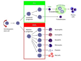
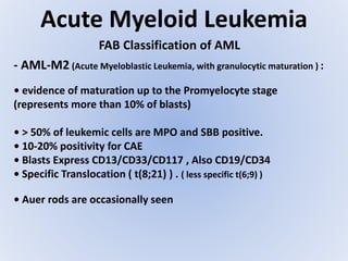
SlideShare a Scribd company logo
Acute Leukemia
Ahmad A. Al-Qudah
Clinical Hematology II LM-754
Acute Leukemia
- Introduction to Leukemia and Acute Leukemia
- Revision of Special Stains and Cytochemistry related to Acute Leukemia Diagnosis .
- Classification of Bone Marrow Cell Types
- AML : - Definition
- Sign and Symptoms
- Pathophysiology & Lab Findings
- Classifications : - FAB - WHO
- ALL :
- Definition
- sign and symptoms
- Pathophysiology & Lab Findings
- Classifications : - FAB -WHO
- Is a group of malignant (neoplastic) disorders , characterized
by the clonal expansion and accumulation of one or more blood
cell line(s) , with eventual involvement of all hematopoietic
organs and other organs.
Leukemia
• Prolonged life (immortal) resistant to apoptosis
• Growth factor independent growth
• Insensitivity to growth-inhibitory signals
• Ability to invade and metastasize
• Blockage of intracellular differentiation
Tow or more Mutations within the genome of HSC or multipotential
progenitors/precursors
Activation of specific proto-oncogene
De-activation of tumor suppressor genes
Leukemia
Clone of cells with characteristics of a malignant cell
Leukemia
Based on the clinical and path-physiologic
characteristics:
• Acute: - Characterized by the clonal expansion and accumulation of
immature leukocytes (blood cells) in hematopoietic organs .
• Chronic:- Clonal expansion and accumulation of mature, but
abnormal leukocytes in hematopoietic organs .
Based on the affected blood cell lines:
• Myelogenous: Myeloid cell line
• Lymphocytic: Lymphoid cell line
Leukemia
Acute Chronic
Myeloid
origin
Lymphoid
origin
Acute Myeloid
Leukemia (AML)
Acute Lymphoblastic
Leukemia (ALL)
Chronic Myeloid Leukemia (CML)
Chronic Lymphocytic Leukemia
(CLL)
Leukemia
• Symptoms reflects B.M. failure to produce normally functioning
blood cells :
- Features of anemia: Weakness, shortness of breath and pallor .
- Features of leukopenia: infections (septicemia, pneumonitis)
- Features of thrombocytopenia: Bleeding and increased hemorrhagic
tendency .
• Symptoms reflects B.M. hyperplasia and excessive accumulation,
infiltration and proliferation of malignant cells :
- Bone and Joint pain
- Hepatosplenomegaly
- Lymphadenopathy
- Gingival hypertrophy (hyperplasia) and oral lesions
Leukemia
Leukemia
Leukemia
Leukemia
Leukemia
• CBC and blood film examination :
- Normocytic normochromic anemia (mild to severe)
- Platelets count: reduced to normal
- WBC’s count: decreased mature leukocytes
- The appearance of abnormally circulating blood cells
(malignant cells)
• BM aspirate and/or biopsy
- B.M. hyperactivation and hyperplasia
- Blasts : represents greater than 20 - 30% depends on the type
of Classification and type of Leukemia .
- BM Cell Types : ANC , EC , NEC
Leukemia
• Cytochemistry:
Blasts Identified
Cellular Element
Stained
Cytochemical Reaction
Myeloblasts strong positive;
monoblasts faint positive
Lymphoblast Negative
Neutrophil primary
granules
Myeloperoxidase (MPO)
Myeloblasts strong positive;
monoblasts faint positive
Lymphoblast Negative
PhospholipidsSudan Black B (SBB)
Promyelocyte stage positiveCellular enzymeSpecific esterase
Monoblasts strong positive
Others Negative
Cellular enzyme
Nonspecific esterase
(NSE)
lymphoblast's and pronormoblasts
Negative to Positive .
Myeloblasts usually negative.
Metamyelocyte & PMN Strong +ve
Glycogen and
related substances
Periodic acid-Schiff
Leukemia
•Immunological markers (Surface, Cytoplasmic & Nuclear )
- TdT: Terminal Deoxynucleotidyl Transferase , differentiate Myeloblasts
from lymphoblasts (B & T precursors)
- CD markers : e.g. AML: CD11/CD13/CD33/CD117
ALL (B-Cells): CD10/CD19/CD20/CD22
ALL (T-Cells): CD2/CD3/CD5/CD7
• Cytogenetics: specific Chromosomal abnormalities
- Philadelphia chromosome (t(9;22)): common in CML
- t(8;21): AML-M2
- t(1;19), t(8;22), t(11,14) ALL
Acute Myeloid Leukemia
- Malignant neoplastic proliferation and accumulation
of immature and nonfunctional myeloid line of blood
cells in the bone marrow .
- Also known as acute myelogenous leukemia or acute
nonlymphocytic leukemia (ANLL)
- Most common Acute Leukemia affecting adults.
- As an Acute Leukemia, AML progresses rapidly and is typically
fatal within weeks or months if left untreated .
Hematopoietic
stem cell
Neutrophils
Eosinophils
Basophils
Monocytes
Platelets
Red cells
Myeloid
progenitor
Lymphoid
progenitor
B-lymphocytes
T-lymphocytes
Plasma
cells
naïve
ALL
AML
Acute Myeloid Leukemia
Pathophysiology
- In AML, a single myeloblast genetic changes which "freeze"
the cell in its immature state and prevent differentiation.
- In normal hematopoiesis, the myeloblast is an immature
precursor of myeloid white blood cells; a normal myeloblast
will gradually mature into a mature white blood cell .
- When such a "differentiation arrest" is combined with other
mutations which disrupt genes controlling proliferation, the
result is an uncontrolled growth of an immature clone of cells,
leading to the clinical entity of AML .
Acute Myeloid Leukemia
- The clinical signs and symptoms of AML result from the
growth of leukemic clone cells, which tends to displace or
interfere with the development of normal blood cells in
the bone marrow .
- This leads to neutropenia, anemia, and thrombocytopenia.
The symptoms of AML are often due to the low numbers of
these normal blood elements.
Pathophysiology
Acute Myeloid Leukemia
*Specific Signs and Symptoms :
- Most signs and symptoms of AML are general ( Same common
signs of all Leukemia ) :
- Features related to Pancytopenia .
- Features related to infiltration and accumulation .
- Some Specific Features :
- Chloroma : a solid leukemic mass or tumor developed
outside of the bone marrow .
Acute Myeloid Leukemia
A
B
C
Chloromas
NEJM 1998
Acute Myeloid Leukemia
Laboratory findings:
1- Peripheral Blood:
- The leukocyte count ranges from < 1X10⁹ to >100X109 .
- Presence of blast on the blood smear.
- Neoplastic blast have few granules in the RNA rich
cytoplasm. According to the FAB:
1- Type I: typical blast features without granules.
2- Type II: has < 20 granules.
3- Type III: has numerous granules.
Acute Myeloid Leukemia
- Erythrocyte decreased, Hb less than 10g/dl
- Slightly Macrocytic because of the inability to compete
the neoplastic cell for folate and vitB12 or early release of
Retic cells.
2- Bone marrow:
- Typically, hypocellular with increased fat content .
Laboratory findings:
- Special Stains and Percentage of Blasts aid in the Diagnosis .
Acute Myeloid Leukemia
Classification :
1- FAB Group Classification , Based on :
- The Morphological criteria of PB and BM .
- The Immunophenotyping of Leukemic cells .
- The cytochemistry of Leukemic Cells .
- For Diagnosis, Blast count in PB or BM is >30% .
2- WHO Classification :
- Molecular/Genetic Features of Malignancies .
- FAB Class. Incorporated into WHO Class.
- For Diagnosis, Blast count in PB or BM is >20% .
FAB Classification of AML
Acute Myeloid Leukemia
- AML-M0 (Acute Myeloblastic Leukemia without differentiation ):
• AML with no evident morphological differentiation.
( no characteristic myeloid features )
• Bone marrow contains 30 to 90% blast forms with clear cytoplasm
(granules free ).
• The blasts seen in AML and ALL are very similar; with a high nuclear
cytoplasmic ratio, immature chromatin, nucleoli, and a variable
amount of cytoplasm .
• <3% of blasts are MPO and SBB positive
• Blasts are positive for CD13 and CD33 markers
Acute Myeloid Leukemia
FAB Classification of AML
- AML-M1 (Acute Myeloblastic Leukemia, without maturation ):
• Poorly differentiated myeloblasts (more than 90% of NEB)
• Blasts are Type I ( Agranular ) and Type II ( Granular )
• MPO & SBB are positive (>3 but < 50%)
• CAE is with at least 10-20% positivity
• Blasts Express CD13/CD33/CD117
• Auer rods (may present but rarely seen)
Acute Myeloid Leukemia
FAB Classification of AML
- AML-M2 (Acute Myeloblastic Leukemia, with granulocytic maturation ) :
• evidence of maturation up to the Promyelocyte stage
(represents more than 10% of blasts)
• > 50% of leukemic cells are MPO and SBB positive.
• 10-20% positivity for CAE
• Blasts Express CD13/CD33/CD117 , Also CD19/CD34
• Specific Translocation ( t(8;21) ) . ( less specific t(6;9) )
• Auer rods are occasionally seen
Acute Myeloid Leukemia
FAB Classification of AML
- AML-M3 (Acute Promyelocytic leukemia )
• Promyelocytes with heavy granulation . ( >30% of NEC )
• Auer rods frequently seen (Faggot cells: cells with bundles of AR)
• Cells are strongly positive with MPO and SBB
• Negative with CAE
• t(15;17) is a unique and common feature of AML-M3.
• AML-M3 variant: showing the characteristic bilobed
hypogranular Promyelocytes .
• AML-M3 and DIC ( Thromboplastic Substances )
Acute Myeloid Leukemia
FAB Classification of AML
- AML-M4 (Acute Myelomonocytic leukemia ) :
• Cells are with granulocytic and monocytic differentiation
• 20-80% of cells are positive for MPO, SBB and NSE.
• Differential diagnosis: serum lysozyme level > 3 times
•16q deletion or inversion are common.
• AML-M4 with Eosinophilia
- Eosinophils represents > 5% of nonerythroid cells
- immature eosinophils positive for SE and PAS
Acute Myeloid Leukemia
FAB Classification of AML
- AML-M5 (Acute Monoblastic leukemia ):
• > 80% of NEC are monocytic lineage
• > 80% of cells are positive for NSE
• Cells are negative for MPO and SBB (weakly positive in
monoblast).
• del (11q), t(9;11), t(11;19)
• Two subclasses: Poorly (M5a) & well (M5b) differentiated
- M5a: Predominance of monoblasts
- M5b: Predominance of promonocytes and monocytes
Acute Myeloid Leukemia
FAB Classification of AML
- AML-M6 (Acute Eythroblastic leukemia ) :
• Hypercellularity of B.M. with marked erythroid hyperplasia .
• > 50% of ANC are of erythroid lineage .
• Blasts commonly show megaloblastoid characterestic
features including multi-nucleation, cytoplasm vaculation.
• Most erythroid precursors are positive for PAS (Fine staining)
• Negative to weakly positive for MPO, SBB and SE
• AML (M6) against MDS :
- >50% or <50% Erythroblast of ANC:
- With >30% Blast of ANC ---> AML (M6)
- With <30% Blast of ANC ---> MDS
Acute Myeloid Leukemia
FAB Classification of AML
AML-M7 (Acute Megakaryoblastic leukemia ) :
• Uncommon form
• Extensive proliferation of Megakaryoblasts and ~cyte.
• >50% of blasts are megakaryoblasts
• Blasts identified by platelets peroxidase
• Negative for MPO and SBB, while positive for PAS.
• Blasts Express CD41/CD42
• t(1;22) is common .
Acute Myeloid Leukemia
WHO Classification of AML
1- AML with recurrent Cytogenetic abnormalities
- usually translocation, in most cases the chromosomal
rearrangement create a fusion gene, encoding a novel
fusion protein.
I. AML with t(8;21)(q22;q22);(ETO/AML1):
- Present morphological as AML with maturation.
- Core binding factor (CBF) also called AML1 is a transcription
factor critical for hematopoietic development.
- It is believed that HSC is the origin of leukemia
Acute Myeloid Leukemia
WHO Classification of AML
- The t(8;21) translocation result in a chimeric protein
containing the N- terminal portion of CBF (chromosome 21)
and most of the ETO protein from chromosome 8 ( nuclear
protein involved in the regulation of transcription).
- The fusion protein blocks the normal function of CBF, and
induce abnormal gene activation and gene repression, this
will lead to increase proliferation with blocked
differentiation.
Acute Myeloid Leukemia
WHO Classification of AML
II. AML with abnormal B.M eosinphils
inv (16)(p13;q22) or t(16;16)(p13;q22)(CBFB/MYH11):
- Present morphology as AML with monocytic and granulocytic
maturation and presence of abnormal eosinphils in B.M.
- Combination of acute myelomonocytic leukemia (AMML) with
abnormal eosinphilis is morphologically AMML Eo.
- Abnormal immature ( basophilic ) granules in the eosinphils,
promyelocyte, and myelocyte stages.
- The inv (16)(p13;q22) and t(16;16)(p13;q22) both result in the fusion
of the CBFB gene (16q22) to the smooth muscle mysoin heavy chain
gene SMMHC (MYH11)at (16p13).
- The CBFB/SMMHC fusion proetin binds to AML1/CBFa and represses
it is function as transcription factor.
Acute Myeloid Leukemia
WHO Classification of AML
III . AML with t(15;17)(q22;q12)(PML/RARa) and variants:
- it is acute promyelocytic leukemia(APL), an AML in which abnormal
promyelocyte predominate.
- Both hypergranular ( typical APL) and hypogranular or microgranular
are seen.
- The presenting signs are DIC and bleeding .
- The typical t(15;17) gene rearrangement result in the fusion of
(PML/RARa) gene and reciprocal ( RARa/PML) gene.
- (PML/RARa) mRNA has been identified in all APL patient while (
RARa/PML) mRNA in two third.
- The RARa protein is a nuclear hormone receptor that bind to specific
DNA sequence and controls transcription.
Acute Myeloid Leukemia
WHO Classification of AML
- PML is growth suppressor nuclear protein normally found in
complex macromolecular structure.
- The PML/RARa fusion protein leads to formation of co-repressor
complex molecules that enhance the oncogenesis of APL .
- It is believed that HSC is the origin of leukemia
Acute Myeloid Leukemia
WHO Classification of AML
IV. AML with 11q23(MLL) abnormalities:
- These leukemia associated with monocytic features ( monoblasts
and promonocyte).
- Monoblast and promonocyte can have scattered azurophilic
granules and vacuoles.
- The MLL gene (11q13) is involved in a number of leukemia
associated translocation with different partner chromosome.
- The MLL protein is a DNA binding protein that interact with other
nuclear protein and permits the association of transcription factor
which regulate transcription.
Acute Myeloid Leukemia
WHO Classification of AML
2- AML with multilinage Dysplasia ( with or without prior MDS)
- It is an Acute leukemia ( >20% blast) with dysplasia in more
than 50% of the cells in two or more myeloid cell lines.
- It is occurs with or following MDS / MPD.
- examples of dysplasia include: hypogranular PMNs, psuedo-
pelger-Huet anomaly, megaloblastic erythrocyte, ringed
sideroblast.
- The cytogenetics abnormalities are variable and are similar to
these in MDS.
- HSC is the origin of leukemia
- poor prognosis.
Acute Myeloid Leukemia
WHO Classification of AML
3- AML and Myelodysplastic syndrome, therapy releated:
- These disorder arise as a result of cytotoxic chemotherapy
and / or radiation therapy.
Two major subtypes:
I. Alkylating agent/ radiation treatment: initially it start with
MDS and eventually evolving AML.
II. Topoisomerase II inhibitor treatment.
Acute Myeloid Leukemia
WHO Classification of AML
4- Acute Myeloid leukemias not otherwise categorized:
- Include all AML cases that not fulfill criteria for any of other
described.
- The subtypes of this AML are classified according to
differentiated on morphology and cytochemical features.
I. AML Minimally Differentiated
II. AML without Maturation
III. AML with Maturation
IV. Acute Myelomonocytic leukemia (AMML)
V. Acute monoblastic leukemia and Acute monocytic leukemia
VI. Acute Erythroid leukemia (AEL)
VII. Acute Megakaryoblastic leukemia
Acute Myeloid Leukemia
WHO Classification of AML
VIII. Acute Basophilic leukemia
- The most characteristic feature by cytochemistry is metachromatic
positivity with toluindine blue.
IX. Acute panmyelosis with mylofibrosis
- it is occur with or following chemo and radio therapy.
X. Myeloid Sarcoma
- a tumor of myeloblast or immature myeloid cells occures in the
extramedullary site or in the bone.
Acute Lymphoid Leukemia
Malignant neoplastic proliferation and accumulation
of immature and nonfunctional Lymphoid line of
blood cells in the bone marrow .
The "lymphocytic" in acute lymphocytic leukemia refers
to the white blood cells called lymphocytes, which ALL
affects. Acute lymphocytic leukemia is also known as
acute lymphoblastic leukemia.
Acute lymphocytic leukemia is the most common type
of cancer in children
Acute Lymphoid Leukemia
*The signs and symptoms of ALL are variable but follow from bone
marrow replacement and/or organ infiltration :
- Generalized weakness and fatigue
- Anemia
- Frequent or unexplained fever and infection
- Weight loss and/or loss of appetite
- Bone pain, joint pain (caused by the spread of "blast" cells to the
surface of the bone or into the joint from the marrow cavity)
- Breathlessness
- Enlarged lymph nodes, liver and/or spleen
- Pitting edema (swelling) in the lower limbs and/or abdomen
- Petechiae, which are tiny red spots or lines in the skin due to
low platelet levels
Acute Lymphoid Leukemia
- Damage to DNA that leads to uncontrolled cellular growth and
spread throughout the body.
Pathophysiology
- This damage may be caused by environmental factors such as
chemicals, drugs or radiation.
- Some evidence suggests that secondary leukemia can develop
in individuals treated for other cancers with radiation and
chemotherapy as a result of that treatment
- Damage can be caused through the formation of fusion genes,
as well as the dysregulation.
Initial Laboratory finding characteristic of ALL
Peripheral Blood: 1- Leukocyte count usually increased but may be normal or decreased.
2- Neutropenia
3- Lymphoblast
4- Thrombocytopenia
Bone Marrow: 1- Hypercellular
2- >20% lymphoblast ( WHO)
Other laboratory finding: 1- associated with increased cellular metabolism and turn over such:
Hyperuricemia.
incrased serum LD.
hypercalacemia due to increased BM resorption.
2- Renal failure.
3- increased CSF lymphblast.
Acute Lymphoid Leukemia
- Terminal Deoxynucleotidyl transferase (TdT):
- Lymphocyte can identified by immunophenotying and certain
intracellular enzyme.
- TdT, is the most important enzyme that is helpful in the
identifying cellular subtypes.
- TdT is a DNA polymerase found in cell nuclei , this enzyme not
present in the normal mature lymphocyte but can be found in
65% of the thymic population of lymphocyte.
It can be determined by direct enzyme assay and indirect
immunofluorescence.
Acute Lymphoid Leukemia
Classification
- Based on FAB classification, ALL is categorized into three
catergories ALL-L1, ALL-L2 and ALL-L3 .
- Immunohistochemistry and immunophenotyping are
almost always necessary to distinguish ALL from AML.
- On other hand WHO identify of malignant cell as T, B and
on the degree of maturation.
Acute Lymphoid Leukemia
- Blasts in ALL-L1 are with high N/C ratio.
- Delicate diffuse chromatin pattern and small prominent
nucleoli.
- Scant cytoplasm .
- Affects primarily children
FAB classification
ALL-L1
Acute Lymphoid Leukemia
FAB classification
ALL-L2
- The blasts are larger than those of L1, have more plentiful
cytoplasm and are more pleomorphic.
- Abundant cytoplasm, predominant nucleoli, nuclear
clefting .
- Affects adults
Acute Lymphoid Leukemia
FAB classification
ALL-L3 : Burkitt's type
- L3 blasts cells are fairly regular in shape with strong
basophilic cytoplasm and prominent cytoplasmic
vaculation .
Affects adults and children
FAB Classification of ALL
Morphology TdT CD10
ALL-L1 Small lymphocyte scant cytoplasm; + +
Moderately clumped chromatin;
Inconspicuous nucleoli
ALL-L2 Small and medium size lymphblast; + +
Mixed chromatin patterns;
Inconspicuous nucleoli
ALL-L3Burkitt- type Large lymphblast; large nucleus with - +/-
nucleoli; cytoplasm vacuolization;
Intense cytoplasm basophilia
Acute Lymphoid Leukemia
WHO classification
- WHO classification considers ALL and lymphblastic
lymphoma to be single disease with different clinical
presentation.
- Precursor T- and B-cell neoplasm with B.M and
peripheral blood involvement are ALL, while precursor T-
and B-cell neoplasm presenting solid tumors are
lymphoma.
Acute Lymphoid Leukemia
WHO classification
WHO classification defines two subgroups of ALL:
1- Precursor B- and T-cell neoplasm ( leukemia/ lymphoma)
which include L1 and L2 .
2- Burkitt type ALL (L3).
Acute Lymphoid Leukemia
WHO classification
1- Precursor B- cell leukemia:
- it is a neoplasm of lymphoblast committed to the B- cell
linage, involve B.M, peripheral blood
- some cases may present with primary involvement of
lymph node and exranodal site ( lymphoma).
- Rearrangement of immunoglobulin genes can be detected
by molecular testing.
- Additional markers of B linage commitment required for
dignosis. CD10/CD19/CD20/CD22 , TdT = +ve
Acute Lymphoid Leukemia
WHO classification
1- Precursor B- cell leukemia:
- Cytogenetic abnormalities associated with B-ALL include
translocation, hypodiploidy, and hyperdiploidy.
- The most common is t(12;21)(p13;q22)
- Produce TEL-AML1 fusion gene.
- Hyperdiploid B-ALL have a mutation in the receptor
tyrosine Kinase FLT-3 , resulting in constitutive activation of
the receptor.
- t(1;19) PBX1-E2A , t(4;11) AF4-MLL
- Precursor B- cell has good prognosis.
Acute Lymphoid Leukemia
WHO classification
2- Precursor T- cell leukemia:
- It is a neoplasm of lymphoblasts committed to T- cell
linage, involving B.M and peripheral blood.
- High WBC count, lymphoblast and cytochemistry similar to
B-cell, but acid phosphatase shoe intense positivity in T-
ALL.
- T- cell linage can be detected by rearrangement of the T
cell receptor genes by molecular studies.
- There are four TCR genes capable of rearranging, coding
for the a ,B, Y and & chain of the TCR.
Acute Lymphoid Leukemia
WHO classification
- Cytogenetic studies show translocation involving
alpha and Delta TCR loci (14q11.2), the beta locus
(7q35) or gamma locus (7p14-15).
- The translocation involve a variety of partner genes
including transcription factors.
- CD2/CD3/CD5/CD7/CD10 = +ve
2- Precursor T- cell leukemia:
Acute Lymphoid Leukemia
WHO classification
3- ALL- Burkitt type :
- Cell Large in size.
- Nuclear chromatin : fine and homogeneous
- Nuclear shape : Regular, oval to round.
- Nucleoli prominent, one or more.
- Cytoplasm amount : Moderately Abundant.
- Vaculation Often prominent.
- This tumor has a high proliferation rate and many
mitotic figures may be seen in the B.M smear.
Acute Lymphoid Leukemia
WHO classification
- Burkitt's cell show clonal rearrangement of the
immunoglobulin heavy and light chain genes
3- ALL- Burkitt type :
All cases have translocation of the MYC gene (8q14) to
the:
1. heavy chain region on chromosome 14{t(8;14)} or
2. light chain loci on chromosome 2p12{t(2;8)} or
3. chromosome 22q11{t(8;22)}.
AMLALL
Common in adultCommon in childrenAge
Anemia, neutropenia,
thrompocytopenia,
myeloblast, Promyelocyte.
Anemia, neutropenia,
thrompocytopenia,
Lymphoblast, Prolymphcytes
Hematologic prsentation
Medium to large myeloblast
with distinct nucleoli, fine
nuclear chromatin and
abundant basophilic
cytoplasm, Auer rod can be
present
Small to medium lymphoblast,
fine chromatin with scanty to
abundant cytoplasm, indistinct
nucleoli
Prominent cell morphology
PAS negative, Peroxidase and
SBB are positive,TdT may
positive or negative
PAS and TdT are Positive,
Peroxidase and SBB are
negative
Cytochemistry
CD2
CD3
CD5
CD7
CD10CD19
CD20
CD22
CD13
CD33
TdT
---+-AML
-++-+B cell
++/---+T cell
Acute Leukemia
Prognosis :
1- AML :
- Prognosis Varies from Poor to Good According to :
1- type of Cytogenetic abnormalities
2- MDS
3- Prognostic markers ( Serum LD )
4- Expectation of cure
Risk Category Abnormality
Good t(8;21), t(15;17), inv(16)
Intermediate
Normal, +8, +21, +22, del(7q), del(9q), Abnormal
11q23, all other structural or numerical changes
Poor -5, -7, del(5q), Abnormal 3q, Complex cytogenetics
Acute Leukemia
Prognosis :
1- ALL :
- Prognosis Varies from Poor to Good According to :
1- Type of Cytogenetic abnormalities
2- Diploidy , Hyper or Hypo
3- Metastasis
Cytogenetic change Risk category
t(4;11)(q21;q23) Poor prognosis
t(8;14)(q24.1;q32) Poor prognosis
Complex karyotype (more than four
abnormalities)
Poor prognosis
Low hypodiploidy or near triploidy Poor prognosis
High hyperdiploidy (specifically, trisomy 4, 10,
17)
Good prognosis
del(9p) Good prognosis
Acute leukemia lm754

More Related Content

What's hot

Plasma cell dyscrasias
Plasma cell dyscrasias Plasma cell dyscrasias
Plasma cell dyscrasias
Prince Lokwani
 
Myeloproliferative disorders
Myeloproliferative disordersMyeloproliferative disorders
Myeloproliferative disorders
drsapnaharsha
 
Chronic Lymphocytic Leukemia (CLL)
Chronic Lymphocytic Leukemia (CLL)Chronic Lymphocytic Leukemia (CLL)
Chronic Lymphocytic Leukemia (CLL)
Subhash Thakur
 
Acute lymphoblastic leukemia dr narmada
Acute lymphoblastic leukemia dr narmadaAcute lymphoblastic leukemia dr narmada
Acute lymphoblastic leukemia dr narmada
Narmada Tiwari
 
Myelodysplastic syndrome
Myelodysplastic syndromeMyelodysplastic syndrome
Myelodysplastic syndrome
ajayyadav753
 
Myelodysplastic syndrome
Myelodysplastic syndromeMyelodysplastic syndrome
Myelodysplastic syndrome
Dr. Pritika Nehra
 
Essential thrombocytosis
Essential thrombocytosisEssential thrombocytosis
Essential thrombocytosis
sakinah43
 
Myeloproliferative neoplasms for students
Myeloproliferative neoplasms for studentsMyeloproliferative neoplasms for students
Myeloproliferative neoplasms for students
Monkez M Yousif
 
Anemia of Chronic Disease
Anemia of Chronic DiseaseAnemia of Chronic Disease
Anemia of Chronic Disease
Subhash Thakur
 
Myelodysplastic Syndromes (MDS)
Myelodysplastic Syndromes (MDS)Myelodysplastic Syndromes (MDS)
Myelodysplastic Syndromes (MDS)
Ahmed Makboul
 
Acute Lymphoblastic Leukaemia
Acute Lymphoblastic LeukaemiaAcute Lymphoblastic Leukaemia
AML management
AML managementAML management
AML management
Abhishek Soni
 
Plasma cell disorders
Plasma cell disordersPlasma cell disorders
Plasma cell disorders
Vijay Shankar
 
Chronic Leukaemia
Chronic LeukaemiaChronic Leukaemia
Chronic Leukaemia
Dr. Sandesh Shrestha
 
Benign White blood cell (WBC) Disorders
Benign White blood cell (WBC) DisordersBenign White blood cell (WBC) Disorders
Benign White blood cell (WBC) Disorders
Dr. Varughese George
 
Haemolytic anemia
Haemolytic anemia Haemolytic anemia
Genetic Markers in AML
Genetic Markers in AMLGenetic Markers in AML
Genetic Markers in AML
Ferdie Fatiga
 
Acute leukemia
Acute leukemia Acute leukemia
Acute leukemia
anil kumar g
 
An introduction to haematological malignancies
An introduction to haematological malignanciesAn introduction to haematological malignancies
An introduction to haematological malignancies
meducationdotnet
 
Myeloproliferative Neoplasms
Myeloproliferative NeoplasmsMyeloproliferative Neoplasms
Myeloproliferative Neoplasms
Ayaz Ahmed
 

What's hot (20)

Plasma cell dyscrasias
Plasma cell dyscrasias Plasma cell dyscrasias
Plasma cell dyscrasias
 
Myeloproliferative disorders
Myeloproliferative disordersMyeloproliferative disorders
Myeloproliferative disorders
 
Chronic Lymphocytic Leukemia (CLL)
Chronic Lymphocytic Leukemia (CLL)Chronic Lymphocytic Leukemia (CLL)
Chronic Lymphocytic Leukemia (CLL)
 
Acute lymphoblastic leukemia dr narmada
Acute lymphoblastic leukemia dr narmadaAcute lymphoblastic leukemia dr narmada
Acute lymphoblastic leukemia dr narmada
 
Myelodysplastic syndrome
Myelodysplastic syndromeMyelodysplastic syndrome
Myelodysplastic syndrome
 
Myelodysplastic syndrome
Myelodysplastic syndromeMyelodysplastic syndrome
Myelodysplastic syndrome
 
Essential thrombocytosis
Essential thrombocytosisEssential thrombocytosis
Essential thrombocytosis
 
Myeloproliferative neoplasms for students
Myeloproliferative neoplasms for studentsMyeloproliferative neoplasms for students
Myeloproliferative neoplasms for students
 
Anemia of Chronic Disease
Anemia of Chronic DiseaseAnemia of Chronic Disease
Anemia of Chronic Disease
 
Myelodysplastic Syndromes (MDS)
Myelodysplastic Syndromes (MDS)Myelodysplastic Syndromes (MDS)
Myelodysplastic Syndromes (MDS)
 
Acute Lymphoblastic Leukaemia
Acute Lymphoblastic LeukaemiaAcute Lymphoblastic Leukaemia
Acute Lymphoblastic Leukaemia
 
AML management
AML managementAML management
AML management
 
Plasma cell disorders
Plasma cell disordersPlasma cell disorders
Plasma cell disorders
 
Chronic Leukaemia
Chronic LeukaemiaChronic Leukaemia
Chronic Leukaemia
 
Benign White blood cell (WBC) Disorders
Benign White blood cell (WBC) DisordersBenign White blood cell (WBC) Disorders
Benign White blood cell (WBC) Disorders
 
Haemolytic anemia
Haemolytic anemia Haemolytic anemia
Haemolytic anemia
 
Genetic Markers in AML
Genetic Markers in AMLGenetic Markers in AML
Genetic Markers in AML
 
Acute leukemia
Acute leukemia Acute leukemia
Acute leukemia
 
An introduction to haematological malignancies
An introduction to haematological malignanciesAn introduction to haematological malignancies
An introduction to haematological malignancies
 
Myeloproliferative Neoplasms
Myeloproliferative NeoplasmsMyeloproliferative Neoplasms
Myeloproliferative Neoplasms
 

Viewers also liked

Ophthalmic manifestations of leukemia
Ophthalmic manifestations of leukemiaOphthalmic manifestations of leukemia
Ophthalmic manifestations of leukemia
Devdutta Nayak
 
Presentation1.pptx, radiological imaging of bronchiectasis.
Presentation1.pptx, radiological imaging of bronchiectasis.Presentation1.pptx, radiological imaging of bronchiectasis.
Presentation1.pptx, radiological imaging of bronchiectasis.
Abdellah Nazeer
 
Lymphoma
LymphomaLymphoma
Lymphoma
raj kumar
 
Copy of pneumonia
Copy of pneumoniaCopy of pneumonia
Copy of pneumonia
kcmct20
 
Lung Abscess 2010
Lung Abscess 2010Lung Abscess 2010
Lung Abscess 2010
Ibrahim Moursi
 
lymphoid leukemia overview
lymphoid leukemia overviewlymphoid leukemia overview
lymphoid leukemia overview
derosaMSKCC
 
Acute leukemia
Acute leukemiaAcute leukemia
Acute leukemia
Dr Vijay Raturi
 
Introduction to leukemia
Introduction to leukemiaIntroduction to leukemia
Introduction to leukemia
madurai
 
Leucaemias, lymphomas
Leucaemias, lymphomasLeucaemias, lymphomas
Leucaemias, lymphomas
nizhgma.ru
 
Blood cancer webinar 2013
Blood cancer webinar 2013Blood cancer webinar 2013
Blood cancer webinar 2013
Hinduja Hospital
 
Foreign students acute leukemia-zhou
Foreign students acute leukemia-zhouForeign students acute leukemia-zhou
Foreign students acute leukemia-zhou
internalmed
 
14 wbc
14 wbc14 wbc
14 wbc
Reach Na
 
Chronic leukemia
Chronic leukemiaChronic leukemia
Chronic leukemia
dream10f
 
Acute leukemia
Acute leukemia Acute leukemia
Acute leukemia
Saudamini Sharma
 
PATHOGENESIS OF BRONCHIECTASIS BY DR BASHIR AHMED DAR ASSOCIATE PROFESSOR MED...
PATHOGENESIS OF BRONCHIECTASIS BY DR BASHIR AHMED DAR ASSOCIATE PROFESSOR MED...PATHOGENESIS OF BRONCHIECTASIS BY DR BASHIR AHMED DAR ASSOCIATE PROFESSOR MED...
PATHOGENESIS OF BRONCHIECTASIS BY DR BASHIR AHMED DAR ASSOCIATE PROFESSOR MED...
Prof Dr Bashir Ahmed Dar
 
leukemias
leukemiasleukemias
leukemias
drsapnaharsha
 
Leukaemia for bds
Leukaemia for bdsLeukaemia for bds
Leukaemia for bds
Sunil Rao
 
Bronchiectasis
BronchiectasisBronchiectasis
Bronchiectasis
Hytham Nafady
 
Leukemia
LeukemiaLeukemia
Leukemia
vijaypj17
 

Viewers also liked (19)

Ophthalmic manifestations of leukemia
Ophthalmic manifestations of leukemiaOphthalmic manifestations of leukemia
Ophthalmic manifestations of leukemia
 
Presentation1.pptx, radiological imaging of bronchiectasis.
Presentation1.pptx, radiological imaging of bronchiectasis.Presentation1.pptx, radiological imaging of bronchiectasis.
Presentation1.pptx, radiological imaging of bronchiectasis.
 
Lymphoma
LymphomaLymphoma
Lymphoma
 
Copy of pneumonia
Copy of pneumoniaCopy of pneumonia
Copy of pneumonia
 
Lung Abscess 2010
Lung Abscess 2010Lung Abscess 2010
Lung Abscess 2010
 
lymphoid leukemia overview
lymphoid leukemia overviewlymphoid leukemia overview
lymphoid leukemia overview
 
Acute leukemia
Acute leukemiaAcute leukemia
Acute leukemia
 
Introduction to leukemia
Introduction to leukemiaIntroduction to leukemia
Introduction to leukemia
 
Leucaemias, lymphomas
Leucaemias, lymphomasLeucaemias, lymphomas
Leucaemias, lymphomas
 
Blood cancer webinar 2013
Blood cancer webinar 2013Blood cancer webinar 2013
Blood cancer webinar 2013
 
Foreign students acute leukemia-zhou
Foreign students acute leukemia-zhouForeign students acute leukemia-zhou
Foreign students acute leukemia-zhou
 
14 wbc
14 wbc14 wbc
14 wbc
 
Chronic leukemia
Chronic leukemiaChronic leukemia
Chronic leukemia
 
Acute leukemia
Acute leukemia Acute leukemia
Acute leukemia
 
PATHOGENESIS OF BRONCHIECTASIS BY DR BASHIR AHMED DAR ASSOCIATE PROFESSOR MED...
PATHOGENESIS OF BRONCHIECTASIS BY DR BASHIR AHMED DAR ASSOCIATE PROFESSOR MED...PATHOGENESIS OF BRONCHIECTASIS BY DR BASHIR AHMED DAR ASSOCIATE PROFESSOR MED...
PATHOGENESIS OF BRONCHIECTASIS BY DR BASHIR AHMED DAR ASSOCIATE PROFESSOR MED...
 
leukemias
leukemiasleukemias
leukemias
 
Leukaemia for bds
Leukaemia for bdsLeukaemia for bds
Leukaemia for bds
 
Bronchiectasis
BronchiectasisBronchiectasis
Bronchiectasis
 
Leukemia
LeukemiaLeukemia
Leukemia
 

Similar to Acute leukemia lm754

Acute Myeloid Leukemia
Acute Myeloid LeukemiaAcute Myeloid Leukemia
acuteleukemiacomplt-161017163342.pdf
acuteleukemiacomplt-161017163342.pdfacuteleukemiacomplt-161017163342.pdf
acuteleukemiacomplt-161017163342.pdf
VandanaChandan1
 
acuteleukemiacomplt-161017163342 (1).pptx
acuteleukemiacomplt-161017163342 (1).pptxacuteleukemiacomplt-161017163342 (1).pptx
acuteleukemiacomplt-161017163342 (1).pptx
vandana thakur
 
ACUTE LEUKEMIA
ACUTE LEUKEMIAACUTE LEUKEMIA
ACUTE LEUKEMIA
Suraj Dhara
 
Adult Myelodysplastic/myeloproliferative neoplasms
Adult Myelodysplastic/myeloproliferative neoplasmsAdult Myelodysplastic/myeloproliferative neoplasms
Adult Myelodysplastic/myeloproliferative neoplasms
Nilay Nishith
 
LEUKEMIC DISEASES AND THE EYE.pptx. This talks about the ocular manifestation...
LEUKEMIC DISEASES AND THE EYE.pptx. This talks about the ocular manifestation...LEUKEMIC DISEASES AND THE EYE.pptx. This talks about the ocular manifestation...
LEUKEMIC DISEASES AND THE EYE.pptx. This talks about the ocular manifestation...
BARNABASMUGABI
 
acute myeloid leukemia - neoplastic disease.pptx
acute myeloid leukemia - neoplastic disease.pptxacute myeloid leukemia - neoplastic disease.pptx
acute myeloid leukemia - neoplastic disease.pptx
MeethuRappai1
 
Aml l6
Aml l6Aml l6
MDS/MPN (2021)
MDS/MPN (2021)MDS/MPN (2021)
MDS/MPN (2021)
Ahmed Makboul
 
AML vs ALL 5th Semester
AML vs ALL 5th SemesterAML vs ALL 5th Semester
AML vs ALL 5th Semester
MLT LECTURES BY TANVEER TARA
 
Acute myeloid leukemia
Acute myeloid leukemiaAcute myeloid leukemia
Acute myeloid leukemia
DR RML DELHI
 
leukemia: Aml and all by asif
leukemia: Aml and all by asifleukemia: Aml and all by asif
leukemia: Aml and all by asif
Ihsan Ullah
 
Leukemia,mds,myeloproliferative
Leukemia,mds,myeloproliferativeLeukemia,mds,myeloproliferative
Leukemia,mds,myeloproliferative
HasiburRahman82
 
acute and chronic Leukemia therapy by irfan hamid
 acute and chronic Leukemia  therapy by irfan hamid acute and chronic Leukemia  therapy by irfan hamid
acute and chronic Leukemia therapy by irfan hamid
ayeshahmed786
 
Leukemia
LeukemiaLeukemia
Leukemias-basic pathology
Leukemias-basic pathologyLeukemias-basic pathology
Leukemias-basic pathology
jaiminmanek4
 
Acute Myeloid Leukemia
Acute Myeloid LeukemiaAcute Myeloid Leukemia
Acute Myeloid Leukemia
vigneshS354
 
Acute leukemia; imtiaz
Acute leukemia; imtiazAcute leukemia; imtiaz
Acute leukemia; imtiaz
MD Patholgoy, AFMC
 
leukemia.pptx
leukemia.pptxleukemia.pptx
leukemia.pptx
Lawrenceshamboko
 
Chronic leukemias basic overview .ppt
Chronic leukemias basic overview    .pptChronic leukemias basic overview    .ppt
Chronic leukemias basic overview .ppt
PayalDesai29
 

Similar to Acute leukemia lm754 (20)

Acute Myeloid Leukemia
Acute Myeloid LeukemiaAcute Myeloid Leukemia
Acute Myeloid Leukemia
 
acuteleukemiacomplt-161017163342.pdf
acuteleukemiacomplt-161017163342.pdfacuteleukemiacomplt-161017163342.pdf
acuteleukemiacomplt-161017163342.pdf
 
acuteleukemiacomplt-161017163342 (1).pptx
acuteleukemiacomplt-161017163342 (1).pptxacuteleukemiacomplt-161017163342 (1).pptx
acuteleukemiacomplt-161017163342 (1).pptx
 
ACUTE LEUKEMIA
ACUTE LEUKEMIAACUTE LEUKEMIA
ACUTE LEUKEMIA
 
Adult Myelodysplastic/myeloproliferative neoplasms
Adult Myelodysplastic/myeloproliferative neoplasmsAdult Myelodysplastic/myeloproliferative neoplasms
Adult Myelodysplastic/myeloproliferative neoplasms
 
LEUKEMIC DISEASES AND THE EYE.pptx. This talks about the ocular manifestation...
LEUKEMIC DISEASES AND THE EYE.pptx. This talks about the ocular manifestation...LEUKEMIC DISEASES AND THE EYE.pptx. This talks about the ocular manifestation...
LEUKEMIC DISEASES AND THE EYE.pptx. This talks about the ocular manifestation...
 
acute myeloid leukemia - neoplastic disease.pptx
acute myeloid leukemia - neoplastic disease.pptxacute myeloid leukemia - neoplastic disease.pptx
acute myeloid leukemia - neoplastic disease.pptx
 
Aml l6
Aml l6Aml l6
Aml l6
 
MDS/MPN (2021)
MDS/MPN (2021)MDS/MPN (2021)
MDS/MPN (2021)
 
AML vs ALL 5th Semester
AML vs ALL 5th SemesterAML vs ALL 5th Semester
AML vs ALL 5th Semester
 
Acute myeloid leukemia
Acute myeloid leukemiaAcute myeloid leukemia
Acute myeloid leukemia
 
leukemia: Aml and all by asif
leukemia: Aml and all by asifleukemia: Aml and all by asif
leukemia: Aml and all by asif
 
Leukemia,mds,myeloproliferative
Leukemia,mds,myeloproliferativeLeukemia,mds,myeloproliferative
Leukemia,mds,myeloproliferative
 
acute and chronic Leukemia therapy by irfan hamid
 acute and chronic Leukemia  therapy by irfan hamid acute and chronic Leukemia  therapy by irfan hamid
acute and chronic Leukemia therapy by irfan hamid
 
Leukemia
LeukemiaLeukemia
Leukemia
 
Leukemias-basic pathology
Leukemias-basic pathologyLeukemias-basic pathology
Leukemias-basic pathology
 
Acute Myeloid Leukemia
Acute Myeloid LeukemiaAcute Myeloid Leukemia
Acute Myeloid Leukemia
 
Acute leukemia; imtiaz
Acute leukemia; imtiazAcute leukemia; imtiaz
Acute leukemia; imtiaz
 
leukemia.pptx
leukemia.pptxleukemia.pptx
leukemia.pptx
 
Chronic leukemias basic overview .ppt
Chronic leukemias basic overview    .pptChronic leukemias basic overview    .ppt
Chronic leukemias basic overview .ppt
 

More from Ahmad Qudah

The role of peptid and DNA vaccines in myeloid leukemia immunotherapy
The role of peptid and DNA vaccines in myeloid leukemia immunotherapyThe role of peptid and DNA vaccines in myeloid leukemia immunotherapy
The role of peptid and DNA vaccines in myeloid leukemia immunotherapy
Ahmad Qudah
 
Stem cells
Stem cellsStem cells
Stem cells
Ahmad Qudah
 
Polymerase Chain Reaction - PCR
Polymerase Chain Reaction - PCRPolymerase Chain Reaction - PCR
Polymerase Chain Reaction - PCR
Ahmad Qudah
 
Metabolic acidosis and alkalosis -
Metabolic acidosis and alkalosis - Metabolic acidosis and alkalosis -
Metabolic acidosis and alkalosis -
Ahmad Qudah
 
Leukocytes - Advance Hematology
Leukocytes - Advance Hematology Leukocytes - Advance Hematology
Leukocytes - Advance Hematology
Ahmad Qudah
 
Human papilloma virus in oropharyngeal cancers
Human papilloma virus in oropharyngeal cancersHuman papilloma virus in oropharyngeal cancers
Human papilloma virus in oropharyngeal cancers
Ahmad Qudah
 
Hemolytic anemia I
Hemolytic anemia IHemolytic anemia I
Hemolytic anemia I
Ahmad Qudah
 
Genetics of hemophilia A
Genetics of hemophilia AGenetics of hemophilia A
Genetics of hemophilia A
Ahmad Qudah
 
Factor v leiden in abortion
Factor v leiden  in abortion Factor v leiden  in abortion
Factor v leiden in abortion
Ahmad Qudah
 
Cross transfusion and compatibility
Cross transfusion and compatibilityCross transfusion and compatibility
Cross transfusion and compatibility
Ahmad Qudah
 

More from Ahmad Qudah (10)

The role of peptid and DNA vaccines in myeloid leukemia immunotherapy
The role of peptid and DNA vaccines in myeloid leukemia immunotherapyThe role of peptid and DNA vaccines in myeloid leukemia immunotherapy
The role of peptid and DNA vaccines in myeloid leukemia immunotherapy
 
Stem cells
Stem cellsStem cells
Stem cells
 
Polymerase Chain Reaction - PCR
Polymerase Chain Reaction - PCRPolymerase Chain Reaction - PCR
Polymerase Chain Reaction - PCR
 
Metabolic acidosis and alkalosis -
Metabolic acidosis and alkalosis - Metabolic acidosis and alkalosis -
Metabolic acidosis and alkalosis -
 
Leukocytes - Advance Hematology
Leukocytes - Advance Hematology Leukocytes - Advance Hematology
Leukocytes - Advance Hematology
 
Human papilloma virus in oropharyngeal cancers
Human papilloma virus in oropharyngeal cancersHuman papilloma virus in oropharyngeal cancers
Human papilloma virus in oropharyngeal cancers
 
Hemolytic anemia I
Hemolytic anemia IHemolytic anemia I
Hemolytic anemia I
 
Genetics of hemophilia A
Genetics of hemophilia AGenetics of hemophilia A
Genetics of hemophilia A
 
Factor v leiden in abortion
Factor v leiden  in abortion Factor v leiden  in abortion
Factor v leiden in abortion
 
Cross transfusion and compatibility
Cross transfusion and compatibilityCross transfusion and compatibility
Cross transfusion and compatibility
 

Recently uploaded

Travel Clinic Cardiff: Health Advice for International Travelers
Travel Clinic Cardiff: Health Advice for International TravelersTravel Clinic Cardiff: Health Advice for International Travelers
Travel Clinic Cardiff: Health Advice for International Travelers
NX Healthcare
 
Cosmetology and Trichology Courses at Kosmoderma Academy PRP (Hair), DR Growt...
Cosmetology and Trichology Courses at Kosmoderma Academy PRP (Hair), DR Growt...Cosmetology and Trichology Courses at Kosmoderma Academy PRP (Hair), DR Growt...
Cosmetology and Trichology Courses at Kosmoderma Academy PRP (Hair), DR Growt...
Kosmoderma Academy Of Aesthetic Medicine
 
REGULATION FOR COMBINATION PRODUCTS AND MEDICAL DEVICES.pptx
REGULATION FOR COMBINATION PRODUCTS AND MEDICAL DEVICES.pptxREGULATION FOR COMBINATION PRODUCTS AND MEDICAL DEVICES.pptx
REGULATION FOR COMBINATION PRODUCTS AND MEDICAL DEVICES.pptx
LaniyaNasrink
 
CHEMOTHERAPY_RDP_CHAPTER 4_ANTI VIRAL DRUGS.pdf
CHEMOTHERAPY_RDP_CHAPTER 4_ANTI VIRAL DRUGS.pdfCHEMOTHERAPY_RDP_CHAPTER 4_ANTI VIRAL DRUGS.pdf
CHEMOTHERAPY_RDP_CHAPTER 4_ANTI VIRAL DRUGS.pdf
rishi2789
 
K CỔ TỬ CUNG.pdf tự ghi chép, chữ hơi xấu
K CỔ TỬ CUNG.pdf tự ghi chép, chữ hơi xấuK CỔ TỬ CUNG.pdf tự ghi chép, chữ hơi xấu
K CỔ TỬ CUNG.pdf tự ghi chép, chữ hơi xấu
HongBiThi1
 
The Nervous and Chemical Regulation of Respiration
The Nervous and Chemical Regulation of RespirationThe Nervous and Chemical Regulation of Respiration
The Nervous and Chemical Regulation of Respiration
MedicoseAcademics
 
CHEMOTHERAPY_RDP_CHAPTER 6_Anti Malarial Drugs.pdf
CHEMOTHERAPY_RDP_CHAPTER 6_Anti Malarial Drugs.pdfCHEMOTHERAPY_RDP_CHAPTER 6_Anti Malarial Drugs.pdf
CHEMOTHERAPY_RDP_CHAPTER 6_Anti Malarial Drugs.pdf
rishi2789
 
Osteoporosis - Definition , Evaluation and Management .pdf
Osteoporosis - Definition , Evaluation and Management .pdfOsteoporosis - Definition , Evaluation and Management .pdf
Osteoporosis - Definition , Evaluation and Management .pdf
Jim Jacob Roy
 
CHEMOTHERAPY_RDP_CHAPTER 3_ANTIFUNGAL AGENT.pdf
CHEMOTHERAPY_RDP_CHAPTER 3_ANTIFUNGAL AGENT.pdfCHEMOTHERAPY_RDP_CHAPTER 3_ANTIFUNGAL AGENT.pdf
CHEMOTHERAPY_RDP_CHAPTER 3_ANTIFUNGAL AGENT.pdf
rishi2789
 
Vestibulocochlear Nerve by Dr. Rabia Inam Gandapore.pptx
Vestibulocochlear Nerve by Dr. Rabia Inam Gandapore.pptxVestibulocochlear Nerve by Dr. Rabia Inam Gandapore.pptx
Vestibulocochlear Nerve by Dr. Rabia Inam Gandapore.pptx
Dr. Rabia Inam Gandapore
 
Ketone bodies and metabolism-biochemistry
Ketone bodies and metabolism-biochemistryKetone bodies and metabolism-biochemistry
Ketone bodies and metabolism-biochemistry
Dhayanithi C
 
Complementary feeding in infant IAP PROTOCOLS
Complementary feeding in infant IAP PROTOCOLSComplementary feeding in infant IAP PROTOCOLS
Complementary feeding in infant IAP PROTOCOLS
chiranthgowda16
 
Post-Menstrual Smell- When to Suspect Vaginitis.pptx
Post-Menstrual Smell- When to Suspect Vaginitis.pptxPost-Menstrual Smell- When to Suspect Vaginitis.pptx
Post-Menstrual Smell- When to Suspect Vaginitis.pptx
FFragrant
 
Clinic ^%[+27633867063*Abortion Pills For Sale In Tembisa Central
Clinic ^%[+27633867063*Abortion Pills For Sale In Tembisa CentralClinic ^%[+27633867063*Abortion Pills For Sale In Tembisa Central
Clinic ^%[+27633867063*Abortion Pills For Sale In Tembisa Central
19various
 
June 2024 Oncology Cartoons By Dr Kanhu Charan Patro
June 2024 Oncology Cartoons By Dr Kanhu Charan PatroJune 2024 Oncology Cartoons By Dr Kanhu Charan Patro
June 2024 Oncology Cartoons By Dr Kanhu Charan Patro
Kanhu Charan
 
Physical demands in sports - WCSPT Oslo 2024
Physical demands in sports - WCSPT Oslo 2024Physical demands in sports - WCSPT Oslo 2024
Physical demands in sports - WCSPT Oslo 2024
Torstein Dalen-Lorentsen
 
Abortion PG Seminar Power point presentation
Abortion PG Seminar Power point presentationAbortion PG Seminar Power point presentation
Abortion PG Seminar Power point presentation
AksshayaRajanbabu
 
Tests for analysis of different pharmaceutical.pptx
Tests for analysis of different pharmaceutical.pptxTests for analysis of different pharmaceutical.pptx
Tests for analysis of different pharmaceutical.pptx
taiba qazi
 
Top Travel Vaccinations in Manchester
Top Travel Vaccinations in ManchesterTop Travel Vaccinations in Manchester
Top Travel Vaccinations in Manchester
NX Healthcare
 
Muscles of Mastication by Dr. Rabia Inam Gandapore.pptx
Muscles of Mastication by Dr. Rabia Inam Gandapore.pptxMuscles of Mastication by Dr. Rabia Inam Gandapore.pptx
Muscles of Mastication by Dr. Rabia Inam Gandapore.pptx
Dr. Rabia Inam Gandapore
 

Recently uploaded (20)

Travel Clinic Cardiff: Health Advice for International Travelers
Travel Clinic Cardiff: Health Advice for International TravelersTravel Clinic Cardiff: Health Advice for International Travelers
Travel Clinic Cardiff: Health Advice for International Travelers
 
Cosmetology and Trichology Courses at Kosmoderma Academy PRP (Hair), DR Growt...
Cosmetology and Trichology Courses at Kosmoderma Academy PRP (Hair), DR Growt...Cosmetology and Trichology Courses at Kosmoderma Academy PRP (Hair), DR Growt...
Cosmetology and Trichology Courses at Kosmoderma Academy PRP (Hair), DR Growt...
 
REGULATION FOR COMBINATION PRODUCTS AND MEDICAL DEVICES.pptx
REGULATION FOR COMBINATION PRODUCTS AND MEDICAL DEVICES.pptxREGULATION FOR COMBINATION PRODUCTS AND MEDICAL DEVICES.pptx
REGULATION FOR COMBINATION PRODUCTS AND MEDICAL DEVICES.pptx
 
CHEMOTHERAPY_RDP_CHAPTER 4_ANTI VIRAL DRUGS.pdf
CHEMOTHERAPY_RDP_CHAPTER 4_ANTI VIRAL DRUGS.pdfCHEMOTHERAPY_RDP_CHAPTER 4_ANTI VIRAL DRUGS.pdf
CHEMOTHERAPY_RDP_CHAPTER 4_ANTI VIRAL DRUGS.pdf
 
K CỔ TỬ CUNG.pdf tự ghi chép, chữ hơi xấu
K CỔ TỬ CUNG.pdf tự ghi chép, chữ hơi xấuK CỔ TỬ CUNG.pdf tự ghi chép, chữ hơi xấu
K CỔ TỬ CUNG.pdf tự ghi chép, chữ hơi xấu
 
The Nervous and Chemical Regulation of Respiration
The Nervous and Chemical Regulation of RespirationThe Nervous and Chemical Regulation of Respiration
The Nervous and Chemical Regulation of Respiration
 
CHEMOTHERAPY_RDP_CHAPTER 6_Anti Malarial Drugs.pdf
CHEMOTHERAPY_RDP_CHAPTER 6_Anti Malarial Drugs.pdfCHEMOTHERAPY_RDP_CHAPTER 6_Anti Malarial Drugs.pdf
CHEMOTHERAPY_RDP_CHAPTER 6_Anti Malarial Drugs.pdf
 
Osteoporosis - Definition , Evaluation and Management .pdf
Osteoporosis - Definition , Evaluation and Management .pdfOsteoporosis - Definition , Evaluation and Management .pdf
Osteoporosis - Definition , Evaluation and Management .pdf
 
CHEMOTHERAPY_RDP_CHAPTER 3_ANTIFUNGAL AGENT.pdf
CHEMOTHERAPY_RDP_CHAPTER 3_ANTIFUNGAL AGENT.pdfCHEMOTHERAPY_RDP_CHAPTER 3_ANTIFUNGAL AGENT.pdf
CHEMOTHERAPY_RDP_CHAPTER 3_ANTIFUNGAL AGENT.pdf
 
Vestibulocochlear Nerve by Dr. Rabia Inam Gandapore.pptx
Vestibulocochlear Nerve by Dr. Rabia Inam Gandapore.pptxVestibulocochlear Nerve by Dr. Rabia Inam Gandapore.pptx
Vestibulocochlear Nerve by Dr. Rabia Inam Gandapore.pptx
 
Ketone bodies and metabolism-biochemistry
Ketone bodies and metabolism-biochemistryKetone bodies and metabolism-biochemistry
Ketone bodies and metabolism-biochemistry
 
Complementary feeding in infant IAP PROTOCOLS
Complementary feeding in infant IAP PROTOCOLSComplementary feeding in infant IAP PROTOCOLS
Complementary feeding in infant IAP PROTOCOLS
 
Post-Menstrual Smell- When to Suspect Vaginitis.pptx
Post-Menstrual Smell- When to Suspect Vaginitis.pptxPost-Menstrual Smell- When to Suspect Vaginitis.pptx
Post-Menstrual Smell- When to Suspect Vaginitis.pptx
 
Clinic ^%[+27633867063*Abortion Pills For Sale In Tembisa Central
Clinic ^%[+27633867063*Abortion Pills For Sale In Tembisa CentralClinic ^%[+27633867063*Abortion Pills For Sale In Tembisa Central
Clinic ^%[+27633867063*Abortion Pills For Sale In Tembisa Central
 
June 2024 Oncology Cartoons By Dr Kanhu Charan Patro
June 2024 Oncology Cartoons By Dr Kanhu Charan PatroJune 2024 Oncology Cartoons By Dr Kanhu Charan Patro
June 2024 Oncology Cartoons By Dr Kanhu Charan Patro
 
Physical demands in sports - WCSPT Oslo 2024
Physical demands in sports - WCSPT Oslo 2024Physical demands in sports - WCSPT Oslo 2024
Physical demands in sports - WCSPT Oslo 2024
 
Abortion PG Seminar Power point presentation
Abortion PG Seminar Power point presentationAbortion PG Seminar Power point presentation
Abortion PG Seminar Power point presentation
 
Tests for analysis of different pharmaceutical.pptx
Tests for analysis of different pharmaceutical.pptxTests for analysis of different pharmaceutical.pptx
Tests for analysis of different pharmaceutical.pptx
 
Top Travel Vaccinations in Manchester
Top Travel Vaccinations in ManchesterTop Travel Vaccinations in Manchester
Top Travel Vaccinations in Manchester
 
Muscles of Mastication by Dr. Rabia Inam Gandapore.pptx
Muscles of Mastication by Dr. Rabia Inam Gandapore.pptxMuscles of Mastication by Dr. Rabia Inam Gandapore.pptx
Muscles of Mastication by Dr. Rabia Inam Gandapore.pptx
 

Acute leukemia lm754

  • 1. Acute Leukemia Ahmad A. Al-Qudah Clinical Hematology II LM-754
  • 2. Acute Leukemia - Introduction to Leukemia and Acute Leukemia - Revision of Special Stains and Cytochemistry related to Acute Leukemia Diagnosis . - Classification of Bone Marrow Cell Types - AML : - Definition - Sign and Symptoms - Pathophysiology & Lab Findings - Classifications : - FAB - WHO - ALL : - Definition - sign and symptoms - Pathophysiology & Lab Findings - Classifications : - FAB -WHO
  • 3. - Is a group of malignant (neoplastic) disorders , characterized by the clonal expansion and accumulation of one or more blood cell line(s) , with eventual involvement of all hematopoietic organs and other organs. Leukemia
  • 4. • Prolonged life (immortal) resistant to apoptosis • Growth factor independent growth • Insensitivity to growth-inhibitory signals • Ability to invade and metastasize • Blockage of intracellular differentiation Tow or more Mutations within the genome of HSC or multipotential progenitors/precursors Activation of specific proto-oncogene De-activation of tumor suppressor genes Leukemia Clone of cells with characteristics of a malignant cell
  • 5. Leukemia Based on the clinical and path-physiologic characteristics: • Acute: - Characterized by the clonal expansion and accumulation of immature leukocytes (blood cells) in hematopoietic organs . • Chronic:- Clonal expansion and accumulation of mature, but abnormal leukocytes in hematopoietic organs . Based on the affected blood cell lines: • Myelogenous: Myeloid cell line • Lymphocytic: Lymphoid cell line
  • 6. Leukemia Acute Chronic Myeloid origin Lymphoid origin Acute Myeloid Leukemia (AML) Acute Lymphoblastic Leukemia (ALL) Chronic Myeloid Leukemia (CML) Chronic Lymphocytic Leukemia (CLL)
  • 7. Leukemia • Symptoms reflects B.M. failure to produce normally functioning blood cells : - Features of anemia: Weakness, shortness of breath and pallor . - Features of leukopenia: infections (septicemia, pneumonitis) - Features of thrombocytopenia: Bleeding and increased hemorrhagic tendency . • Symptoms reflects B.M. hyperplasia and excessive accumulation, infiltration and proliferation of malignant cells : - Bone and Joint pain - Hepatosplenomegaly - Lymphadenopathy - Gingival hypertrophy (hyperplasia) and oral lesions
  • 12. Leukemia • CBC and blood film examination : - Normocytic normochromic anemia (mild to severe) - Platelets count: reduced to normal - WBC’s count: decreased mature leukocytes - The appearance of abnormally circulating blood cells (malignant cells) • BM aspirate and/or biopsy - B.M. hyperactivation and hyperplasia - Blasts : represents greater than 20 - 30% depends on the type of Classification and type of Leukemia . - BM Cell Types : ANC , EC , NEC
  • 13. Leukemia • Cytochemistry: Blasts Identified Cellular Element Stained Cytochemical Reaction Myeloblasts strong positive; monoblasts faint positive Lymphoblast Negative Neutrophil primary granules Myeloperoxidase (MPO) Myeloblasts strong positive; monoblasts faint positive Lymphoblast Negative PhospholipidsSudan Black B (SBB) Promyelocyte stage positiveCellular enzymeSpecific esterase Monoblasts strong positive Others Negative Cellular enzyme Nonspecific esterase (NSE) lymphoblast's and pronormoblasts Negative to Positive . Myeloblasts usually negative. Metamyelocyte & PMN Strong +ve Glycogen and related substances Periodic acid-Schiff
  • 14. Leukemia •Immunological markers (Surface, Cytoplasmic & Nuclear ) - TdT: Terminal Deoxynucleotidyl Transferase , differentiate Myeloblasts from lymphoblasts (B & T precursors) - CD markers : e.g. AML: CD11/CD13/CD33/CD117 ALL (B-Cells): CD10/CD19/CD20/CD22 ALL (T-Cells): CD2/CD3/CD5/CD7 • Cytogenetics: specific Chromosomal abnormalities - Philadelphia chromosome (t(9;22)): common in CML - t(8;21): AML-M2 - t(1;19), t(8;22), t(11,14) ALL
  • 15. Acute Myeloid Leukemia - Malignant neoplastic proliferation and accumulation of immature and nonfunctional myeloid line of blood cells in the bone marrow . - Also known as acute myelogenous leukemia or acute nonlymphocytic leukemia (ANLL) - Most common Acute Leukemia affecting adults. - As an Acute Leukemia, AML progresses rapidly and is typically fatal within weeks or months if left untreated .
  • 17. Acute Myeloid Leukemia Pathophysiology - In AML, a single myeloblast genetic changes which "freeze" the cell in its immature state and prevent differentiation. - In normal hematopoiesis, the myeloblast is an immature precursor of myeloid white blood cells; a normal myeloblast will gradually mature into a mature white blood cell . - When such a "differentiation arrest" is combined with other mutations which disrupt genes controlling proliferation, the result is an uncontrolled growth of an immature clone of cells, leading to the clinical entity of AML .
  • 18. Acute Myeloid Leukemia - The clinical signs and symptoms of AML result from the growth of leukemic clone cells, which tends to displace or interfere with the development of normal blood cells in the bone marrow . - This leads to neutropenia, anemia, and thrombocytopenia. The symptoms of AML are often due to the low numbers of these normal blood elements. Pathophysiology
  • 19. Acute Myeloid Leukemia *Specific Signs and Symptoms : - Most signs and symptoms of AML are general ( Same common signs of all Leukemia ) : - Features related to Pancytopenia . - Features related to infiltration and accumulation . - Some Specific Features : - Chloroma : a solid leukemic mass or tumor developed outside of the bone marrow .
  • 21. Acute Myeloid Leukemia Laboratory findings: 1- Peripheral Blood: - The leukocyte count ranges from < 1X10⁹ to >100X109 . - Presence of blast on the blood smear. - Neoplastic blast have few granules in the RNA rich cytoplasm. According to the FAB: 1- Type I: typical blast features without granules. 2- Type II: has < 20 granules. 3- Type III: has numerous granules.
  • 22. Acute Myeloid Leukemia - Erythrocyte decreased, Hb less than 10g/dl - Slightly Macrocytic because of the inability to compete the neoplastic cell for folate and vitB12 or early release of Retic cells. 2- Bone marrow: - Typically, hypocellular with increased fat content . Laboratory findings: - Special Stains and Percentage of Blasts aid in the Diagnosis .
  • 23. Acute Myeloid Leukemia Classification : 1- FAB Group Classification , Based on : - The Morphological criteria of PB and BM . - The Immunophenotyping of Leukemic cells . - The cytochemistry of Leukemic Cells . - For Diagnosis, Blast count in PB or BM is >30% . 2- WHO Classification : - Molecular/Genetic Features of Malignancies . - FAB Class. Incorporated into WHO Class. - For Diagnosis, Blast count in PB or BM is >20% .
  • 24. FAB Classification of AML Acute Myeloid Leukemia - AML-M0 (Acute Myeloblastic Leukemia without differentiation ): • AML with no evident morphological differentiation. ( no characteristic myeloid features ) • Bone marrow contains 30 to 90% blast forms with clear cytoplasm (granules free ). • The blasts seen in AML and ALL are very similar; with a high nuclear cytoplasmic ratio, immature chromatin, nucleoli, and a variable amount of cytoplasm . • <3% of blasts are MPO and SBB positive • Blasts are positive for CD13 and CD33 markers
  • 25.
  • 26.
  • 27. Acute Myeloid Leukemia FAB Classification of AML - AML-M1 (Acute Myeloblastic Leukemia, without maturation ): • Poorly differentiated myeloblasts (more than 90% of NEB) • Blasts are Type I ( Agranular ) and Type II ( Granular ) • MPO & SBB are positive (>3 but < 50%) • CAE is with at least 10-20% positivity • Blasts Express CD13/CD33/CD117 • Auer rods (may present but rarely seen)
  • 28.
  • 29.
  • 30. Acute Myeloid Leukemia FAB Classification of AML - AML-M2 (Acute Myeloblastic Leukemia, with granulocytic maturation ) : • evidence of maturation up to the Promyelocyte stage (represents more than 10% of blasts) • > 50% of leukemic cells are MPO and SBB positive. • 10-20% positivity for CAE • Blasts Express CD13/CD33/CD117 , Also CD19/CD34 • Specific Translocation ( t(8;21) ) . ( less specific t(6;9) ) • Auer rods are occasionally seen
  • 31.
  • 32.
  • 33.
  • 34. Acute Myeloid Leukemia FAB Classification of AML - AML-M3 (Acute Promyelocytic leukemia ) • Promyelocytes with heavy granulation . ( >30% of NEC ) • Auer rods frequently seen (Faggot cells: cells with bundles of AR) • Cells are strongly positive with MPO and SBB • Negative with CAE • t(15;17) is a unique and common feature of AML-M3. • AML-M3 variant: showing the characteristic bilobed hypogranular Promyelocytes . • AML-M3 and DIC ( Thromboplastic Substances )
  • 35.
  • 36.
  • 37. Acute Myeloid Leukemia FAB Classification of AML - AML-M4 (Acute Myelomonocytic leukemia ) : • Cells are with granulocytic and monocytic differentiation • 20-80% of cells are positive for MPO, SBB and NSE. • Differential diagnosis: serum lysozyme level > 3 times •16q deletion or inversion are common. • AML-M4 with Eosinophilia - Eosinophils represents > 5% of nonerythroid cells - immature eosinophils positive for SE and PAS
  • 38.
  • 39.
  • 40. Acute Myeloid Leukemia FAB Classification of AML - AML-M5 (Acute Monoblastic leukemia ): • > 80% of NEC are monocytic lineage • > 80% of cells are positive for NSE • Cells are negative for MPO and SBB (weakly positive in monoblast). • del (11q), t(9;11), t(11;19) • Two subclasses: Poorly (M5a) & well (M5b) differentiated - M5a: Predominance of monoblasts - M5b: Predominance of promonocytes and monocytes
  • 41.
  • 42.
  • 43. Acute Myeloid Leukemia FAB Classification of AML - AML-M6 (Acute Eythroblastic leukemia ) : • Hypercellularity of B.M. with marked erythroid hyperplasia . • > 50% of ANC are of erythroid lineage . • Blasts commonly show megaloblastoid characterestic features including multi-nucleation, cytoplasm vaculation. • Most erythroid precursors are positive for PAS (Fine staining) • Negative to weakly positive for MPO, SBB and SE • AML (M6) against MDS : - >50% or <50% Erythroblast of ANC: - With >30% Blast of ANC ---> AML (M6) - With <30% Blast of ANC ---> MDS
  • 44.
  • 45.
  • 46. Acute Myeloid Leukemia FAB Classification of AML AML-M7 (Acute Megakaryoblastic leukemia ) : • Uncommon form • Extensive proliferation of Megakaryoblasts and ~cyte. • >50% of blasts are megakaryoblasts • Blasts identified by platelets peroxidase • Negative for MPO and SBB, while positive for PAS. • Blasts Express CD41/CD42 • t(1;22) is common .
  • 47.
  • 48.
  • 49.
  • 50. Acute Myeloid Leukemia WHO Classification of AML 1- AML with recurrent Cytogenetic abnormalities - usually translocation, in most cases the chromosomal rearrangement create a fusion gene, encoding a novel fusion protein. I. AML with t(8;21)(q22;q22);(ETO/AML1): - Present morphological as AML with maturation. - Core binding factor (CBF) also called AML1 is a transcription factor critical for hematopoietic development. - It is believed that HSC is the origin of leukemia
  • 51. Acute Myeloid Leukemia WHO Classification of AML - The t(8;21) translocation result in a chimeric protein containing the N- terminal portion of CBF (chromosome 21) and most of the ETO protein from chromosome 8 ( nuclear protein involved in the regulation of transcription). - The fusion protein blocks the normal function of CBF, and induce abnormal gene activation and gene repression, this will lead to increase proliferation with blocked differentiation.
  • 52. Acute Myeloid Leukemia WHO Classification of AML II. AML with abnormal B.M eosinphils inv (16)(p13;q22) or t(16;16)(p13;q22)(CBFB/MYH11): - Present morphology as AML with monocytic and granulocytic maturation and presence of abnormal eosinphils in B.M. - Combination of acute myelomonocytic leukemia (AMML) with abnormal eosinphilis is morphologically AMML Eo. - Abnormal immature ( basophilic ) granules in the eosinphils, promyelocyte, and myelocyte stages. - The inv (16)(p13;q22) and t(16;16)(p13;q22) both result in the fusion of the CBFB gene (16q22) to the smooth muscle mysoin heavy chain gene SMMHC (MYH11)at (16p13). - The CBFB/SMMHC fusion proetin binds to AML1/CBFa and represses it is function as transcription factor.
  • 53. Acute Myeloid Leukemia WHO Classification of AML III . AML with t(15;17)(q22;q12)(PML/RARa) and variants: - it is acute promyelocytic leukemia(APL), an AML in which abnormal promyelocyte predominate. - Both hypergranular ( typical APL) and hypogranular or microgranular are seen. - The presenting signs are DIC and bleeding . - The typical t(15;17) gene rearrangement result in the fusion of (PML/RARa) gene and reciprocal ( RARa/PML) gene. - (PML/RARa) mRNA has been identified in all APL patient while ( RARa/PML) mRNA in two third. - The RARa protein is a nuclear hormone receptor that bind to specific DNA sequence and controls transcription.
  • 54. Acute Myeloid Leukemia WHO Classification of AML - PML is growth suppressor nuclear protein normally found in complex macromolecular structure. - The PML/RARa fusion protein leads to formation of co-repressor complex molecules that enhance the oncogenesis of APL . - It is believed that HSC is the origin of leukemia
  • 55. Acute Myeloid Leukemia WHO Classification of AML IV. AML with 11q23(MLL) abnormalities: - These leukemia associated with monocytic features ( monoblasts and promonocyte). - Monoblast and promonocyte can have scattered azurophilic granules and vacuoles. - The MLL gene (11q13) is involved in a number of leukemia associated translocation with different partner chromosome. - The MLL protein is a DNA binding protein that interact with other nuclear protein and permits the association of transcription factor which regulate transcription.
  • 56. Acute Myeloid Leukemia WHO Classification of AML 2- AML with multilinage Dysplasia ( with or without prior MDS) - It is an Acute leukemia ( >20% blast) with dysplasia in more than 50% of the cells in two or more myeloid cell lines. - It is occurs with or following MDS / MPD. - examples of dysplasia include: hypogranular PMNs, psuedo- pelger-Huet anomaly, megaloblastic erythrocyte, ringed sideroblast. - The cytogenetics abnormalities are variable and are similar to these in MDS. - HSC is the origin of leukemia - poor prognosis.
  • 57. Acute Myeloid Leukemia WHO Classification of AML 3- AML and Myelodysplastic syndrome, therapy releated: - These disorder arise as a result of cytotoxic chemotherapy and / or radiation therapy. Two major subtypes: I. Alkylating agent/ radiation treatment: initially it start with MDS and eventually evolving AML. II. Topoisomerase II inhibitor treatment.
  • 58. Acute Myeloid Leukemia WHO Classification of AML 4- Acute Myeloid leukemias not otherwise categorized: - Include all AML cases that not fulfill criteria for any of other described. - The subtypes of this AML are classified according to differentiated on morphology and cytochemical features. I. AML Minimally Differentiated II. AML without Maturation III. AML with Maturation IV. Acute Myelomonocytic leukemia (AMML) V. Acute monoblastic leukemia and Acute monocytic leukemia VI. Acute Erythroid leukemia (AEL) VII. Acute Megakaryoblastic leukemia
  • 59. Acute Myeloid Leukemia WHO Classification of AML VIII. Acute Basophilic leukemia - The most characteristic feature by cytochemistry is metachromatic positivity with toluindine blue. IX. Acute panmyelosis with mylofibrosis - it is occur with or following chemo and radio therapy. X. Myeloid Sarcoma - a tumor of myeloblast or immature myeloid cells occures in the extramedullary site or in the bone.
  • 60.
  • 61. Acute Lymphoid Leukemia Malignant neoplastic proliferation and accumulation of immature and nonfunctional Lymphoid line of blood cells in the bone marrow . The "lymphocytic" in acute lymphocytic leukemia refers to the white blood cells called lymphocytes, which ALL affects. Acute lymphocytic leukemia is also known as acute lymphoblastic leukemia. Acute lymphocytic leukemia is the most common type of cancer in children
  • 62. Acute Lymphoid Leukemia *The signs and symptoms of ALL are variable but follow from bone marrow replacement and/or organ infiltration : - Generalized weakness and fatigue - Anemia - Frequent or unexplained fever and infection - Weight loss and/or loss of appetite - Bone pain, joint pain (caused by the spread of "blast" cells to the surface of the bone or into the joint from the marrow cavity) - Breathlessness - Enlarged lymph nodes, liver and/or spleen - Pitting edema (swelling) in the lower limbs and/or abdomen - Petechiae, which are tiny red spots or lines in the skin due to low platelet levels
  • 63. Acute Lymphoid Leukemia - Damage to DNA that leads to uncontrolled cellular growth and spread throughout the body. Pathophysiology - This damage may be caused by environmental factors such as chemicals, drugs or radiation. - Some evidence suggests that secondary leukemia can develop in individuals treated for other cancers with radiation and chemotherapy as a result of that treatment - Damage can be caused through the formation of fusion genes, as well as the dysregulation.
  • 64. Initial Laboratory finding characteristic of ALL Peripheral Blood: 1- Leukocyte count usually increased but may be normal or decreased. 2- Neutropenia 3- Lymphoblast 4- Thrombocytopenia Bone Marrow: 1- Hypercellular 2- >20% lymphoblast ( WHO) Other laboratory finding: 1- associated with increased cellular metabolism and turn over such: Hyperuricemia. incrased serum LD. hypercalacemia due to increased BM resorption. 2- Renal failure. 3- increased CSF lymphblast.
  • 65. Acute Lymphoid Leukemia - Terminal Deoxynucleotidyl transferase (TdT): - Lymphocyte can identified by immunophenotying and certain intracellular enzyme. - TdT, is the most important enzyme that is helpful in the identifying cellular subtypes. - TdT is a DNA polymerase found in cell nuclei , this enzyme not present in the normal mature lymphocyte but can be found in 65% of the thymic population of lymphocyte. It can be determined by direct enzyme assay and indirect immunofluorescence.
  • 66. Acute Lymphoid Leukemia Classification - Based on FAB classification, ALL is categorized into three catergories ALL-L1, ALL-L2 and ALL-L3 . - Immunohistochemistry and immunophenotyping are almost always necessary to distinguish ALL from AML. - On other hand WHO identify of malignant cell as T, B and on the degree of maturation.
  • 67. Acute Lymphoid Leukemia - Blasts in ALL-L1 are with high N/C ratio. - Delicate diffuse chromatin pattern and small prominent nucleoli. - Scant cytoplasm . - Affects primarily children FAB classification ALL-L1
  • 68.
  • 69. Acute Lymphoid Leukemia FAB classification ALL-L2 - The blasts are larger than those of L1, have more plentiful cytoplasm and are more pleomorphic. - Abundant cytoplasm, predominant nucleoli, nuclear clefting . - Affects adults
  • 70.
  • 71. Acute Lymphoid Leukemia FAB classification ALL-L3 : Burkitt's type - L3 blasts cells are fairly regular in shape with strong basophilic cytoplasm and prominent cytoplasmic vaculation . Affects adults and children
  • 72.
  • 73. FAB Classification of ALL Morphology TdT CD10 ALL-L1 Small lymphocyte scant cytoplasm; + + Moderately clumped chromatin; Inconspicuous nucleoli ALL-L2 Small and medium size lymphblast; + + Mixed chromatin patterns; Inconspicuous nucleoli ALL-L3Burkitt- type Large lymphblast; large nucleus with - +/- nucleoli; cytoplasm vacuolization; Intense cytoplasm basophilia
  • 74. Acute Lymphoid Leukemia WHO classification - WHO classification considers ALL and lymphblastic lymphoma to be single disease with different clinical presentation. - Precursor T- and B-cell neoplasm with B.M and peripheral blood involvement are ALL, while precursor T- and B-cell neoplasm presenting solid tumors are lymphoma.
  • 75. Acute Lymphoid Leukemia WHO classification WHO classification defines two subgroups of ALL: 1- Precursor B- and T-cell neoplasm ( leukemia/ lymphoma) which include L1 and L2 . 2- Burkitt type ALL (L3).
  • 76. Acute Lymphoid Leukemia WHO classification 1- Precursor B- cell leukemia: - it is a neoplasm of lymphoblast committed to the B- cell linage, involve B.M, peripheral blood - some cases may present with primary involvement of lymph node and exranodal site ( lymphoma). - Rearrangement of immunoglobulin genes can be detected by molecular testing. - Additional markers of B linage commitment required for dignosis. CD10/CD19/CD20/CD22 , TdT = +ve
  • 77. Acute Lymphoid Leukemia WHO classification 1- Precursor B- cell leukemia: - Cytogenetic abnormalities associated with B-ALL include translocation, hypodiploidy, and hyperdiploidy. - The most common is t(12;21)(p13;q22) - Produce TEL-AML1 fusion gene. - Hyperdiploid B-ALL have a mutation in the receptor tyrosine Kinase FLT-3 , resulting in constitutive activation of the receptor. - t(1;19) PBX1-E2A , t(4;11) AF4-MLL - Precursor B- cell has good prognosis.
  • 78. Acute Lymphoid Leukemia WHO classification 2- Precursor T- cell leukemia: - It is a neoplasm of lymphoblasts committed to T- cell linage, involving B.M and peripheral blood. - High WBC count, lymphoblast and cytochemistry similar to B-cell, but acid phosphatase shoe intense positivity in T- ALL. - T- cell linage can be detected by rearrangement of the T cell receptor genes by molecular studies. - There are four TCR genes capable of rearranging, coding for the a ,B, Y and & chain of the TCR.
  • 79. Acute Lymphoid Leukemia WHO classification - Cytogenetic studies show translocation involving alpha and Delta TCR loci (14q11.2), the beta locus (7q35) or gamma locus (7p14-15). - The translocation involve a variety of partner genes including transcription factors. - CD2/CD3/CD5/CD7/CD10 = +ve 2- Precursor T- cell leukemia:
  • 80. Acute Lymphoid Leukemia WHO classification 3- ALL- Burkitt type : - Cell Large in size. - Nuclear chromatin : fine and homogeneous - Nuclear shape : Regular, oval to round. - Nucleoli prominent, one or more. - Cytoplasm amount : Moderately Abundant. - Vaculation Often prominent. - This tumor has a high proliferation rate and many mitotic figures may be seen in the B.M smear.
  • 81. Acute Lymphoid Leukemia WHO classification - Burkitt's cell show clonal rearrangement of the immunoglobulin heavy and light chain genes 3- ALL- Burkitt type : All cases have translocation of the MYC gene (8q14) to the: 1. heavy chain region on chromosome 14{t(8;14)} or 2. light chain loci on chromosome 2p12{t(2;8)} or 3. chromosome 22q11{t(8;22)}.
  • 82. AMLALL Common in adultCommon in childrenAge Anemia, neutropenia, thrompocytopenia, myeloblast, Promyelocyte. Anemia, neutropenia, thrompocytopenia, Lymphoblast, Prolymphcytes Hematologic prsentation Medium to large myeloblast with distinct nucleoli, fine nuclear chromatin and abundant basophilic cytoplasm, Auer rod can be present Small to medium lymphoblast, fine chromatin with scanty to abundant cytoplasm, indistinct nucleoli Prominent cell morphology PAS negative, Peroxidase and SBB are positive,TdT may positive or negative PAS and TdT are Positive, Peroxidase and SBB are negative Cytochemistry
  • 84. Acute Leukemia Prognosis : 1- AML : - Prognosis Varies from Poor to Good According to : 1- type of Cytogenetic abnormalities 2- MDS 3- Prognostic markers ( Serum LD ) 4- Expectation of cure Risk Category Abnormality Good t(8;21), t(15;17), inv(16) Intermediate Normal, +8, +21, +22, del(7q), del(9q), Abnormal 11q23, all other structural or numerical changes Poor -5, -7, del(5q), Abnormal 3q, Complex cytogenetics
  • 85. Acute Leukemia Prognosis : 1- ALL : - Prognosis Varies from Poor to Good According to : 1- Type of Cytogenetic abnormalities 2- Diploidy , Hyper or Hypo 3- Metastasis Cytogenetic change Risk category t(4;11)(q21;q23) Poor prognosis t(8;14)(q24.1;q32) Poor prognosis Complex karyotype (more than four abnormalities) Poor prognosis Low hypodiploidy or near triploidy Poor prognosis High hyperdiploidy (specifically, trisomy 4, 10, 17) Good prognosis del(9p) Good prognosis