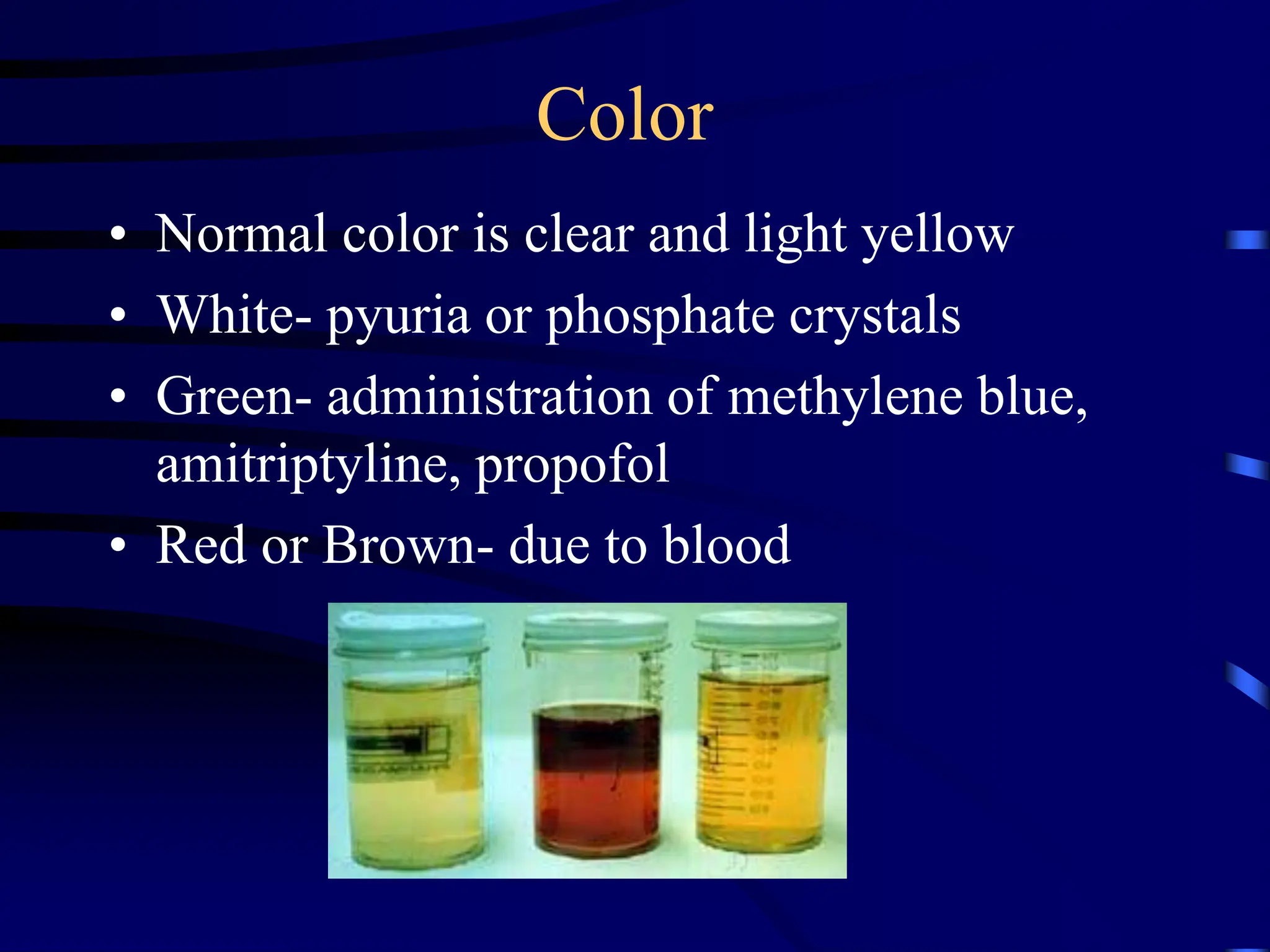
Urinalysis
Belinda Jim, MD
July 17, 2009
Urinalysis
• Major noninvasive tool
• Provide information about disease severity,
though not always a direct relationship
• Specimen should be examined about 30-60
minutes of voiding
• Sample centrifuged at 3000 rpm for 3-5
minutes, supernatant be tested by dipstick
Color
• Normal color is clear and light yellow
• White- pyuria or phosphate crystals
• Green- administration of methylene blue,
amitriptyline, propofol
• Red or Brown- due to blood
Turbidity
Normal urine should be clear
Turbidity indicates:
•cellular material
•bacteria
•protein
•lipids
•crystals
•salt precipitation
Odor
• Normal odor is urinoid
• Ammonia- related to UTI, prolonged
urinary retention
• Sweetish- diabetic ketoacidosis due to
acetone
• Sulfuric- cysteine decomposition
• Drugs/Foods- e.g. antibiotics, asparagus
Test Reagent Strip
Protein
• Detects primarily albumin
• Highly specific, but not sensitive
• Positive only when protein excretion > 300-
500 mg/day
• Not good to detect microalbuminuria or
immunoglobulin light chains
Sulfosalicylic acid test (SSA)
• SSA detects all proteins in the urine
• Especially useful if suspects multiple myeloma in
which immunogloblin light chains form casts that
obstruct the tubules
• A positive SSA test in conjunction with a negative
dipstick indicates presence of non-albumin
proteins in urine
• Mix 1 part urine supernatant with 3 parts SSA
(3%), grade its turbidity
pH
• Reflects degree of acidification of urine
• Urine pH ranges from 4.5 to 8.0
• Major clinical use for patient with metabolic
acidosis
• Appropriate response to increase urinary acid
excretion, with urine pH falling below 5.3
• A higher value indicate presence of RTA or
infection
Ketones
• Detects acetone and acetoacetate using
nitroprusside reaction
• Does not detect b-hydroxybutyrate
• Presence in the urine indicates diabetic
ketoacidosis, starvation ketosis, low carb
diet
Bilirubin/Urobilinogen
• Normal bilirubin not detected in urine
• If increased conjugated bilirubin in urine,
indicates liver dysfunction
• Bilirubin metabolized by bacteria in the
intestines to form urobilins
• Small % absorbed in intestine and appears
in urine
Osmolality and specific gravity
• Osmolality- number of solute particles per
unit volume
• Specific gravity- weight of solution
compared with weight of water
– Generally varies with osmolality, though
presence of large molecules in urine, such as
glucose or radiocontrast media can produce
large changes in specific gravity with little
changes in osmolality
Glucose
• Presence of glucose indicates inability to
reabsorb filtered glucose in the proximal
tubule despite normal plasma levels
• OR urinary spillage due to abnormally high
plasma concentrations
• Generally glucosuria does not occur until
plasma glucose exceeds 180 mg/dL
Hematuria and Pyuria
• Dipsticks very sensitive in detecting rbc’s,
as few as 1 to 2 rbc’s per high power field
• Detect leukocyte esterase (pyuria) and
nitrite (enterobacteriaceae converts nitrate
to nitrite)
• Sterile pyuria: interstitial nephritis, renal
tuberculosis, and nephrolithiasis
Urine sediment
• Crystals
• Bacteria
• Red blood cells (0-2 rbc’s in normal)
• White blood cells (0-4 wbc’s)
• Casts
• Artifact
Crystals
• Calcium phosphate or calcium oxalate
crystals
• Uric acid crystals
• Magnesium ammonium phosphate crystals
• Cystine crystals
Bacteria
• Significance depends on quality of
collection
• More significant in presence of wbc’s and
other elements
• Urine in the bladder should be sterile, but
becomes contaminated as it passes through
bladder
Hematuria
• Transient hematuria - relatively common in
young subjects, not indicative of disease
• Persistant hematuria- gross or microscopic
– Morphology is important, usually uniform and
round with extrarenal bleeding
– Dysmorphic appearance with renal lesions, I.e.
blebs, budding, segmental loss of membrane
Pyuria
• White cells are larger than red cells, granular
cytoplasm and multilobed nuclei
• Pyuria alone usually indicated infection
• Less diagnostic value when seen with other
elements such as cellular casts, and/or proteinuria
• Eosinophils may indicate allergic interstitial
nephritis, cholesterol emboli disease
• Lymphocytes seen in disorders associated with
infiltration of kidney by lymphocytes, such as
chronic tubulointerstitial disease
Epithelial Cells
• May be shed anywhere within the GU tract
• Only renal tubular cells are significant
• Generally 1.5 to 3 times larger than white
cells, contain a round, large nucleus
• May be normal or indicative of ATN,
pyelonephritis, nephrotic syndrome
Casts
• Conform to the shape of the renal tubule in
which they form
• Organic matrix composed mostly of Tamm-
Horsfall mucoprotein
• Types of casts: hyaline, red cell, white cell,
epithelial, granular, waxy, broad
Theory of Casts Genesis
•
Key:
A = Cellular casts
B = Coarsely granular cast
C = Finely granular cast
D = Waxy cast
RBC cast
©2005 UpToDate
®
e-mail this to a colleague
Miscellaneous Findings
Trichomonas Vaginalis
Sperm
Talc
Fiber
Case 1
• A 46 y.o. man with HTN presents with
acute right flank pain that moves around.
He is slightly febrile to 100.4 C. Exam
unremarkable. UA shows hematuria.
Case 2
• A 22 y.o. woman with no significant past
medical history presents with dysuria,
ammonia-smelling urine and increased
urinary frequency for 3 days.
• What will her UA show?
• What do you expect on the urine sediment?
Case 3
• A 57 y.o. woman with h/o poorly controlled
HTN and DM, diabetic retinopathy presents
with creatinine of 1.8 and lower extremity
edema.
• What would you expect your UA to show?
• What would you do next?
Case 4
• A 3-year old boy brought to pediatrician for
increasing facial and peripheral edema of
eight days duration. Urine- 4.2 grams
protein/24 hours, cholesterol 450 mg/dl,
albumin 3.0 g/dl
Case 5
• A 45-year old man hospitalized for
peripheral edema and proteinuria. Urine-6 g
protein/24 hours, albumin 1.6 g/dl.
Cholesterol 380 mg/dl. Creatinine 1.6.
Review of medical records showed he had
1+ proteinuria four years previously.
Case 6
• A 52-year old woman with sudden onset of
edema, “dark urine”, headaches, malaise.
BP: 150/105, urine 900 cc/24 hr, 1.7g
protein/24 hours, creatinine 4.5 mg/dl, BUN
68 mg/dl. Review of records show that she
had normal renal function 1 month ago.
Urinalysis; urine examination in the lab.ppt

Urinalysis; urine examination in the lab.ppt

  • 1.
  • 2.
    Urinalysis • Major noninvasivetool • Provide information about disease severity, though not always a direct relationship • Specimen should be examined about 30-60 minutes of voiding • Sample centrifuged at 3000 rpm for 3-5 minutes, supernatant be tested by dipstick
  • 3.
    Color • Normal coloris clear and light yellow • White- pyuria or phosphate crystals • Green- administration of methylene blue, amitriptyline, propofol • Red or Brown- due to blood
  • 4.
    Turbidity Normal urine shouldbe clear Turbidity indicates: •cellular material •bacteria •protein •lipids •crystals •salt precipitation
  • 5.
    Odor • Normal odoris urinoid • Ammonia- related to UTI, prolonged urinary retention • Sweetish- diabetic ketoacidosis due to acetone • Sulfuric- cysteine decomposition • Drugs/Foods- e.g. antibiotics, asparagus
  • 6.
  • 7.
    Protein • Detects primarilyalbumin • Highly specific, but not sensitive • Positive only when protein excretion > 300- 500 mg/day • Not good to detect microalbuminuria or immunoglobulin light chains
  • 8.
    Sulfosalicylic acid test(SSA) • SSA detects all proteins in the urine • Especially useful if suspects multiple myeloma in which immunogloblin light chains form casts that obstruct the tubules • A positive SSA test in conjunction with a negative dipstick indicates presence of non-albumin proteins in urine • Mix 1 part urine supernatant with 3 parts SSA (3%), grade its turbidity
  • 9.
    pH • Reflects degreeof acidification of urine • Urine pH ranges from 4.5 to 8.0 • Major clinical use for patient with metabolic acidosis • Appropriate response to increase urinary acid excretion, with urine pH falling below 5.3 • A higher value indicate presence of RTA or infection
  • 10.
    Ketones • Detects acetoneand acetoacetate using nitroprusside reaction • Does not detect b-hydroxybutyrate • Presence in the urine indicates diabetic ketoacidosis, starvation ketosis, low carb diet
  • 11.
    Bilirubin/Urobilinogen • Normal bilirubinnot detected in urine • If increased conjugated bilirubin in urine, indicates liver dysfunction • Bilirubin metabolized by bacteria in the intestines to form urobilins • Small % absorbed in intestine and appears in urine
  • 12.
    Osmolality and specificgravity • Osmolality- number of solute particles per unit volume • Specific gravity- weight of solution compared with weight of water – Generally varies with osmolality, though presence of large molecules in urine, such as glucose or radiocontrast media can produce large changes in specific gravity with little changes in osmolality
  • 13.
    Glucose • Presence ofglucose indicates inability to reabsorb filtered glucose in the proximal tubule despite normal plasma levels • OR urinary spillage due to abnormally high plasma concentrations • Generally glucosuria does not occur until plasma glucose exceeds 180 mg/dL
  • 14.
    Hematuria and Pyuria •Dipsticks very sensitive in detecting rbc’s, as few as 1 to 2 rbc’s per high power field • Detect leukocyte esterase (pyuria) and nitrite (enterobacteriaceae converts nitrate to nitrite) • Sterile pyuria: interstitial nephritis, renal tuberculosis, and nephrolithiasis
  • 15.
    Urine sediment • Crystals •Bacteria • Red blood cells (0-2 rbc’s in normal) • White blood cells (0-4 wbc’s) • Casts • Artifact
  • 16.
    Crystals • Calcium phosphateor calcium oxalate crystals • Uric acid crystals • Magnesium ammonium phosphate crystals • Cystine crystals
  • 23.
    Bacteria • Significance dependson quality of collection • More significant in presence of wbc’s and other elements • Urine in the bladder should be sterile, but becomes contaminated as it passes through bladder
  • 25.
    Hematuria • Transient hematuria- relatively common in young subjects, not indicative of disease • Persistant hematuria- gross or microscopic – Morphology is important, usually uniform and round with extrarenal bleeding – Dysmorphic appearance with renal lesions, I.e. blebs, budding, segmental loss of membrane
  • 29.
    Pyuria • White cellsare larger than red cells, granular cytoplasm and multilobed nuclei • Pyuria alone usually indicated infection • Less diagnostic value when seen with other elements such as cellular casts, and/or proteinuria • Eosinophils may indicate allergic interstitial nephritis, cholesterol emboli disease • Lymphocytes seen in disorders associated with infiltration of kidney by lymphocytes, such as chronic tubulointerstitial disease
  • 31.
    Epithelial Cells • Maybe shed anywhere within the GU tract • Only renal tubular cells are significant • Generally 1.5 to 3 times larger than white cells, contain a round, large nucleus • May be normal or indicative of ATN, pyelonephritis, nephrotic syndrome
  • 34.
    Casts • Conform tothe shape of the renal tubule in which they form • Organic matrix composed mostly of Tamm- Horsfall mucoprotein • Types of casts: hyaline, red cell, white cell, epithelial, granular, waxy, broad
  • 35.
    Theory of CastsGenesis • Key: A = Cellular casts B = Coarsely granular cast C = Finely granular cast D = Waxy cast
  • 38.
  • 41.
  • 45.
  • 46.
  • 47.
  • 48.
  • 49.
  • 50.
    Case 1 • A46 y.o. man with HTN presents with acute right flank pain that moves around. He is slightly febrile to 100.4 C. Exam unremarkable. UA shows hematuria.
  • 52.
    Case 2 • A22 y.o. woman with no significant past medical history presents with dysuria, ammonia-smelling urine and increased urinary frequency for 3 days. • What will her UA show? • What do you expect on the urine sediment?
  • 55.
    Case 3 • A57 y.o. woman with h/o poorly controlled HTN and DM, diabetic retinopathy presents with creatinine of 1.8 and lower extremity edema. • What would you expect your UA to show? • What would you do next?
  • 56.
    Case 4 • A3-year old boy brought to pediatrician for increasing facial and peripheral edema of eight days duration. Urine- 4.2 grams protein/24 hours, cholesterol 450 mg/dl, albumin 3.0 g/dl
  • 58.
    Case 5 • A45-year old man hospitalized for peripheral edema and proteinuria. Urine-6 g protein/24 hours, albumin 1.6 g/dl. Cholesterol 380 mg/dl. Creatinine 1.6. Review of medical records showed he had 1+ proteinuria four years previously.
  • 60.
    Case 6 • A52-year old woman with sudden onset of edema, “dark urine”, headaches, malaise. BP: 150/105, urine 900 cc/24 hr, 1.7g protein/24 hours, creatinine 4.5 mg/dl, BUN 68 mg/dl. Review of records show that she had normal renal function 1 month ago.