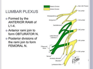
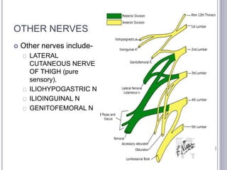
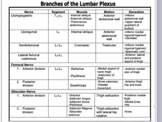
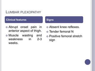
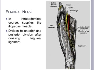
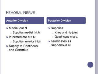
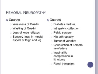
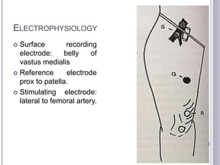
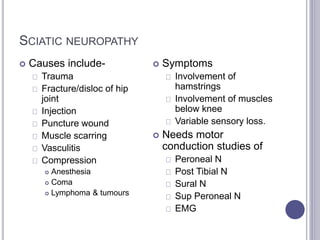
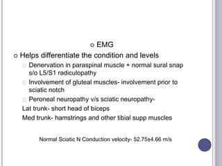
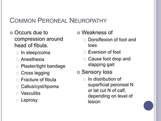
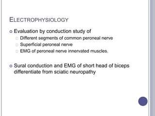
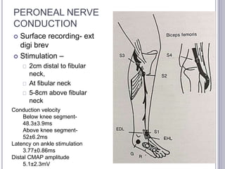
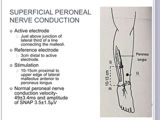
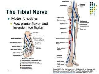
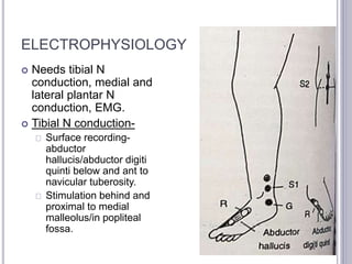
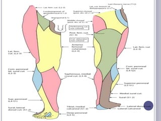
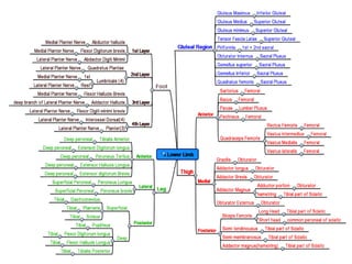
This presentation describes the common conditions, anatomy and the ideal ways to do and perform nerve conduction studies in lower limbs. It is nicely depicted with self explanatory pictures.
3. LUMBAR PLEXUS
Formed by the
ANTERIOR RAMI of
L1-4.
Anterior rami join to
form OBTURATOR N.
Posterior divisions of
the rami join to form
FEMORAL N.
4. OTHER NERVES
Other nerves include-
LATERAL
CUTANEOUS NERVE
OF THIGH (pure
sensory).
ILIOHYPOGASTRIC N
ILIOINGUINAL N
GENITOFEMORAL N
5.
6. LUMBAR PLEXOPATHY
Clinical features Signs
Abrupt onset pain in
anterior aspect of thigh.
Muscle wasting and
weakness in 2-3
weeks.
Absent knee reflexes.
Tender femoral N
Positive femoral stretch
sign
7. Sensory symptoms are partial and seen in 1/3rd
patients.
NCV shows normal nerves- femoral, peroneal, sural
and saphenous N.
May show reduced amplitude.
EMG may show changes of denervation and
renervation.
Recovery may be spontaneous over months-years.
8. Normal femoral conduction velocity – 70.0 ± 5.5 m/S
FEMORAL N
From dorsal portion of anterior rami of L2-L4
Mixed Nerve
9. FEMORAL NERVE
In intraabdominal
course, supplies the
iliopsoas muscle.
Divides to anterior and
posterior division after
crossing Inguinal
ligament.
10. FEMORAL NERVE
Anterior Division Posterior Division
Medial cut N
Supplies medial thigh
Intermediate cut N
Supplies anterior thigh
Supply to Pectineus
and Sartorius
Supplies
Knee and hip joint
Quadriceps musc.
Terminates as
Saphenous N
11. FEMORAL NEUROPATHY
Causes
Weakness of Quadri.
Wasting of Quadri.
Loss of knee reflexes
Sensory loss in medial
aspect of thigh and leg
Causes
Diabetes mellitus
Intrapelvic collection
Pelvic surgery
Hip arthroplasty
Tumor of vertebra
Cannulation of Femoral
vein/artery
Inguinal lig
compression in
lithotomy
Renal transplant
12. ELECTROPHYSIOLOGY
Surface recording
electrode: belly of
vastus medialis
Reference electrode
prox to patella.
Stimulating electrode:
lateral to femoral artery.
13. NCV
Slowing of conduction velocity
Small CMAP amplitude.
Conduction block (if compressed at inguinal lig)
Saphenous vein can be used to evaluate sensory loss.
Normal femoral conduction velocity – 70.0 ± 5.5
m/S
14. Normal Saphenous conduction velocity- 49.03 ± 3.36 m/s.
SNAP Amplitude- 3.54 ± 1.52 μV
SAPHENOUS NERVE
Largest and longest pure sensory
branch of Femoral N .
Supplies skin over medial aspect of
leg and foot.
15. SAPHENOUS N
Arises from posterior
division of Femoral N
Becomes superficial
just above medial
condyle
Continues down to
head of 1st metatarsal
16. SAPHENOUS NEUROPATHY
Uncommon
Follows
Laceration injuries
Entrapment in subsartorial canal
Surgery for varicose veins
Causes sensory impairment in medial aspect of
knee, leg and foot.
17. SAPHENOUS NERVE CONDUCTION
Stimulate 1 cm above
inferior border of
patella between gracilis
and sartorius.
Recording electrode-
15 cm distal on medial
border of tibia.
18. Stimulate between
medial head of
gastrocnemius and
tibia, 12- 14 cm
proximal to med
malleolus
Recording electrode is
placed anterior to
medial malleolus
Normal Saphenous conduction velocity- 49.03 ± 3.36 m/s.
SNAP Amplitude- 3.54 ± 1.52 μV
19. Latency and Amplitude of SNAP
above Inguinal Lig- 2.8±0.4ms
and 6±1.5 μV
LATERAL FEMORAL
CUTANEOUS NERVE
OF THIGH
L2-3.
Sensory supply to
Anterolateral aspect of
thigh.
20. MERALGIA PARESTHETICA
Entrapment of the
nerve at Inguinal tunnel
Seat belts
Obesity
Unknown
Proximal lesions
Psoas abscess
Retroperitoneal tumor
Post op scarring
Pain & paresthesia over
lateral surface of thigh.
Symptoms increase on
standing and prolonged
walking.
Sensory loss is in area
smaller than supplied by
the nerve
NCV- abnormal conduction in Lat Cut N of Thigh
EMG- normal study of paraspinal, iliopsoas, quadriceps.
21. NERVE CONDUCTION-LAT
CUT N OF THIGH
Surface Electrode- 17-
20 cm distal to Ant
Supr Iliac Spine
(ASIS).
Reference electrode- 3
cm distal to surface
electrode.
Antidromic stimulation
above inguinal
ligament 1 cm medial
to ASIS
Latency and Amplitude of SNAP above Inguinal Lig- 2.8±0.4ms
and 6±1.5 μV
24. SACRAL PLEXUS
Branches
Sup Gluteal N(L4-S1)
Gluteus medius
Gluteus minimus
Tensor facsia lata
Inf Gluteal N(L5-S1)
Gluteux maximus
Sciatic N (L4-S3)
25. SACRAL PLEXOPATHY
Abrupt onset pain in
posteror aspect of the
thigh and buttocks.
Weakness of knee
flexor
Absent reflexes.
Sciatic N tenderness
Positive SLR test.
26. Normal Sciatic N Conduction
velocity- 52.75±4.66 m/s
SCIATIC NERVE
L4-S3
Comes out of sciatic notch
Supplies all hamstrings (medial
trunk) except short head of
biceps femoris
All muscles distal to knee
27. SCIATIC NEUROPATHY
Causes include-
Trauma
Fracture/disloc of hip
joint
Injection
Puncture wound
Muscle scarring
Vasculitis
Compression
Anesthesia
Coma
Lymphoma & tumours
Symptoms
Involvement of
hamstrings
Involvement of muscles
below knee
Variable sensory loss.
Needs motor
conduction studies of
Peroneal N
Post Tibial N
Sural N
Sup Peroneal N
EMG
28. SCIATIC N CONDUCTION
NCV
Difficult d/t deep location.
Surface Electrode on
distal peroneal
innervated muscle eg
abd hallucius
Stimulation-
Just below gluteal fold
Medial trunk- apex of
popliteal fossa
Lateral trunk- head of
fibula
29. EMG
Helps differentiate the condition and levels
Denervation in paraspinal muscle + normal sural snap
s/o L5/S1 radiculopathy
Involvement of gluteal muscles- involvement prior to
sciatic notch
Peroneal neuropathy v/s sciatic neuropathy-
Lat trunk- short head of biceps
Med trunk- hamstrings and other tibial supp muscles
Normal Sciatic N Conduction velocity- 52.75±4.66 m/s
31. COURSE & BRANCHES
Branches-
Lat Cut N of Calf
Supplying anterior,
lateral and posterior
surface of leg
Superficial Peroneal N
Also supplies lateral and
dorsal portion of leg and
dorsum of foot.
Deep Peroneal N
32. COMMON PERONEAL NEUROPATHY
Occurs due to
compression around
head of fibula.
In sleep/coma
Anesthesia
Plaster/tight bandage
Cross legging
Fracture of fibula
Callus/cyst/lipoma
Vasculitis
Leprosy
Weakness of
Dorsiflexion of foot and
toes
Eversion of foot
Cause foot drop and
slapping gait
Sensory loss
In distribution of
superficial peroneal N
or lat cut N of calf,
depending on level of
lesion
33. ELECTROPHYSIOLOGY
Evaluation by conduction study of
Different segments of common peroneal nerve
Superficial peroneal nerve
EMG of peroneal nerve innervated muscles.
Sural conduction and EMG of short head of biceps
differentiate from sciatic neuropathy
35. SUPERFICIAL PERONEAL
NERVE CONDUCTION
Active electrode
Just above junction of
lateral third of a line
connecting the malleoli.
Reference electrode
3cm distal to active
electrode.
Stimulation
10-15cm proximal to
upper edge of lateral
malleolus anterior to
peroneus longus
Normal peroneal nerve
conduction velocity-
49±3.4ms and amplitude
of SNAP 3.5±1.5μV
36. In peroneal neuropathy conduction block and
reduction in motor nerve conduction velocity >10ms
across head of fibula localizes the lesion at this site.
In common peroneal neuropathy muscles supplied
by the deep branch are frequently/severely
affected.
Common peroneal nerve and lateral trunk of sciatic
nerve- EMG of short head of biceps are useful
37. Sural N conduction velocity- 50.9±5.4 m/s, amplitude of SNAP 18±10.5μV
SURAL NERVE
S1 and S2
Medial derived from Tibial N
Lateral derived from Peroneal N
Pure sensory N
38. SURAL NEUROPATHY
Uncommon
Part of generalised neuropathies
Compression
Baker’s cyst
Against hard object
Tendon sheath ganglia
Scar tissue
# 5th metatarsal
Presents with
Numbness and paresthesia in supplied region
Low conduction velocity and amplitude in NCV
39. SURAL
Leg should be relaxed
and in lateral position.
Surface Electrode-between
lateral malleolus
and tendoachilles.
Stimulated 10-16 cm
proximal to recording
electrode, distal to lower
border of gastrocnemius
at the junction of middle
and lower third of leg.
Sural N conduction velocity- 50.9±5.4 m/s, amplitude of SNAP 18±10.5μV
42. TIBIAL NEUROPATHY
Damage at popliteal
fossa uncommon.
Causes-
Baker’s cyst
Nerve sheath ganglia
Popliteal A Aneurysm
Leprosy
Weakness of
plantar flexors
Invertors
Intrinsic foot muscles
Sensory loss in sole
43. TARSAL TUNNEL SYNDROME
Rare picture
Pain and paresthesia
of sole
Weakness of intrinsic
foot muscles (rare)
Causes
Ill-fitting footwear
Tight plaster cast
Post traumatic fibrosis
Tenosynovitis
RA
Hypothyroidism
Idiopathic
44. ELECTROPHYSIOLOGY
Needs tibial N
conduction, medial and
lateral plantar N
conduction, EMG.
Tibial N conduction-
Surface recording-abductor
hallucis/abductor digiti
quinti below and ant to
navicular tuberosity.
Stimulation behind and
proximal to medial
malleolus/in popliteal
fossa.
45. Motor conduction of medial and lateral plantar N
Recording electrode (M)- abductor hallucis (belly)
Recording electrode (L)-abductor digiti quinti
Nerve stimulation- behind and above medial malleolus
46. Sensory conduction of
medial and lateral
plantar nerves:
Stimulation- 1st and 5th
toes- M and L
respectively.
Recording electrode-just
below medial
malleolus.
47. In Tarsal Tunnel Syndrome
Conduction block and latency prolongation across tarsal
tunnel
Accurate localisation by inching technique (1cm)-abrupt
prolongation in latency.
Normal conduction velocity of Tibial N-48.3±4.5ms
Motor conduction
Latency for medial plantar nerve-3.8±0.5ms
Latency for lateral plantar nerve-3.9±0.5ms
Sensory conduction for
Latency for medial plantar nerve-2.4±0.2ms, 3.2±0.3ms,
4±0.2ms (10,14 and 18 cm segment).
Latency for lateral plantar nerve-3.2±0.3ms,4±0.3ms (14
and 18 cm segment).