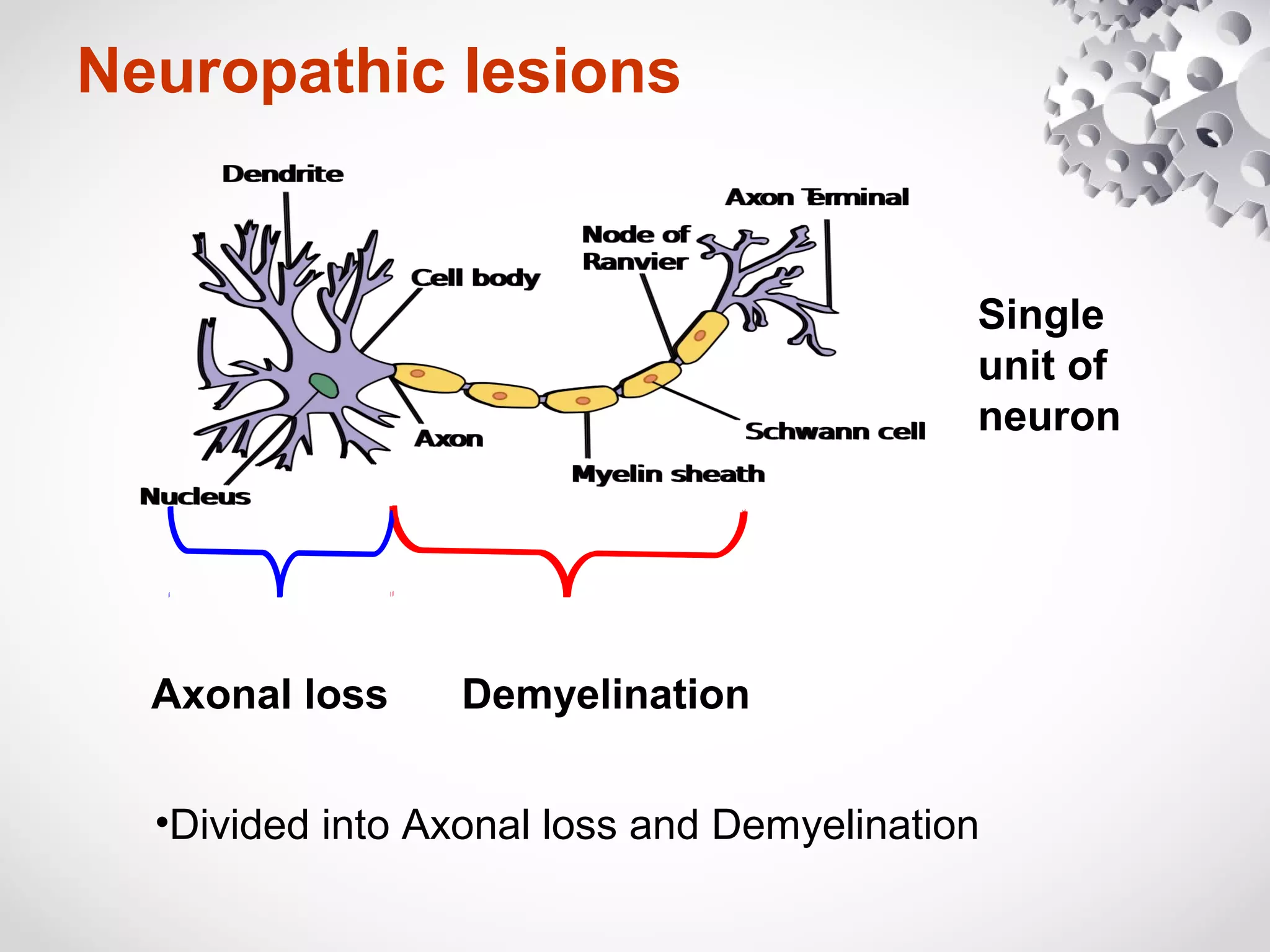
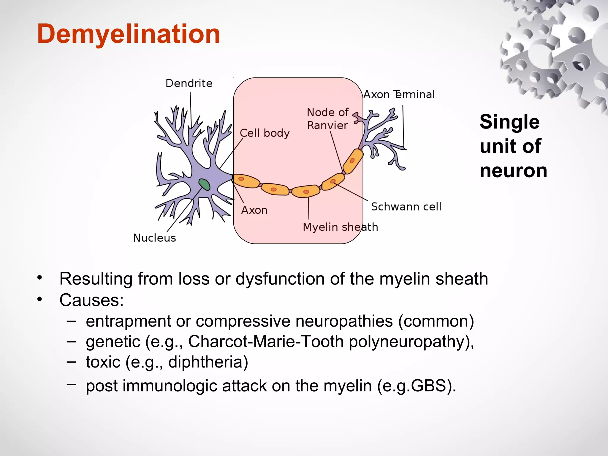
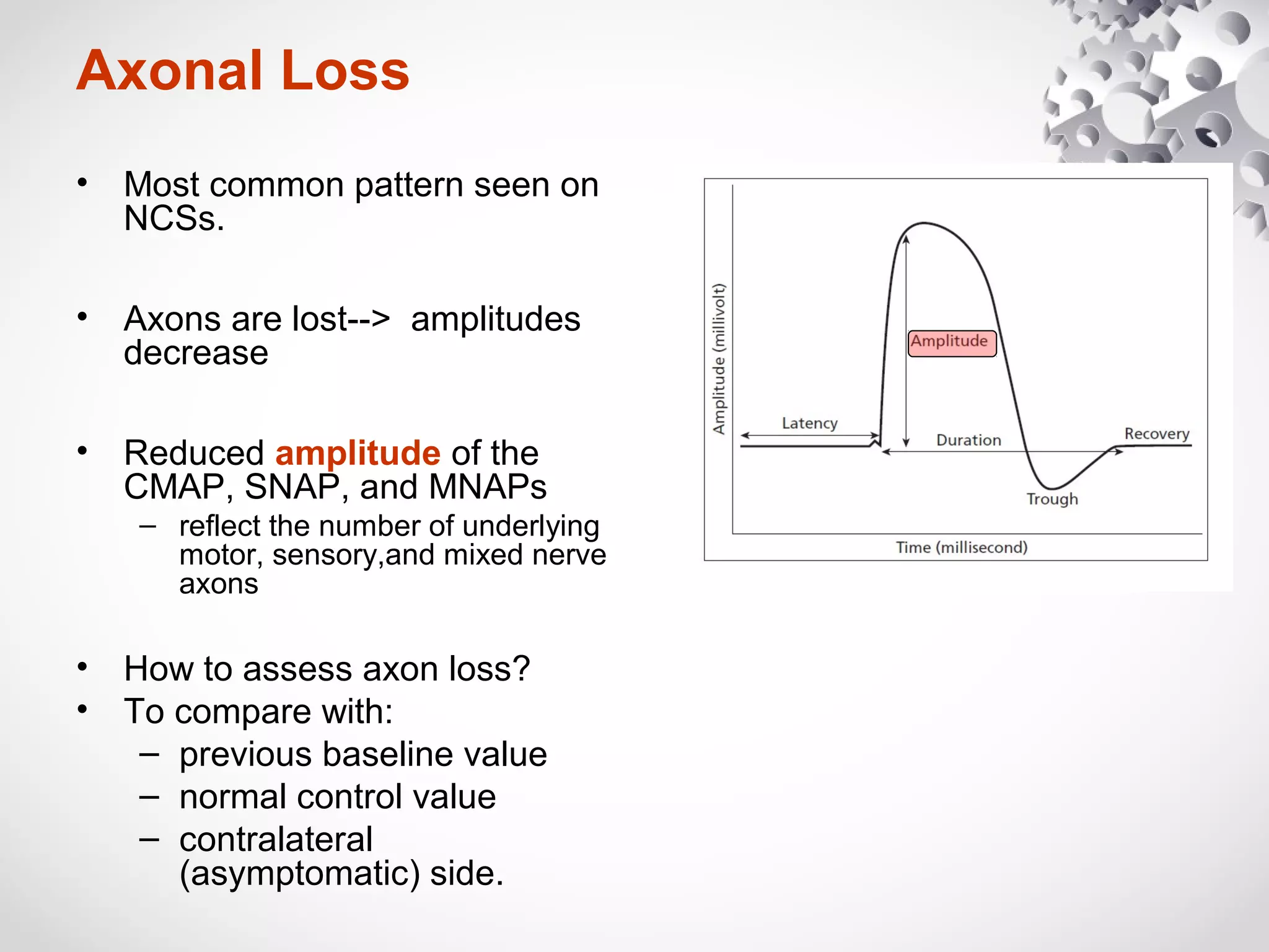
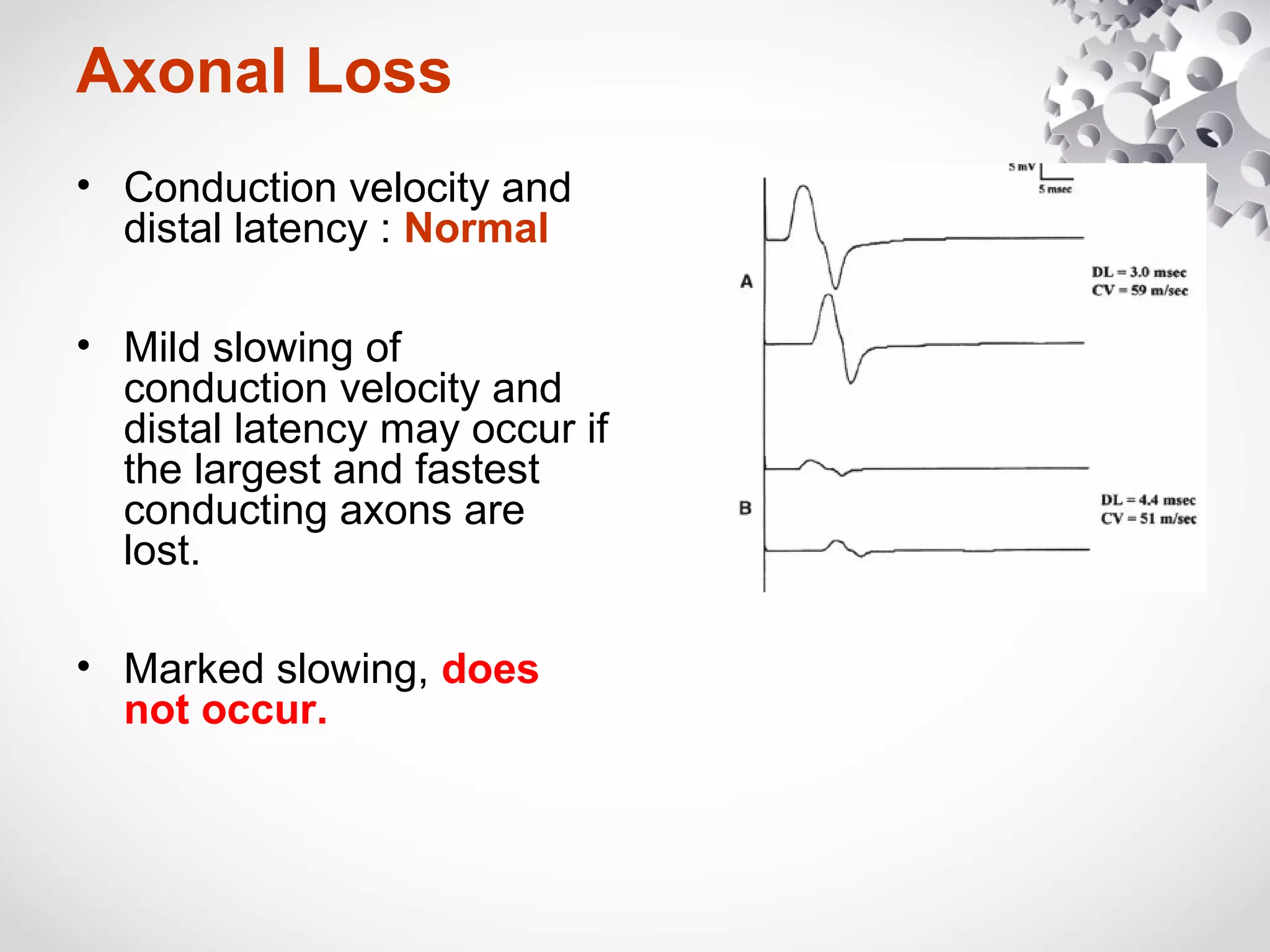
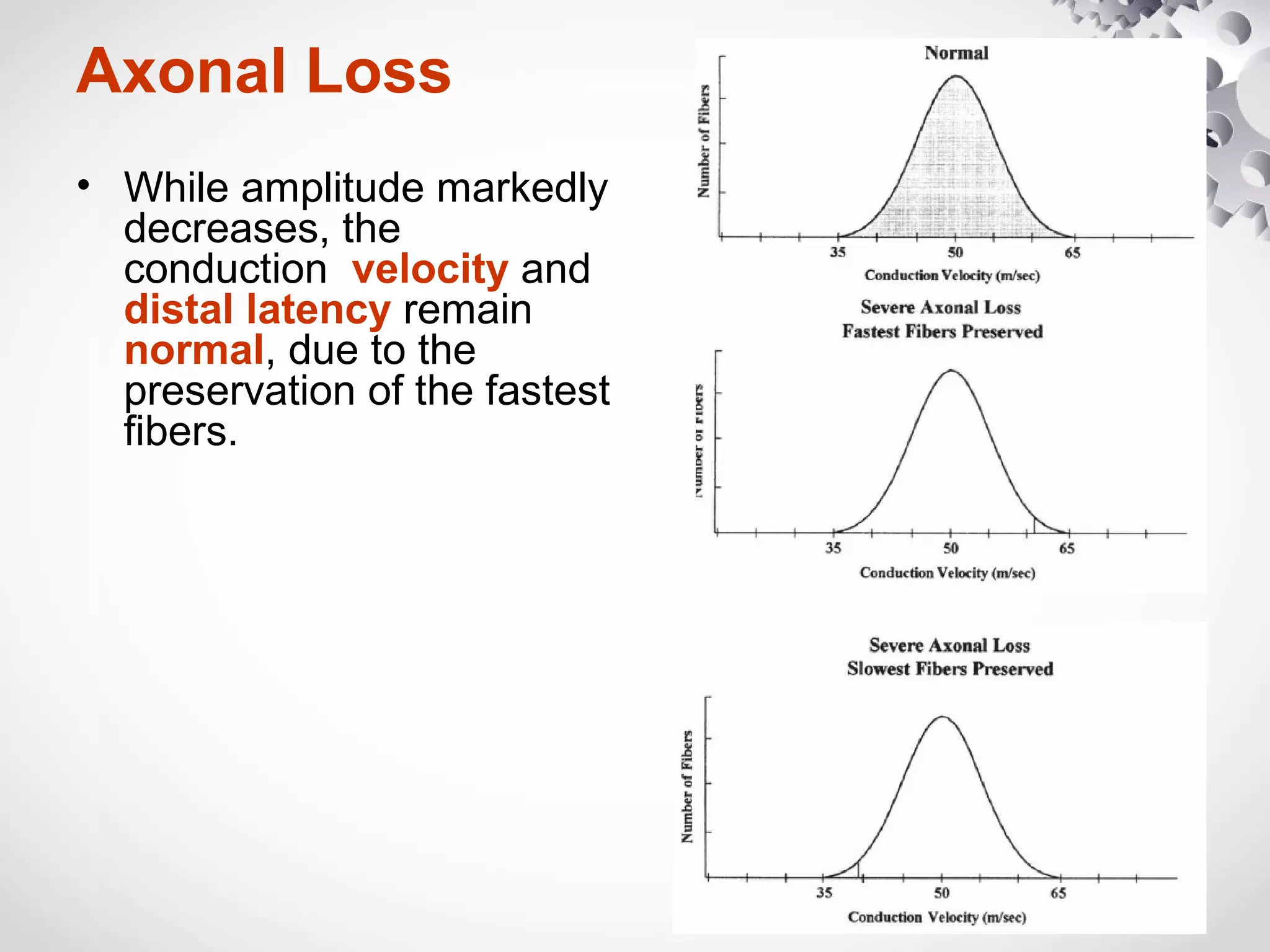
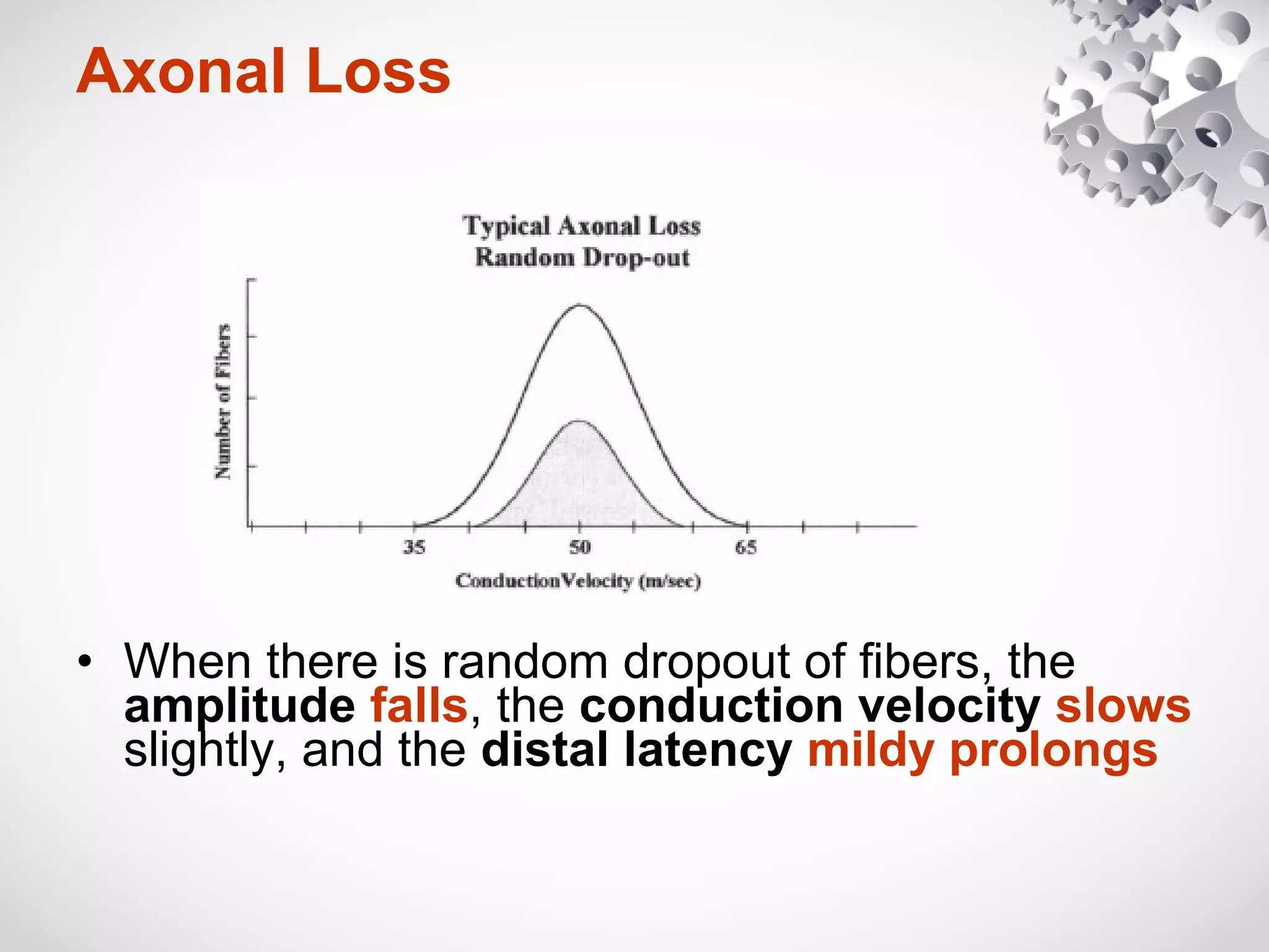
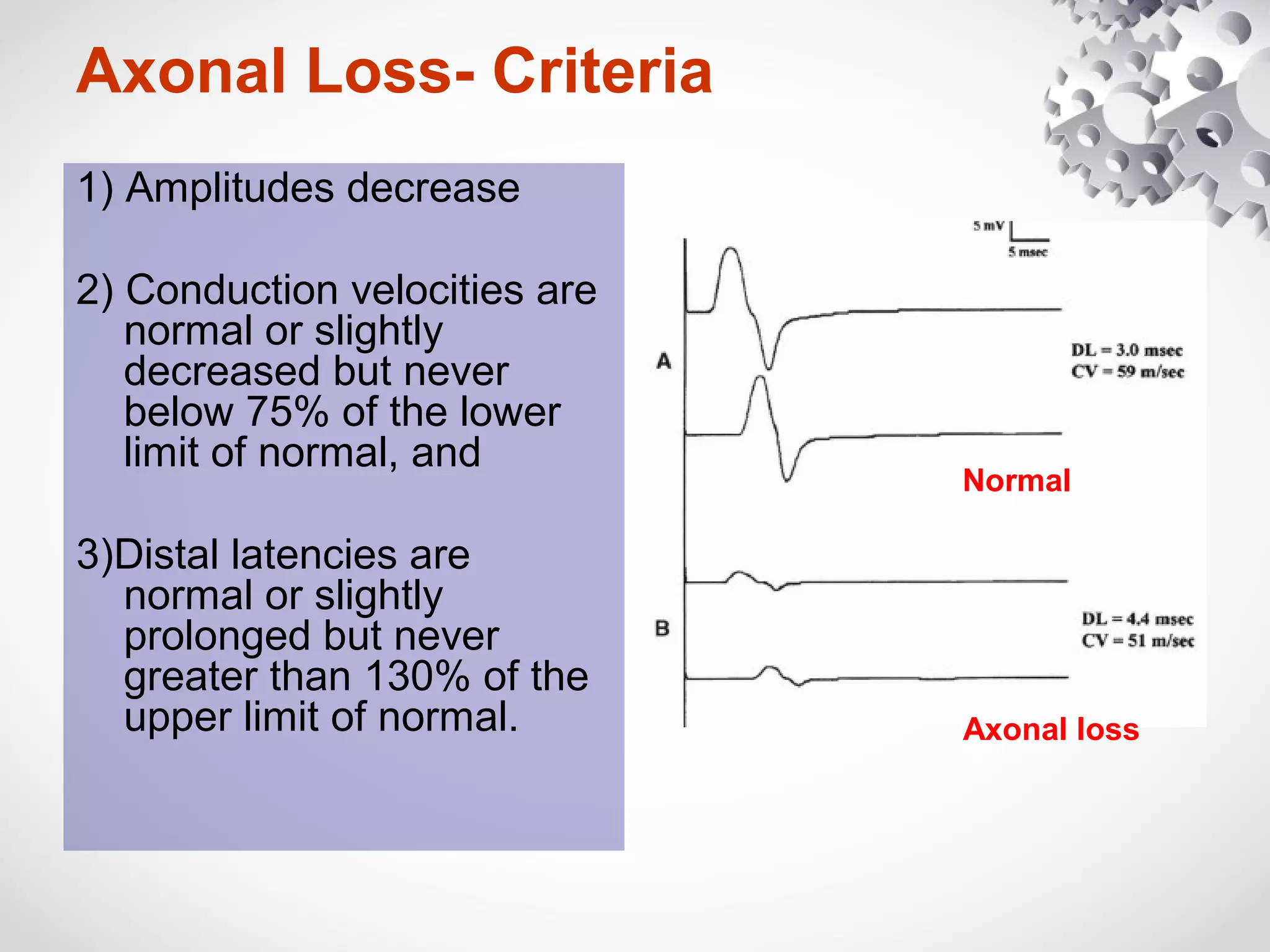
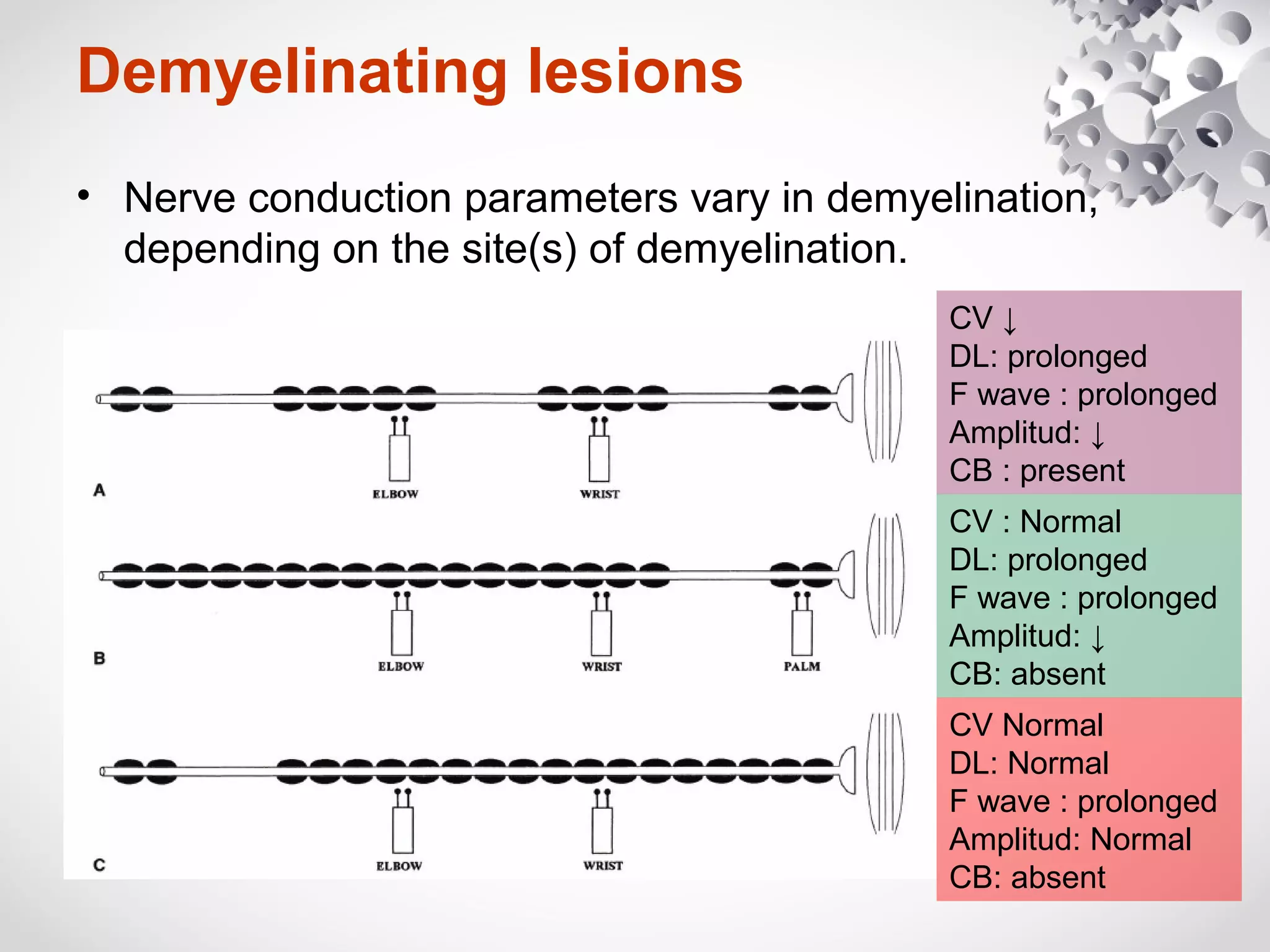
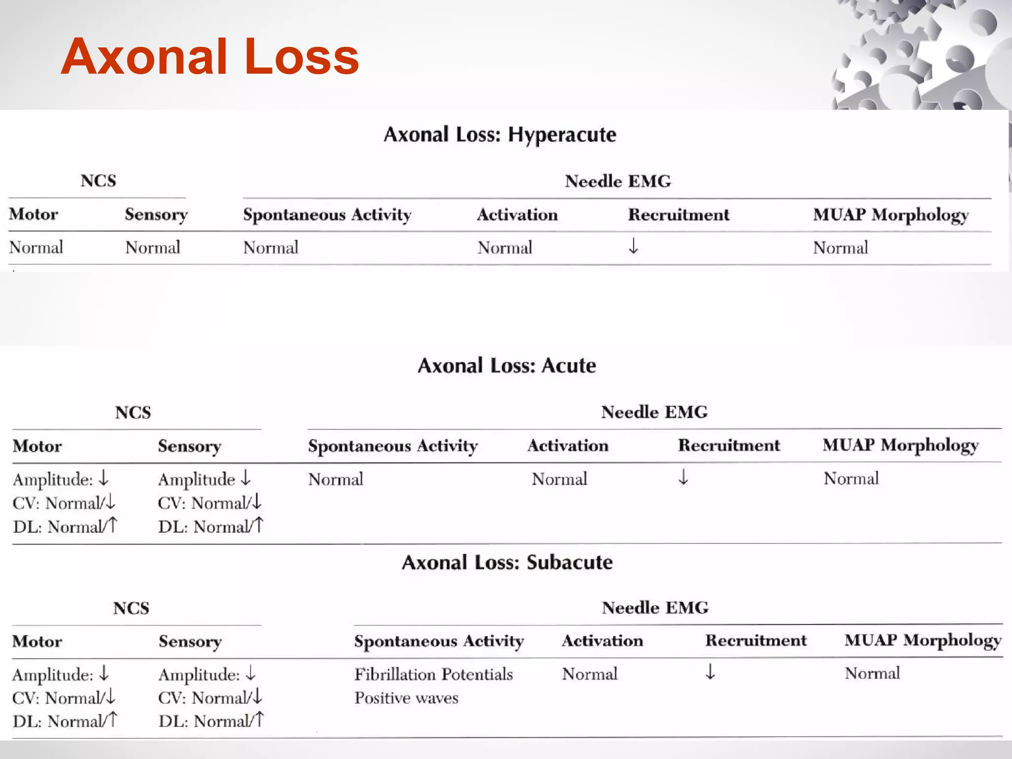
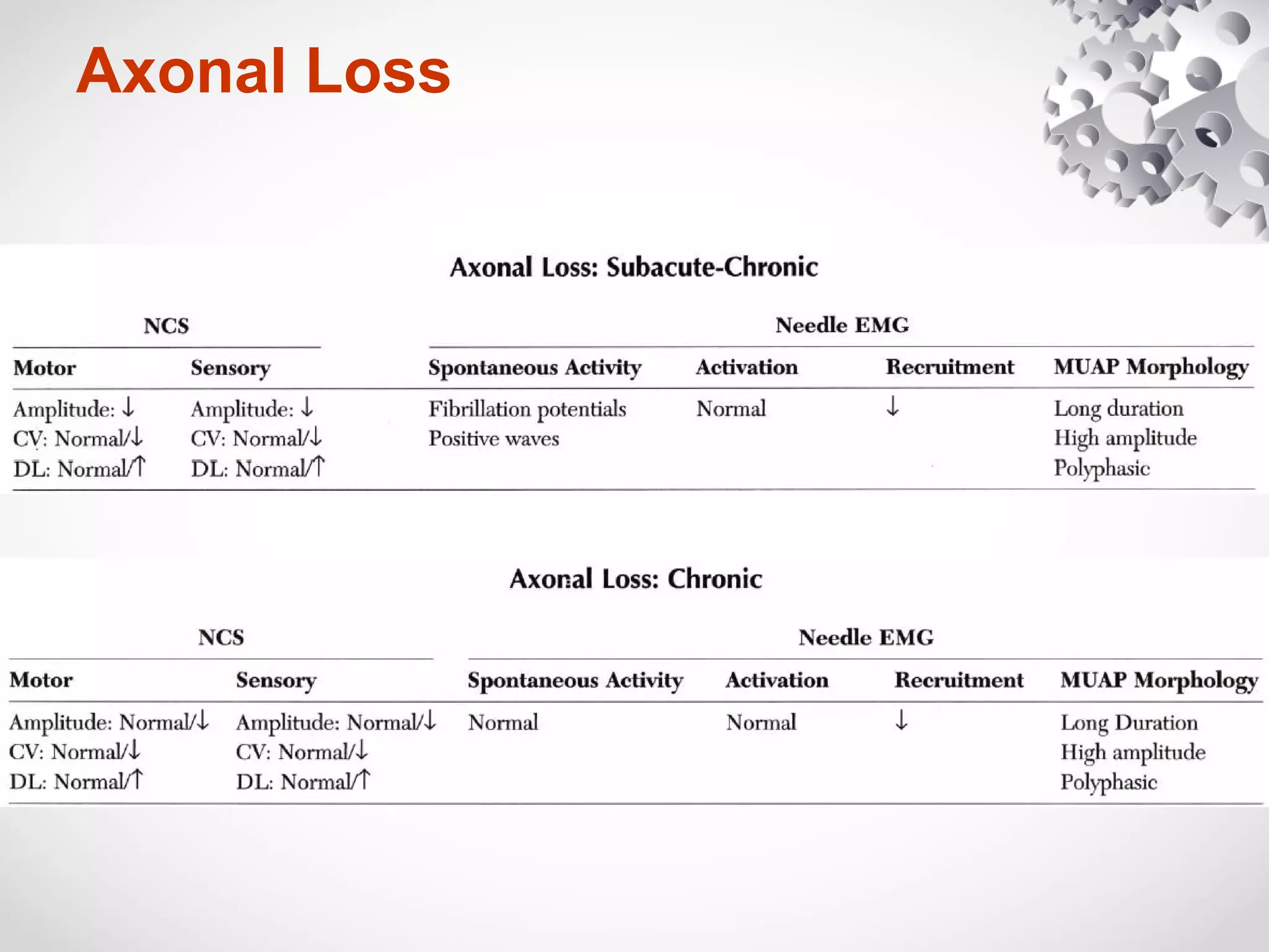
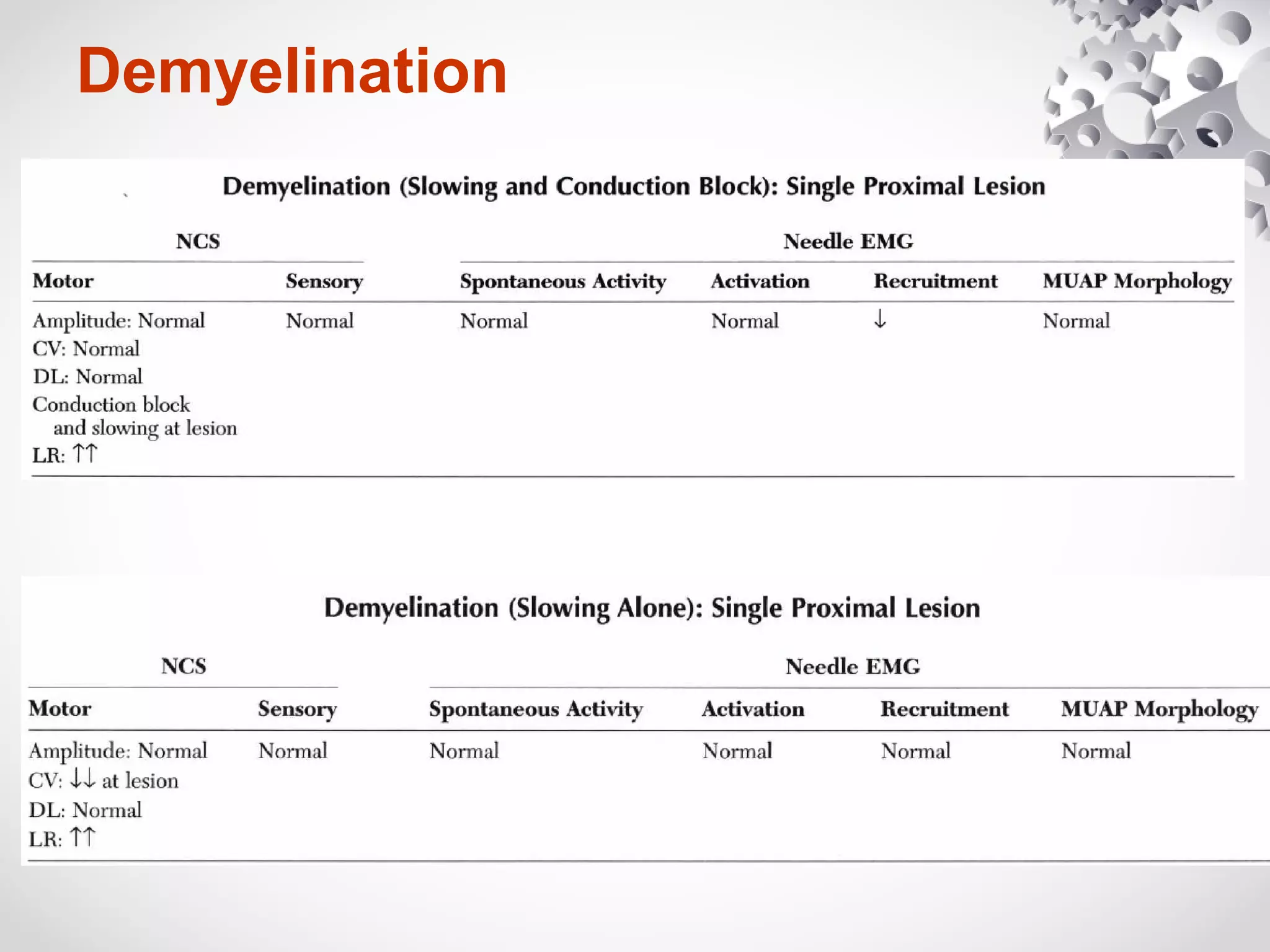
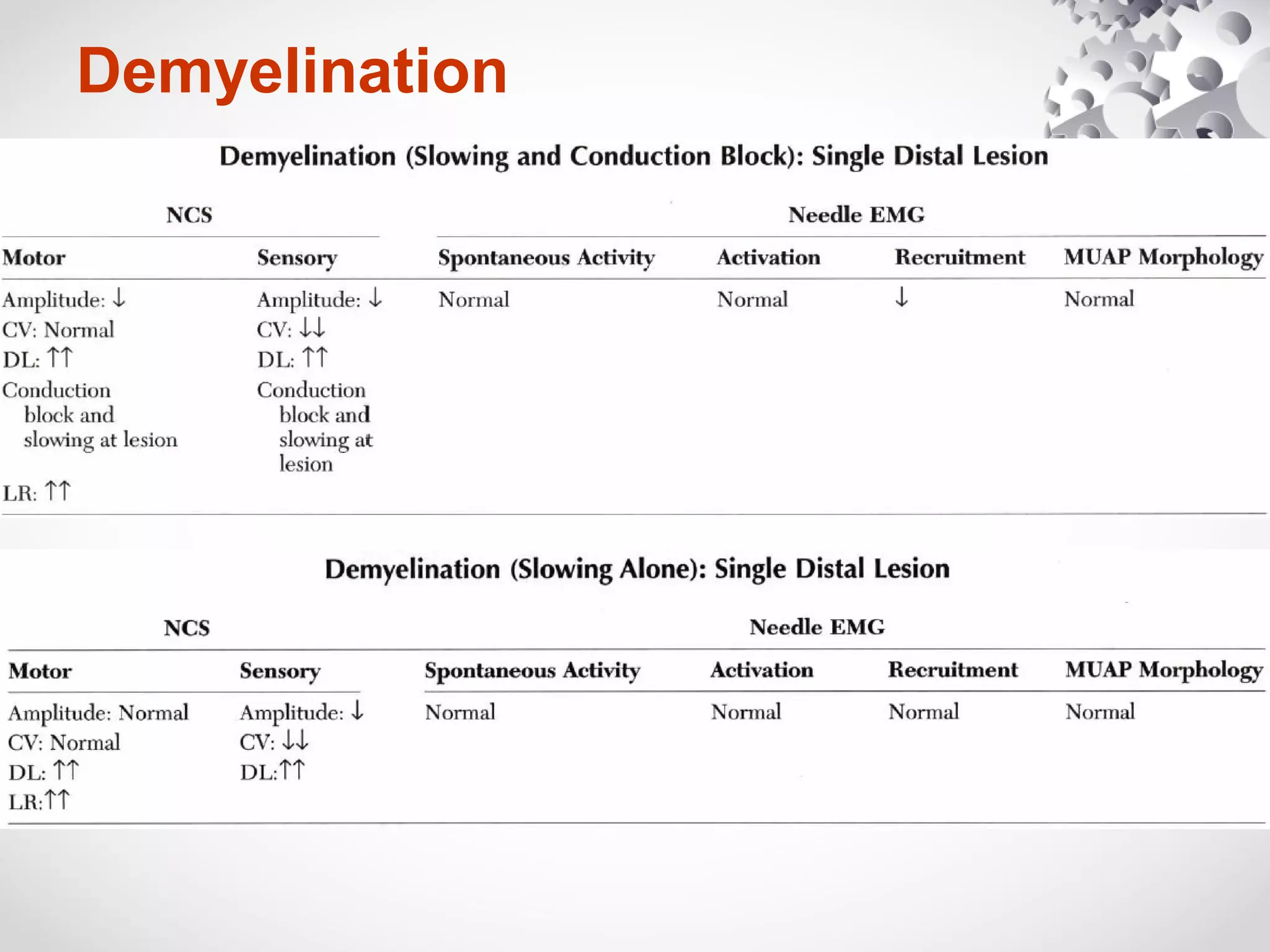
This document summarizes the key differences between axonal loss and demyelination lesions as assessed by nerve conduction studies. Axonal loss lesions typically show decreased amplitudes with normal or slightly slowed conduction velocities and distal latencies. Demyelination lesions are characterized by markedly slowed conduction velocities below 75% of the lower limit of normal or prolonged distal latencies over 130% of the upper limit, indicating primary demyelination. The document discusses various patterns that can be seen on nerve conduction studies for both types of lesions and their clinical implications.
1. Nerves conduction study
Part 2: Axonal loss vs Demyelination
For post basic neurophysiology course
Dr Ahmad Shahir Mawardi
Neurology Departmert
Hospital Kuala Lumpur
21 October 2015
3. • Causes:
– Physical disruption of the nerve
– toxic, metabolic, or genetic conditions
Axonal loss
Single
unit of
neuron
4. • Resulting from loss or dysfunction of the myelin sheath
• Causes:
– entrapment or compressive neuropathies (common)
– genetic (e.g., Charcot-Marie-Tooth polyneuropathy),
– toxic (e.g., diphtheria)
– post immunologic attack on the myelin (e.g.GBS).
Demyelination
Single
unit of
neuron
6. Axonal Loss
• Most common pattern seen on
NCSs.
• Axons are lost--> amplitudes
decrease
• Reduced amplitude of the
CMAP, SNAP, and MNAPs
– reflect the number of underlying
motor, sensory,and mixed nerve
axons
• How to assess axon loss?
• To compare with:
– previous baseline value
– normal control value
– contralateral
(asymptomatic) side.
7. Axonal Loss
• Conduction velocity and
distal latency : Normal
• Mild slowing of
conduction velocity and
distal latency may occur if
the largest and fastest
conducting axons are
lost.
• Marked slowing, does
not occur.
8. Axonal Loss
• While amplitude markedly
decreases, the
conduction velocity and
distal latency remain
normal, due to the
preservation of the fastest
fibers.
9. Axonal Loss
• Axonal loss with abnormal CV only occur in 2
possible extremes:
1.severe loss of axons with only a few of the fastest
fibers remaining
2.all axons are lost except for a few of the normal
slowly conducting fibers (the amplitude also falls
dramatically)
• In general, axonal loss lesions result in a
pattern between these two extremes.
10. Axonal Loss
• However, conduction velocity can drop only as low as 35
m/s (~ 75% of the lower limit of normal).
– normal myelinated fibers do not conduct anything slower than
this.
• Distal latencies generally do not exceed 130% of the
upper limit of normaL
11. Axonal Loss
• When there is random dropout of fibers, the
amplitude falls, the conduction velocity slows
slightly, and the distal latency mildy prolongs
12. Axonal Loss- Criteria
1) Amplitudes decrease
2) Conduction velocities are
normal or slightly
decreased but never
below 75% of the lower
limit of normal, and
3)Distal latencies are
normal or slightly
prolonged but never
greater than 130% of the
upper limit of normal.
Normal
Axonal loss
13. Axonal Loss
• The only exception occurs in hyperacute axonal loss
lesions e.g a nerve transection.
• Day 3 to 4 : NCS normal (provided both stimulation and
recording are done distal to the lesion).
• Days 3 to 10, the process of Wallerian degeneration
occurs: the nerve distal to the transection undergoes
degeneration,resulting in a low amplitude both distally
and proximally.
14. Axonal Loss
• The process of wallerian degeneration for:
– motor fibers (typically days 3-5)
– sensory fibers (typically days 6-10).
• At this point, the typical pattern of axonal loss will be
seen on NCSs.
– simulates conduction block
– best termed pseudo-conduction block.
16. Demyelination
• Myelin is essential for saltatory conduction.
• Without myelin, nerve conduction velocity is either
markedly slowed or blocked
• Demyelination is associated with
– marked slowing of conduction velocity (< than 75% of the lower
limit of normal),
– marked prolongation of distal latency (>130% of the upper limit
of normal), or
– both.
• Conduction velocities and latencies slower than these
cutoff values imply primary demyelination
17. Demyelination
• All pain fibers not stimulated or recorded with routine
NCS
• Any motor, sensory, or mixed nerve conduction velocity
that is slower than 35 m/s in the arms or 30 m/s in the
legs signifies unequivocal demyelination.
• Regenerating nerve fibers after a complete axonal injury
(e.g., nerve transection) velocities can be this slow and
not signify a primary demyelinating lesion (rare).
18. Demyelination
• Boderline slowing--> refer to amplitude of the potential.
– normal amplitude: demyelination
– markedly reduced amplitude : severe axonal loss.
• Example:
demyelination
severe axonal loss
19. Demyelination
• In demyelination, amplitude changes are variable.
• Reduced amplitudes not necessarily a marker of axonal loss
• This is depends on two conditions:
1. whether sensory or motor studies are performed
2. whether or not conduction block is present.
• Sensory amplitudes often are low in demyelinating lesions.
– result from the temporal dispersion and phase cancellation.
(normal processes)
– Exaggerated by demyelinative slowing, which further lowers
sensory amplitudes
20. Temporal dispersion & phase cancellation
Sensory
median nerve
Distal
stimulation
(wrist)
Proximal
stimulation
(elbow)
•longer in duration
•lower in amplitude
and area
*If the SNAP is small at the distal stimulation site, it may be difficult or impossible
to obtain a potential with proximal stimulation.
21. Temporal dispersion & phase cancellation
• present of fast and slow fiber
• at proximal stimulation sites results in the negative phase of the slower fibers
overlapping with the positive trailing phase of fastest fibers.
• These superimposed positive and negative phases cancel each other out (phase
cancellation)
• resulting in a decrease in area and amplitude, beyond the decrease in amplitude and
increase in duration from the effects of temporal dispersion alone.
24. Conduction Block
• Reduced amplitudes in
demyelinating lesions are
seen when conduction
block is present
Normal
Conduction block
stimulation
stimulation
25. Conduction Block
• If a conduction block is present in a demyelinating lesion, the CMAP
amplitude depends on the site of stimulation and the location of
the conduction block
simulate an axonal
loss lesion
RecordingStimulation
26. Conduction Block
• From studies of normal subjects
– CMAP amplitude and area generally do not decrease by more than
20%,
– CMAP duration generally does not increase by more than 15%, when
recorded from the typical distal and proximal stimulation sites
• However, in demyelinating lesions, temporal dispersion and phase
cancellation become more prominent for motor fibers.
Temporal dispersion without
conduction block.
Drop in amplitude in proximal is
mainly due to abnormal temporal
dispersion
27. Conduction Block
• Using computer simulation models,
electrophysiologic conduction block define as:
Drop more than 50% drop in area between
proximal and distal stimulation sites.
28. Demyelination : Clinical implication
1. Entrapment neuropathies
– To detect exact localization by demonstrating focal
demyelination, either by slowing or by conduction block.
1. Prognosis and the time of recovery
– the relative degree of conduction block indicates how much
weakness and sensory loss are due to demyelination rather than
axonal loss.
– e.g conduction block-
demyelination
axonal
loss
good prognosis after
remyelination (weeks)
less complete, longer recovery
29. Demyelination : Clinical implication
3. Presence of conduction block at nonentrapment sites
often can be used to differentiate between acquired and
inherited conditions.
vs
30. • Inherited demyelinating
polyneuropathies (e.g.,
Charcot-Marie-Tooth
polyneuropathy),
– uniform slowing of
conduction velocity without
conduction blocks.
• Acquired demyelinating
polyneuropathies (e.g.,
GBS, CIDP),
– patchy and focal
demyelination , resulting in
conduction block on NCSs
(Figure 3-21).
Demyelination : Clinical implication
CIDP
Conduction block and
temporal dispersion
32. Axonal Loss - time-related changes
• Wallerian degeneration of the nerve does not occur until days 3 to 5
for motor fibers and days 6 to 10 for sensory fibers
• After wallerian degeneration occurs, NCSs become abnormal
-->axonal loss:
– amplitudes decrease,
– with relative preservation of (CVs) and (DLs).
33. Axonal Loss - time-related changes
• After wallerian degeneration occurs:
– Amplitudes for motor studies decline slightly earlier
than sensory nerves
– If the largest and fastest axons lost, there may be
some slowing of CV and DL
34. Axonal Loss - needle EMG
• Onset of the lesion :
– ↓ recruitment of MUAP
• Because some axons and their motor units have been lost,
the only way to increase force is to fire the remaining
available motor units faster
– No spontaneous activity & normal MUAP
• Next several weeks
– Abnormal spontaneous activity (i.e denervating
potentials-fibrillation potentials and PSWs) develops.
35. • Lesion of an L5-SI nerve root
(i. e., the longest distance
between a lesion and the
muscle).
• Fibrillation potentials and
positive sharp waves take:
10 to 14 days to develop in the
paraspinal muscles,
2 to 3 weeks in the proximal thigh,
3 to 4 weeks in the leg
5 to 6 weeks in the distal leg and foot.
Axonal Loss - needle EMG
• Lesion in the distal nerve or
near the NMJ (i. e., the
shortest distance between a
lesion and the muscle, as
occurs in botulism).
• Fibrillation potentials develop
in just a few days.
The time it takes for denervating potentials to develop
depends on
1. the length of nerve between the muscle being studied
2. the site of the lesion.
36. Axonal Loss - needle EMG
• Chronic/Reinnervation : several months.
– MUAPs become longer in duration, higher in
amplitude, and polyphasic
• If reinnervation is successful (months to years),
– spontaneous activity disappears
– leaving only reinnervated MUAPs with decreased
recruitment on needle EMG.
– Motor and sensory amplitudes may improve on NCSs
after successful reinnervation.
37. Demyelinating lesions
• The pattern of abnormalities is depends on the degree
of demyelination.
• Demyelination results in marked slowing of conduction
velocity and if severe enough conduction block
• Wallerian degeneration does not occur.
• Motor: pure slowing therefore does not result in any fixed
weakness.
• Sensory: pure slowing may result in depressed or absent
reflexes and a perception of altered sensation, but not in
fixed numbness.
38. Demyelinating lesions
• Nerve conduction parameters vary in demyelination,
depending on the site(s) of demyelination.
CV ↓
DL: prolonged
F wave : prolonged
Amplitud: ↓
CB : present
CV : Normal
DL: prolonged
F wave : prolonged
Amplitud: ↓
CB: absent
CV Normal
DL: Normal
F wave : prolonged
Amplitud: Normal
CB: absent
39. Conduction block- implications
1.It implies that the clinical deficit (weakness,
numbness) is secondary to demyelination that
recovery can occur with remyelination.
2.Can be used to localize the lesion in entrapment
neuropathies (e.g., radial neuropathy at the
spiral groove, median neuropathy at the carpal
tunnel)
3.Help to differentiates acquired from inherited
demyelinating neuropathy conditions.
– GBS (CB) vc CMT (uniform slowing)
40. Demyelinating lesions- Conduction block
• When a demyelinating lesion results in CB, clinical
numbness and weakness develop acutely.
• Distal to the CB, the nerve continues to conduct normally
– distal NCSs remain normal (acute axonal loss lesions)
• Wallerian degeneration never occurs.
• However, if the nerve is stimulated above the lesion,
electrophysiologic evidence of focal demyelination (Le.,
marked CV slowing, conduction block, or both) will be
seen.
41. Demyelinating lesions: Pseudo-conduction block
• Conduction block may be seen in an axonal loss lesion
ONLY in nerves transection
If the studies are repeated after 1 week, the distal nerve will have
degenerated and the apparent block will no longer be present.
42. Demyelinating lesions- EMG
• Pure demyelinating lesion with conduction block :
– reduced recruitment
• Demyelination (results only in slowing), without
conduction block,
– Normal EMG
Pure demyelinating lesions are uncommon.
Most demyelinating lesions have some secondary axonal
loss, whether they are inherited or acquired, with
conduction block or with slowing alone