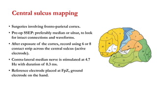
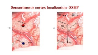
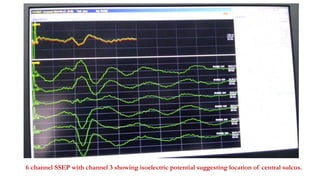
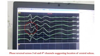
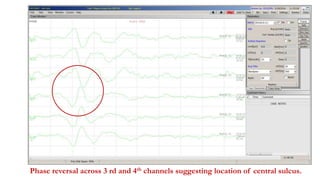
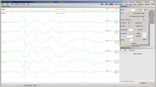
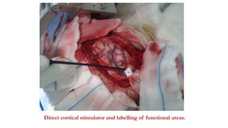
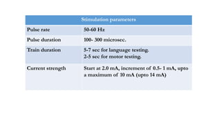
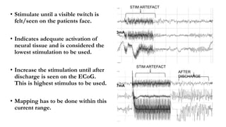
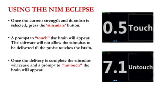
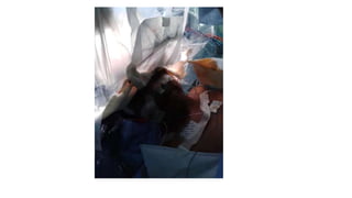
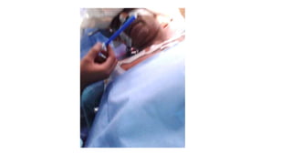
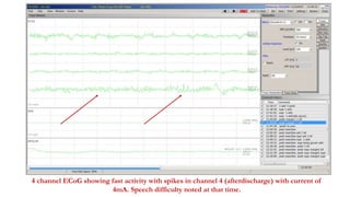
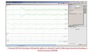
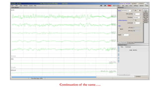
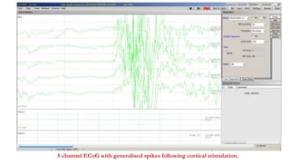
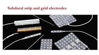
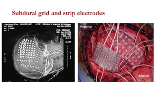
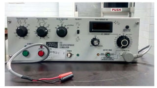
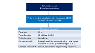
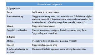
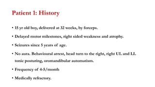
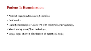
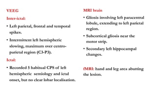
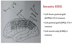
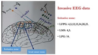
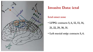
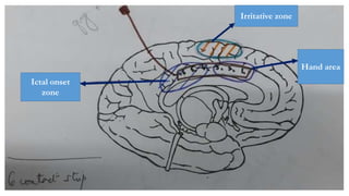
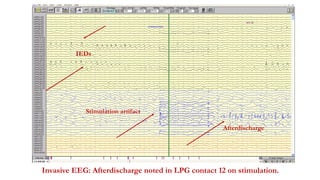
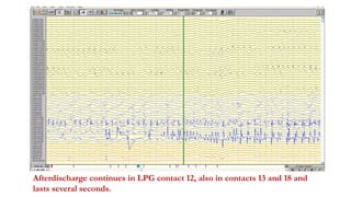
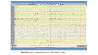
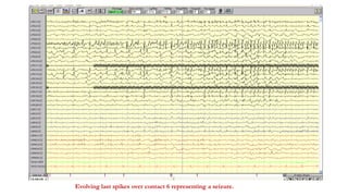
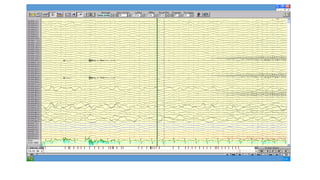
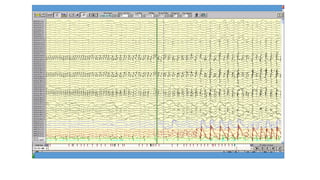
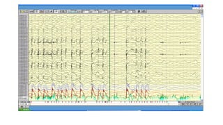
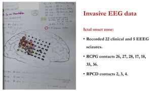
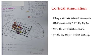
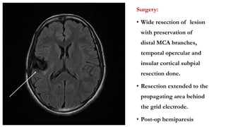
Cortical Stimulation and Mapping
Dr Pramod Krishnan
Consultant Neurologist and Epileptologist
Manipal Hospital, Bengluru
Introduction
• Direct cortical stimulation is used to localise functional cortex.
• Needs special instrumentation and precautions.
Two methods:
• Intraoperative stimulation using a bipolar stimulation wand or probe.
• Extraoperative stimulation through grid or strip electrodes, usually in
epilepsy monitoring units.
Central sulcus mapping
• Surgeries involving fronto-parietal cortex.
• Pre-op SSEP: preferably median or ulnar, to look
for intact connections and waveforms.
• After exposure of the cortex, record using 6 or 8
contact strip across the central sulcus (active
electrode).
• Contra-lateral median nerve is stimulated at 4.7
Hz with duration of 0.3 ms.
• Reference electrode placed at FpZ, ground
electrode on the hand.
P20
N20
Sensorimotor cortex localization -SSEP
P20
N20
6 channel SSEP with channel 3 showing isoelectric potential suggesting location of central sulcus.
Phase reversal across 3 rd and 4th channels suggesting location of central sulcus.
Intraoperative cortical stimulation
Stimuli- bipolar and biphasic
Bipolar: electric current runs between two adjacent active electrodes.
Biphasic: current switches polarity half way through each stimulus pulse.
• Biphasic stimulation does not leave a net electrical polarisation at either
metal-cortex contacts and is desirable.
Polarisation has to be avoided as:
• It can cause metal ions to move onto the brain leading to irritation.
• It alters the electrical sensitivity of the underlying cortex to subsequent
stimulus pulses.
Stimulator probe
30 cm long, with 2 round metal ball tips separated by few millimeters
Direct cortical stimulator and labelling of functional areas.
Stimulation parameters
Pulse rate 50-60 Hz
Pulse duration 100- 300 microsec.
Train duration 5-7 sec for language testing.
2-5 sec for motor testing.
Current strength Start at 2.0 mA, increment of 0.5- 1 mA, upto
a maximum of 10 mA (upto 14 mA)
First channel: EMG, 2nd channel: stimulation. ECoG: normal recording noted in lower 5 channels.
Normal ECoG with excess fast activity (awake state)
4 channel ECoG showing spikes with stimulation at 4mA. No clinical change
Sensory and language mapping
• Can be done only during awake surgery.
• Baseline assessment using the same protocol should be done pre-op.
• Consistent responses should be obtained during baseline testing.
• Patient cooperation throughout the testing is imperative.
• A Neuropsychologist can be involved in language testing.
• A typical aura may be elicited if the seizure focus is stimulated.
Language mapping
1. Visual object naming: naming objects presented as line drawings
from the Boston Naming Test.
2. Sentence reading
3. Word generation: list of words that begin with a certain letter or
category.
4. Auditory response naming
5. Visual response naming
• Language function is often localised to a 1-2 sqcm region.
20
• Stimulate until a visible twitch is
felt/seen on the patients face.
• Indicates adequate activation of
neural tissue and is considered the
lowest stimulation to be used.
• Increase the stimulation until after
discharge is seen on the ECoG.
This is highest stimulus to be used.
• Mapping has to be done within this
current range.
STIM ARTEFACT
STIM ARTEFACT
AFTER
DISCHARGE
3mA
7mA
USING THE NIM ECLIPSE
• Once the current strength and duration is
selected, press the ‘stimulate’ button.
• A prompt to “touch” the brain will appear.
The software will not allow the stimulus to
be delivered til the probe touches the brain.
• Once the delivery is complete the stimulus
will cease and a prompt to “untouch” the
brain will appear.
4 channel ECoG showing fast activity with spikes in channel 4 (afterdischarge) with current of
4mA. Speech difficulty noted at that time.
Stimulation artifact Afterdischarge
5 channel ECoG with stimulation artefact and afterdischarge. Speech difficulty noted at that time.
Motor mapping
• Can be done during awake surgery.
• Can also be done after lightening of anesthesia.
• Neuromuscular blockade should be avoided.
• Look for movements visually or by EMG.
• In awake surgery, patient can be asked to perform some action.
• Exact location of eloquent area shows considerable variation.
5 channel ECoG with afterdischarge. Right hand numbness noted at that time.
Afterdischarges
• During cortical stimulation ECoG is recorded from nearby electrodes.
• This is to look for epileptic-like discharges related to the stimulation.
• When such discharges last longer than the stimulation, they are called
afterdischarges.
• Rhythmic, repetitive, high-voltage spikes or polyspike wave complexes
that last seconds to minutes.
• Usually subclinical, but may be associated with subtle clinical changes.
5 channel ECoG showing evolving fast spikes in channel 1 and 2, following stimulation leading to
clinical seizures (GTCS)
Continuation of the same…..
Afterdischarges
• Afterdischarges indicate spread of stimulation to nearby areas.
• Such stimulation trials should not be considered for mapping.
• Afterdischarges may herald seizures on further stimulation at that site
or intensity.
• If seizures occur during stimulation, irrigation with cold Ringers lactate
solution rapidly stops partial seizures.
5 channel ECoG with generalised spikes following cortical stimulation.
Extra operative cortical stimulation
and mapping.
Subdural strip and grid electrodes
Subdural grid and strip electrodes
Informed consent
Explain the procedure
Perform cortical stimulation after completing VEEG
Do under the cover of AEDs
Stimulation parameters
Pulse rate 50Hz
Pulse duration 0.2 millisec (0.1-0.3)
Train duration 2 sec (1-5 sec)
Current strength Start at 2.0 mA, increment of 0.5 to 1 mA, upto a
maximum of 10 mA (sometimes upto 15 mA).
Channels stimulated Bipolar, between two neighbouring electrodes.
Stimulation end points
1. Symptoms
Aura Indicates ictal onset zone
Somato sensory Suggests sensory area, but stimulate at 0.5-1.0 mA higher
current to see if it is motor area, unless the sensation is
intolerable or afterdischarge has already occurred.
Visual Suggests visual areas.
Cognitive- affective Uncommon, may suggest limbic areas, or may be a
‘psychological reaction’
2. Signs
Motor Negative (loss of tone) or positive (twitch)
Speech Suggests language area
3. After-discharge or
seizure
Do not stimulate again at same strength same site.
Patient 1: History
• 15 yr old boy, delivered at 32 weeks, by forceps.
• Delayed motor milestones, right sided weakness and atrophy.
• Seizures since 5 years of age.
• No aura. Behavioural arrest, head turn to the right, right UL and LL
tonic posturing, oromandibular automatism.
• Frequency of 4-5/month
• Medically refractory.
Patient 1: Examination
• Normal cognitive, language, behaviour.
• Left handed.
• Right hemiparesis of Grade 4/5 with moderate grip weakness.
• Visual acuity was 6/5 on both sides.
• Visual fields showed constriction of peripheral fields.
VEEG
Inter-ictal:
• Left parietal, frontal and temporal
spikes.
• Intermittent left hemispheric
slowing, maximum over centro-
parietal region (C3-P3).
Ictal:
• Recorded 5 habitual CPS of left
hemispheric semiology and ictal
onset, but no clear lobar localisation.
MRI brain
• Gliosis involving left paracentral
lobule, extending to left parietal
region.
• Subcortical gliosis near the
motor strip.
• Secondary left hippocampal
changes.
fMRI: hand and leg area abutting
the lesion.
Hypothesis
• Medically refractory epilepsy of left hemispheric ictal onset close to the
motor strip (eloquent area).
• Semiology correlates with the radiological substrate.
• However, ictal onset zone was unclear on VEEG.
• Relationship of ictal onset zone to motor cortex not established.
• Significance of left hippocampal changes ?
• Planned for invasive EEG.
Invasive EEG
• Left fronto parietal grid
(LFPG): 8 X 4 contacts
• Left parietal grid (LPG): 5 X 4
contacts
• Left mesial strip (LMS): 6
contacts
Invasive EEG data
Irritative zone:
• LFPG: 4,5,12,13,14,20,21.
• LMS: 4,5.
• LPG: 14.
Ictal onset zone
Irritative zone
Invasive Data: ictal
Ictal onset zone
• LFPG: contacts 5, 6, 12, 13, 14,
21, 22, 29, 30, 31.
• Left mesial strip: contacts 5, 6.
Ictal onset
zone
Hand area
Irritative zone
Invasive EEG: Afterdischarge noted in LPG contact 12 on stimulation.
Stimulation artifact
Afterdischarge
IEDs
Afterdischarge continues in LPG contact 12, also in contacts 13 and 18 and
lasts several seconds.
Hand movement on stimulation: no EEG changes noted.
Evolving last spikes over contact 6 representing a seizure.
Surgery
• Resection of left frontal gliotic area.
• Post- resection ECoG did not show any spikes, fast activity.
• No new deficits were noted.
• HPE: reactive gliosis and localised inflammation.
MRI brain T2F axial and coronal sequences showing post-resection changes.
Patient 2
• 15 year old girl.
• Normal birth and development.
• Seizures since 4.5 years of age.
• Semiology 1: abrupt LOC and falls.
• Semiology 2: Left UL paresthesia, stare, head turn to left, falls and tonic
posturing of all limbs lasting 30 sec, post-ictal left sided Todds palsy.
• Frequent secondary generalised seizures.
• Medically refractory.
• No deficits.
VEEG:
Interictal: right centro-parietal,
right temporal, right hemispheric
spikes.
Ictal: Recorded multiple seizures
of right hemispheric semiology
with central ictal onset.
MRI brain:
Right insular, right inferior frontal
FCD. Also, ?right superior
temporal gyrus involvement.
fMRI:
Motor area away from the lesion.
Plan…
• Good clinical- electrical and radiological concordance.
• Poor localisation of ictal onset zone on VEEG.
• Radiological lesion is close to the eloquent cortex. Posterior extent not
clear in relation to the eloquent cortex.
• Invasive EEG planned to delineate the ictal onset zone and its relation
to the eloquent cortex.
Invasive EEG:
1. Right centro-parietal grid (RCPG):
6 X 8 contact grid placed such that
the lower row was on the STG, and
2/3 rd of the grid was anterior to the
central sulcus and 1/3rd was
posterior to it.
2. RACD:
Anterior insular depth electrode.
3. RPCD
Posterior insular depth electrode.
Invasive data
Irritative zone (spikes):
• RCPG contacts 25, 26, 27, 28, 9,
10.
• Posterior part of the grid
overlying the parietal lobe.
Ictal onset zone:
• Recorded 22 clinical and 5 EEEG
seizures.
• RCPG contacts 26, 27, 28, 17, 18,
35, 36.
• RPCD contacts 2, 3, 4.
Invasive EEG data
Irritative zone (spikes):
• RCPG contacts 25, 26, 27, 28, 9,
10.
• Posterior part of the grid
overlying the parietal lobe.
Invasive EEG data
Ictal onset zone:
• Recorded 22 clinical and 5 EEEG
seizures.
• RCPG contacts 26, 27, 28, 17, 18,
35, 36.
• RPCD contacts 2, 3, 4.
Cortical stimulation
• Eloquent cortex (hand area) over
RCPG contacts 9, 17, 18, 25, 26.
• 9,17, 18: left thumb sensory.
• 17, 18, 25, 26: left thumb jerking.
Surgery:
• Wide resection of lesion
with preservation of
distal MCA branches,
temporal opercular and
insular cortical subpial
resection done.
• Resection extended to the
propagating area behind
the grid electrode.
• Post-op hemiparesis
THANK YOU

Cortical stimulation and mapping

  • 1.
    Cortical Stimulation andMapping Dr Pramod Krishnan Consultant Neurologist and Epileptologist Manipal Hospital, Bengluru
  • 2.
    Introduction • Direct corticalstimulation is used to localise functional cortex. • Needs special instrumentation and precautions. Two methods: • Intraoperative stimulation using a bipolar stimulation wand or probe. • Extraoperative stimulation through grid or strip electrodes, usually in epilepsy monitoring units.
  • 3.
  • 4.
    Central sulcus mapping •Surgeries involving fronto-parietal cortex. • Pre-op SSEP: preferably median or ulnar, to look for intact connections and waveforms. • After exposure of the cortex, record using 6 or 8 contact strip across the central sulcus (active electrode). • Contra-lateral median nerve is stimulated at 4.7 Hz with duration of 0.3 ms. • Reference electrode placed at FpZ, ground electrode on the hand.
  • 5.
  • 6.
    6 channel SSEPwith channel 3 showing isoelectric potential suggesting location of central sulcus.
  • 7.
    Phase reversal across3 rd and 4th channels suggesting location of central sulcus.
  • 8.
    Phase reversal across3 rd and 4th channels suggesting location of central sulcus.
  • 10.
  • 11.
    Stimuli- bipolar andbiphasic Bipolar: electric current runs between two adjacent active electrodes. Biphasic: current switches polarity half way through each stimulus pulse. • Biphasic stimulation does not leave a net electrical polarisation at either metal-cortex contacts and is desirable. Polarisation has to be avoided as: • It can cause metal ions to move onto the brain leading to irritation. • It alters the electrical sensitivity of the underlying cortex to subsequent stimulus pulses.
  • 12.
    Stimulator probe 30 cmlong, with 2 round metal ball tips separated by few millimeters
  • 13.
    Direct cortical stimulatorand labelling of functional areas.
  • 14.
    Stimulation parameters Pulse rate50-60 Hz Pulse duration 100- 300 microsec. Train duration 5-7 sec for language testing. 2-5 sec for motor testing. Current strength Start at 2.0 mA, increment of 0.5- 1 mA, upto a maximum of 10 mA (upto 14 mA)
  • 15.
    First channel: EMG,2nd channel: stimulation. ECoG: normal recording noted in lower 5 channels.
  • 16.
    Normal ECoG withexcess fast activity (awake state)
  • 17.
    4 channel ECoGshowing spikes with stimulation at 4mA. No clinical change
  • 18.
    Sensory and languagemapping • Can be done only during awake surgery. • Baseline assessment using the same protocol should be done pre-op. • Consistent responses should be obtained during baseline testing. • Patient cooperation throughout the testing is imperative. • A Neuropsychologist can be involved in language testing. • A typical aura may be elicited if the seizure focus is stimulated.
  • 19.
    Language mapping 1. Visualobject naming: naming objects presented as line drawings from the Boston Naming Test. 2. Sentence reading 3. Word generation: list of words that begin with a certain letter or category. 4. Auditory response naming 5. Visual response naming • Language function is often localised to a 1-2 sqcm region.
  • 20.
    20 • Stimulate untila visible twitch is felt/seen on the patients face. • Indicates adequate activation of neural tissue and is considered the lowest stimulation to be used. • Increase the stimulation until after discharge is seen on the ECoG. This is highest stimulus to be used. • Mapping has to be done within this current range. STIM ARTEFACT STIM ARTEFACT AFTER DISCHARGE 3mA 7mA
  • 21.
    USING THE NIMECLIPSE • Once the current strength and duration is selected, press the ‘stimulate’ button. • A prompt to “touch” the brain will appear. The software will not allow the stimulus to be delivered til the probe touches the brain. • Once the delivery is complete the stimulus will cease and a prompt to “untouch” the brain will appear.
  • 24.
    4 channel ECoGshowing fast activity with spikes in channel 4 (afterdischarge) with current of 4mA. Speech difficulty noted at that time.
  • 25.
    Stimulation artifact Afterdischarge 5channel ECoG with stimulation artefact and afterdischarge. Speech difficulty noted at that time.
  • 26.
    Motor mapping • Canbe done during awake surgery. • Can also be done after lightening of anesthesia. • Neuromuscular blockade should be avoided. • Look for movements visually or by EMG. • In awake surgery, patient can be asked to perform some action. • Exact location of eloquent area shows considerable variation.
  • 27.
    5 channel ECoGwith afterdischarge. Right hand numbness noted at that time.
  • 28.
    Afterdischarges • During corticalstimulation ECoG is recorded from nearby electrodes. • This is to look for epileptic-like discharges related to the stimulation. • When such discharges last longer than the stimulation, they are called afterdischarges. • Rhythmic, repetitive, high-voltage spikes or polyspike wave complexes that last seconds to minutes. • Usually subclinical, but may be associated with subtle clinical changes.
  • 29.
    5 channel ECoGshowing evolving fast spikes in channel 1 and 2, following stimulation leading to clinical seizures (GTCS)
  • 30.
  • 31.
    Afterdischarges • Afterdischarges indicatespread of stimulation to nearby areas. • Such stimulation trials should not be considered for mapping. • Afterdischarges may herald seizures on further stimulation at that site or intensity. • If seizures occur during stimulation, irrigation with cold Ringers lactate solution rapidly stops partial seizures.
  • 32.
    5 channel ECoGwith generalised spikes following cortical stimulation.
  • 33.
    Extra operative corticalstimulation and mapping.
  • 34.
    Subdural strip andgrid electrodes
  • 35.
    Subdural grid andstrip electrodes
  • 37.
    Informed consent Explain theprocedure Perform cortical stimulation after completing VEEG Do under the cover of AEDs Stimulation parameters Pulse rate 50Hz Pulse duration 0.2 millisec (0.1-0.3) Train duration 2 sec (1-5 sec) Current strength Start at 2.0 mA, increment of 0.5 to 1 mA, upto a maximum of 10 mA (sometimes upto 15 mA). Channels stimulated Bipolar, between two neighbouring electrodes.
  • 38.
    Stimulation end points 1.Symptoms Aura Indicates ictal onset zone Somato sensory Suggests sensory area, but stimulate at 0.5-1.0 mA higher current to see if it is motor area, unless the sensation is intolerable or afterdischarge has already occurred. Visual Suggests visual areas. Cognitive- affective Uncommon, may suggest limbic areas, or may be a ‘psychological reaction’ 2. Signs Motor Negative (loss of tone) or positive (twitch) Speech Suggests language area 3. After-discharge or seizure Do not stimulate again at same strength same site.
  • 39.
    Patient 1: History •15 yr old boy, delivered at 32 weeks, by forceps. • Delayed motor milestones, right sided weakness and atrophy. • Seizures since 5 years of age. • No aura. Behavioural arrest, head turn to the right, right UL and LL tonic posturing, oromandibular automatism. • Frequency of 4-5/month • Medically refractory.
  • 40.
    Patient 1: Examination •Normal cognitive, language, behaviour. • Left handed. • Right hemiparesis of Grade 4/5 with moderate grip weakness. • Visual acuity was 6/5 on both sides. • Visual fields showed constriction of peripheral fields.
  • 41.
    VEEG Inter-ictal: • Left parietal,frontal and temporal spikes. • Intermittent left hemispheric slowing, maximum over centro- parietal region (C3-P3). Ictal: • Recorded 5 habitual CPS of left hemispheric semiology and ictal onset, but no clear lobar localisation. MRI brain • Gliosis involving left paracentral lobule, extending to left parietal region. • Subcortical gliosis near the motor strip. • Secondary left hippocampal changes. fMRI: hand and leg area abutting the lesion.
  • 42.
    Hypothesis • Medically refractoryepilepsy of left hemispheric ictal onset close to the motor strip (eloquent area). • Semiology correlates with the radiological substrate. • However, ictal onset zone was unclear on VEEG. • Relationship of ictal onset zone to motor cortex not established. • Significance of left hippocampal changes ? • Planned for invasive EEG.
  • 43.
    Invasive EEG • Leftfronto parietal grid (LFPG): 8 X 4 contacts • Left parietal grid (LPG): 5 X 4 contacts • Left mesial strip (LMS): 6 contacts
  • 44.
    Invasive EEG data Irritativezone: • LFPG: 4,5,12,13,14,20,21. • LMS: 4,5. • LPG: 14. Ictal onset zone Irritative zone
  • 45.
    Invasive Data: ictal Ictalonset zone • LFPG: contacts 5, 6, 12, 13, 14, 21, 22, 29, 30, 31. • Left mesial strip: contacts 5, 6.
  • 46.
  • 47.
    Invasive EEG: Afterdischargenoted in LPG contact 12 on stimulation. Stimulation artifact Afterdischarge IEDs
  • 48.
    Afterdischarge continues inLPG contact 12, also in contacts 13 and 18 and lasts several seconds.
  • 49.
    Hand movement onstimulation: no EEG changes noted.
  • 50.
    Evolving last spikesover contact 6 representing a seizure.
  • 51.
    Surgery • Resection ofleft frontal gliotic area. • Post- resection ECoG did not show any spikes, fast activity. • No new deficits were noted. • HPE: reactive gliosis and localised inflammation.
  • 52.
    MRI brain T2Faxial and coronal sequences showing post-resection changes.
  • 53.
    Patient 2 • 15year old girl. • Normal birth and development. • Seizures since 4.5 years of age. • Semiology 1: abrupt LOC and falls. • Semiology 2: Left UL paresthesia, stare, head turn to left, falls and tonic posturing of all limbs lasting 30 sec, post-ictal left sided Todds palsy. • Frequent secondary generalised seizures. • Medically refractory. • No deficits.
  • 55.
    VEEG: Interictal: right centro-parietal, righttemporal, right hemispheric spikes. Ictal: Recorded multiple seizures of right hemispheric semiology with central ictal onset. MRI brain: Right insular, right inferior frontal FCD. Also, ?right superior temporal gyrus involvement. fMRI: Motor area away from the lesion.
  • 56.
    Plan… • Good clinical-electrical and radiological concordance. • Poor localisation of ictal onset zone on VEEG. • Radiological lesion is close to the eloquent cortex. Posterior extent not clear in relation to the eloquent cortex. • Invasive EEG planned to delineate the ictal onset zone and its relation to the eloquent cortex.
  • 57.
    Invasive EEG: 1. Rightcentro-parietal grid (RCPG): 6 X 8 contact grid placed such that the lower row was on the STG, and 2/3 rd of the grid was anterior to the central sulcus and 1/3rd was posterior to it. 2. RACD: Anterior insular depth electrode. 3. RPCD Posterior insular depth electrode.
  • 61.
    Invasive data Irritative zone(spikes): • RCPG contacts 25, 26, 27, 28, 9, 10. • Posterior part of the grid overlying the parietal lobe. Ictal onset zone: • Recorded 22 clinical and 5 EEEG seizures. • RCPG contacts 26, 27, 28, 17, 18, 35, 36. • RPCD contacts 2, 3, 4.
  • 62.
    Invasive EEG data Irritativezone (spikes): • RCPG contacts 25, 26, 27, 28, 9, 10. • Posterior part of the grid overlying the parietal lobe.
  • 63.
    Invasive EEG data Ictalonset zone: • Recorded 22 clinical and 5 EEEG seizures. • RCPG contacts 26, 27, 28, 17, 18, 35, 36. • RPCD contacts 2, 3, 4.
  • 64.
    Cortical stimulation • Eloquentcortex (hand area) over RCPG contacts 9, 17, 18, 25, 26. • 9,17, 18: left thumb sensory. • 17, 18, 25, 26: left thumb jerking.
  • 65.
    Surgery: • Wide resectionof lesion with preservation of distal MCA branches, temporal opercular and insular cortical subpial resection done. • Resection extended to the propagating area behind the grid electrode. • Post-op hemiparesis
  • 66.