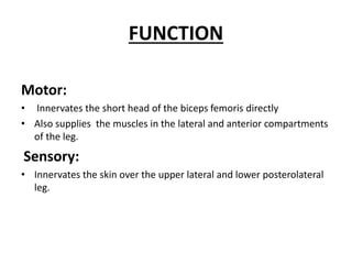
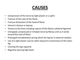
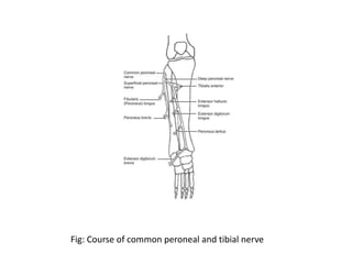
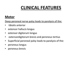
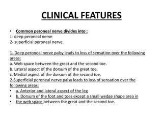
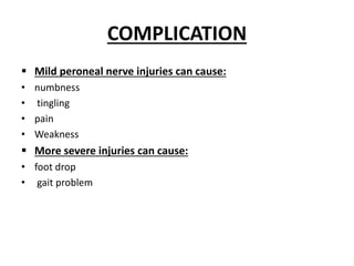
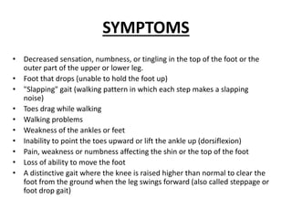
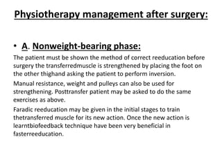
COMMON PERONEAL
NERVE PALSY
INTRODUCTION
The common peroneal nerve is a nerve in the lower leg that provides
sensation over the posterolateral part of the leg and the knee joint.
It branches from the sciatic nerve and provides sensation to the front
and sides of the legs and to the top of the feet.
This nerve also controls the muscles in the leg that lift the ankle and
toes upward.
It divides at the knee into two terminal branches:
1- the superficial peroneal nerve and deep peroneal nerve, which
innervate the muscles of the lateral
2- anterior compartments of the leg respectively
Fig:Common peroneal (fibular) nerve
STRUCTURE
• The common peroneal nerve is the smaller terminal branch of the
sciatic nerve.
• The common peroneal nerve has root values of L4, L5, S1, and S2.
• It arises from the superior angle of the popliteal fossa and extends
to the lateral angle of the popliteal fossa, along the medial border
of the biceps femoris.
• It then winds around the neck of the fibula to pierce the fibularis
longus and divides into terminal branches of superficial peroneal
nerve and deep peroneal nerve.
STRUCTURE
Cutaneous branches
• Lateral sural cutaneous nerve (lateral cutaneous nerve of calf) -
supplies the skin of the upper two-thirds of the lateral side of leg.
• sural communicating nerve - it runs on the posterolateral aspect of
the calf and joins the sural nerve.
STRUCTURE
Articular branches
• Superior lateral genicular nerve - accompanies artery of the same
name and lies above the lateral femoral condyle.
• Inferior lateral genicular nerve - accompanies artery of the same
name and lies just above the head of the fibula.
• Recurrent genicular nerve - It arises from the point of division of the
common peroneal nerve; then ascends anterior to the knee joint
together with the anterior recurrent tibial artery to supply the knee
joint and the tibialis anterior muscle.
• Motor branches
• There is only one motor branch that arises directly from the
common peroneal nerve, the nerve to the short head of the biceps
femoris muscle.
FUNCTION
Motor:
• Innervates the short head of the biceps femoris directly
• Also supplies the muscles in the lateral and anterior compartments
of the leg.
Sensory:
• Innervates the skin over the upper lateral and lower posterolateral
leg.
CAUSES
• Compression of the nerve by tight plaster or a splint
• Fracture of the neck of the fibula
• Fracture dislocation of the head of fibula
• Hansen’s disease or leprosy
• Trauma to the knee including rupture of the fibular collateral ligament
• Entrapped, compressed or irritated nerve by fibrous arch as it winds
around the neck of fibula
• Prolonged immobilization during which the leg lies in external rotation.
• Use of a tight plaster cast (or other long-term constriction) of the lower
leg
• Crossing the legs regularly
• Regularly wearing high boots
ETIOLOGY
Trauma or injury to the knee
 Knee dislocation
o Common peroneal nerve injury can occur in up to 40% of patients
 A direct impact, penetrating trauma, or laceration
o Injury predisposition as the nerve courses around the fibular head/neck
 Fracture of the fibula, especially at the proximal fibula
Anatomic risk factors
 Common peroneal nerve entrapment can occur secondary to a
fibrous band at the origin of the peroneus longus
ETIOLOGY
 External compression sources
 Tight splint/cast
 Compression wrapping/bandage
 Habitual leg crossing
 Prolonged bed rest
 Positioning during anesthesia and surgery
o Important to always pad bony prominences, including the area of the fibular head
and neck at the lateral aspect of the knee
 Intraneural ganglion
 Peripheral nerve tumor
 Iatrogenic injury following surgery about the hip, knee, and
ankle
ETIOLOGY
Several systemic illnesses can cause compressive peroneal neuropathy
and injury to the common peroneal nerve, including:
• Diabetes mellitus
• Inflammatory conditions
• Motor neuron diseases (Charcot-Marie-Tooth disease)
• Anorexia nervosa (exceedingly thin states) due to loss of
subcutaneous fat at this level causing compression of the
nerve
Fig: Course of common peroneal and tibial nerve
CLINICAL FEATURES
• s
• Sensory
Common peroneal nerve by itself is relatively short having only
two sensory branches and no motor branches.
The loss of sensation is as follows:
A. Skin along the lateral aspect of the knee in the proximal third of
the calf (lateral cutaneous sural nerve).
B.Skin over the posterolateral aspect of the calf and over the lateral
malleolus, lateral aspect of the foot and fourth and fifth toes (sural
nerve).
CLINICAL FEATURES
Motor
Deep peroneal nerve palsy leads to paralysis of the:
• tibialis anterior
• extensor hallucis longus
• extensor digitorum longus
• extensordigitorum brevis and peroneus tertius
• Superficial peroneal palsy leads to paralysis of the:
• peroneus longus
• peroneus brevis
CLINICAL FEATURES
• Common peroneal nerve divides into :
1- deep peroneal nerve
2- superficial peroneal nerve.
1- Deep peroneal nerve palsy leads to loss of sensation over the following
areas:
a. Web space between the great and the second toe.
b. Lateral aspect of the dorsum of the great toe.
c. Medial aspect of the dorsum of the second toe.
2-Superficial peroneal nerve palsy leads to loss of sensation over the
following areas:
• a. Anterior and lateral aspect of the leg
• b. Dorsum of the foot and toes except a small wedge shape area in
• the web space between the great and the second toe.
DEFORMITY
• Equino varus deformity results due to over action of the posterior
• compartment muscles and the invertors.
COMPLICATION
 Mild peroneal nerve injuries can cause:
• numbness
• tingling
• pain
• Weakness
 More severe injuries can cause:
• foot drop
• gait problem
SYMPTOMS
• Decreased sensation, numbness, or tingling in the top of the foot or the
outer part of the upper or lower leg.
• Foot that drops (unable to hold the foot up)
• "Slapping" gait (walking pattern in which each step makes a slapping
noise)
• Toes drag while walking
• Walking problems
• Weakness of the ankles or feet
• Inability to point the toes upward or lift the ankle up (dorsiflexion)
• Pain, weakness or numbness affecting the shin or the top of the foot
• Loss of ability to move the foot
• A distinctive gait where the knee is raised higher than normal to clear the
foot from the ground when the leg swings forward (also called steppage or
foot drop gait)
DIAGNOSIS
• Electromyography:
which measures ongoing muscle activity and response to a nerve’s
stimulation of the muscle.
• Nerve conduction study:
which measures the amount and speed of conduction of an electrical
impulse through a nerve.
• imaging techniques:
 CT scan
 Ultrasound
 MRI
 MR neurography:
An MRI that uses specific settings or sequences that provide enhanced
images of nerves From the patient’s perspective, the experience is the same
as undergoing a regular MRI.
TREATMENTS
• Conservative treatment
• IG stimulation of the paralyzed muscles
• Passive movements to maintain the joint range
• Stretching of the Tendoachilles
• Splints or Orthosis: The commonly used orthosis aims to maintain
the ankle in neutral position preventing equinous hence either a
caliper with dorsiflexion stop or plastic ankle foot orthosis in the
form of shoe insert may be prescribed.
TREATMENTS
Surgical management
• Nerve repair
• Nerve grafting
• Nerve transfer
• Tendon transfer:
tendon transfer wherein the tibialis posterior is used to substitute for the lost muscles
The tibialis posterior transfer may be done in two ways:
A) Circumtibial route: The tibialis posterior is detached from its
insertion circles around tibia and is divided into two clips—one
going to tibialis anterior and extensor hallucies longus whereas
the other go to extensor digitorum longus. This procedure is more
commonly done but adhesions are likely to occur which are
treated with : US, laser and kneading technique Decompression surgery
B) Interosseous route:
Insertions are the same as above but the transfer is done by piercing
the interosseus membrance.
• tendoAchilles lengthening: to get maximum range of dorsiflexion.
• Immobilization following tendon transfer can be done in two
ways:
• Total period of immobilization is six weeks. For the first three
weeks the knee is in flexion and the ankle is in full dorsiflexion.
In the next three weeks the knee is kept free but the ankle is still
maintained in full dorsiflexion. This method gives a better range
of dorsiflexion.
• The ankle is immobilized in full dorsiflexion for a period of six
weeks
Physiotherapy management after surgery:
• A. Nonweight-bearing phase:
The patient must be shown the method of correct reeducation before
surgery the transferredmuscle is strengthened by placing the foot on
the other thighand asking the patient to perform inversion.
Manual resistance, weight and pulleys can also be used for
strengthening. Posttransfer patient may be asked to do the same
exercises as above.
Faradic reeducation may be given in the initial stages to train
thetransferred muscle for its new action. Once the new action is
learntbiofeedback technique have been very beneficial in
fasterreeducation.
B: Weight-bearing phase:
Take a weighing scale and the patient is asked to bear weight on the
affected limb and at the same time carry out dorsiflexion.
Initially the patient should take only 10 kg of weight which is indicated
by the pointer. This goes on till patient can take more and more weight
on the affected limb till he eventually starts taking almost 2/3rd of his
body weight (the weight progression is by 10 kg). Then blind fold the
patient to see whether the patient knows how much weight is to be
taken by the affected limb. This can be checked by the weighing scale.
Then progress to walking on the parallel bars, then even ground,
uneven ground and eventually on stairs. The therapist should all the
time take care that the patient does not slap the foot on the ground
and places it gently. Exercises on the tilt board or vestibular ball has
been found to be quite affective in proprioceptive training of the
affected foot

COMMON PERONEAL NERVE PALSY.pptx full ppt

  • 1.
  • 2.
    INTRODUCTION The common peronealnerve is a nerve in the lower leg that provides sensation over the posterolateral part of the leg and the knee joint. It branches from the sciatic nerve and provides sensation to the front and sides of the legs and to the top of the feet. This nerve also controls the muscles in the leg that lift the ankle and toes upward. It divides at the knee into two terminal branches: 1- the superficial peroneal nerve and deep peroneal nerve, which innervate the muscles of the lateral 2- anterior compartments of the leg respectively
  • 3.
  • 4.
    STRUCTURE • The commonperoneal nerve is the smaller terminal branch of the sciatic nerve. • The common peroneal nerve has root values of L4, L5, S1, and S2. • It arises from the superior angle of the popliteal fossa and extends to the lateral angle of the popliteal fossa, along the medial border of the biceps femoris. • It then winds around the neck of the fibula to pierce the fibularis longus and divides into terminal branches of superficial peroneal nerve and deep peroneal nerve.
  • 5.
    STRUCTURE Cutaneous branches • Lateralsural cutaneous nerve (lateral cutaneous nerve of calf) - supplies the skin of the upper two-thirds of the lateral side of leg. • sural communicating nerve - it runs on the posterolateral aspect of the calf and joins the sural nerve.
  • 6.
    STRUCTURE Articular branches • Superiorlateral genicular nerve - accompanies artery of the same name and lies above the lateral femoral condyle. • Inferior lateral genicular nerve - accompanies artery of the same name and lies just above the head of the fibula. • Recurrent genicular nerve - It arises from the point of division of the common peroneal nerve; then ascends anterior to the knee joint together with the anterior recurrent tibial artery to supply the knee joint and the tibialis anterior muscle. • Motor branches • There is only one motor branch that arises directly from the common peroneal nerve, the nerve to the short head of the biceps femoris muscle.
  • 7.
    FUNCTION Motor: • Innervates theshort head of the biceps femoris directly • Also supplies the muscles in the lateral and anterior compartments of the leg. Sensory: • Innervates the skin over the upper lateral and lower posterolateral leg.
  • 8.
    CAUSES • Compression ofthe nerve by tight plaster or a splint • Fracture of the neck of the fibula • Fracture dislocation of the head of fibula • Hansen’s disease or leprosy • Trauma to the knee including rupture of the fibular collateral ligament • Entrapped, compressed or irritated nerve by fibrous arch as it winds around the neck of fibula • Prolonged immobilization during which the leg lies in external rotation. • Use of a tight plaster cast (or other long-term constriction) of the lower leg • Crossing the legs regularly • Regularly wearing high boots
  • 9.
    ETIOLOGY Trauma or injuryto the knee  Knee dislocation o Common peroneal nerve injury can occur in up to 40% of patients  A direct impact, penetrating trauma, or laceration o Injury predisposition as the nerve courses around the fibular head/neck  Fracture of the fibula, especially at the proximal fibula Anatomic risk factors  Common peroneal nerve entrapment can occur secondary to a fibrous band at the origin of the peroneus longus
  • 10.
    ETIOLOGY  External compressionsources  Tight splint/cast  Compression wrapping/bandage  Habitual leg crossing  Prolonged bed rest  Positioning during anesthesia and surgery o Important to always pad bony prominences, including the area of the fibular head and neck at the lateral aspect of the knee  Intraneural ganglion  Peripheral nerve tumor  Iatrogenic injury following surgery about the hip, knee, and ankle
  • 11.
    ETIOLOGY Several systemic illnessescan cause compressive peroneal neuropathy and injury to the common peroneal nerve, including: • Diabetes mellitus • Inflammatory conditions • Motor neuron diseases (Charcot-Marie-Tooth disease) • Anorexia nervosa (exceedingly thin states) due to loss of subcutaneous fat at this level causing compression of the nerve
  • 12.
    Fig: Course ofcommon peroneal and tibial nerve
  • 13.
    CLINICAL FEATURES • s •Sensory Common peroneal nerve by itself is relatively short having only two sensory branches and no motor branches. The loss of sensation is as follows: A. Skin along the lateral aspect of the knee in the proximal third of the calf (lateral cutaneous sural nerve). B.Skin over the posterolateral aspect of the calf and over the lateral malleolus, lateral aspect of the foot and fourth and fifth toes (sural nerve).
  • 14.
    CLINICAL FEATURES Motor Deep peronealnerve palsy leads to paralysis of the: • tibialis anterior • extensor hallucis longus • extensor digitorum longus • extensordigitorum brevis and peroneus tertius • Superficial peroneal palsy leads to paralysis of the: • peroneus longus • peroneus brevis
  • 15.
    CLINICAL FEATURES • Commonperoneal nerve divides into : 1- deep peroneal nerve 2- superficial peroneal nerve. 1- Deep peroneal nerve palsy leads to loss of sensation over the following areas: a. Web space between the great and the second toe. b. Lateral aspect of the dorsum of the great toe. c. Medial aspect of the dorsum of the second toe. 2-Superficial peroneal nerve palsy leads to loss of sensation over the following areas: • a. Anterior and lateral aspect of the leg • b. Dorsum of the foot and toes except a small wedge shape area in • the web space between the great and the second toe.
  • 16.
    DEFORMITY • Equino varusdeformity results due to over action of the posterior • compartment muscles and the invertors.
  • 17.
    COMPLICATION  Mild peronealnerve injuries can cause: • numbness • tingling • pain • Weakness  More severe injuries can cause: • foot drop • gait problem
  • 18.
    SYMPTOMS • Decreased sensation,numbness, or tingling in the top of the foot or the outer part of the upper or lower leg. • Foot that drops (unable to hold the foot up) • "Slapping" gait (walking pattern in which each step makes a slapping noise) • Toes drag while walking • Walking problems • Weakness of the ankles or feet • Inability to point the toes upward or lift the ankle up (dorsiflexion) • Pain, weakness or numbness affecting the shin or the top of the foot • Loss of ability to move the foot • A distinctive gait where the knee is raised higher than normal to clear the foot from the ground when the leg swings forward (also called steppage or foot drop gait)
  • 19.
    DIAGNOSIS • Electromyography: which measuresongoing muscle activity and response to a nerve’s stimulation of the muscle. • Nerve conduction study: which measures the amount and speed of conduction of an electrical impulse through a nerve. • imaging techniques:  CT scan  Ultrasound  MRI  MR neurography: An MRI that uses specific settings or sequences that provide enhanced images of nerves From the patient’s perspective, the experience is the same as undergoing a regular MRI.
  • 20.
    TREATMENTS • Conservative treatment •IG stimulation of the paralyzed muscles • Passive movements to maintain the joint range • Stretching of the Tendoachilles • Splints or Orthosis: The commonly used orthosis aims to maintain the ankle in neutral position preventing equinous hence either a caliper with dorsiflexion stop or plastic ankle foot orthosis in the form of shoe insert may be prescribed.
  • 21.
    TREATMENTS Surgical management • Nerverepair • Nerve grafting • Nerve transfer • Tendon transfer: tendon transfer wherein the tibialis posterior is used to substitute for the lost muscles The tibialis posterior transfer may be done in two ways: A) Circumtibial route: The tibialis posterior is detached from its insertion circles around tibia and is divided into two clips—one going to tibialis anterior and extensor hallucies longus whereas the other go to extensor digitorum longus. This procedure is more commonly done but adhesions are likely to occur which are treated with : US, laser and kneading technique Decompression surgery
  • 22.
    B) Interosseous route: Insertionsare the same as above but the transfer is done by piercing the interosseus membrance. • tendoAchilles lengthening: to get maximum range of dorsiflexion. • Immobilization following tendon transfer can be done in two ways: • Total period of immobilization is six weeks. For the first three weeks the knee is in flexion and the ankle is in full dorsiflexion. In the next three weeks the knee is kept free but the ankle is still maintained in full dorsiflexion. This method gives a better range of dorsiflexion. • The ankle is immobilized in full dorsiflexion for a period of six weeks
  • 23.
    Physiotherapy management aftersurgery: • A. Nonweight-bearing phase: The patient must be shown the method of correct reeducation before surgery the transferredmuscle is strengthened by placing the foot on the other thighand asking the patient to perform inversion. Manual resistance, weight and pulleys can also be used for strengthening. Posttransfer patient may be asked to do the same exercises as above. Faradic reeducation may be given in the initial stages to train thetransferred muscle for its new action. Once the new action is learntbiofeedback technique have been very beneficial in fasterreeducation.
  • 24.
    B: Weight-bearing phase: Takea weighing scale and the patient is asked to bear weight on the affected limb and at the same time carry out dorsiflexion. Initially the patient should take only 10 kg of weight which is indicated by the pointer. This goes on till patient can take more and more weight on the affected limb till he eventually starts taking almost 2/3rd of his body weight (the weight progression is by 10 kg). Then blind fold the patient to see whether the patient knows how much weight is to be taken by the affected limb. This can be checked by the weighing scale. Then progress to walking on the parallel bars, then even ground, uneven ground and eventually on stairs. The therapist should all the time take care that the patient does not slap the foot on the ground and places it gently. Exercises on the tilt board or vestibular ball has been found to be quite affective in proprioceptive training of the affected foot