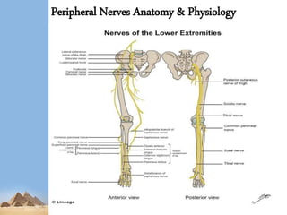
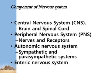
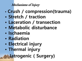
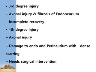
5. • The nervous system is the 1st
system to differentiate.
• At 3rd week as a slipper-shaped
plate of thickened ectoderm
• It receives and interprets stimuli &
transmits impulses to the effector
organs *
6. Component of Nervous system
• Central Nervous System (CNS).
–Brain and Spinal Cord
• Peripheral Nervous System (PNS)
–Nerves and Receptors
• Autonomic nervous system
–Sympathetic and
parasympathetic systems
• Enteric nervous system
7. • The peripheral nervous system is formed
by the cranial
and spinal nerves carrying the somatic and
autonomic nerve fibres.
Preganglionic fibres from the T11–L2 spinal
segments
synapse in the lumbar and sacral ganglia,
and the postganglionic fibres are
distributed to the limb through
the lumbosacral plexus.
9. Sunderland
1951 I II III IV V
Focal
conduction
block
NO Walleria
n
degeneratio
n
Axonal
Disruption
Axon
+
Endoneuriu
m
Disruption
Axon
+
Endoneuriu
m +
Perineurium
Disruption
Axon
+
Endoneurium
+
Perineurium
+
Epineurium
Disruption
Axon
+
Endoneurium
+
Perineurium
+
Epineurium
Disruption
Seddon
BMJ
1942
Classification Of Nerve Injuries
Neurapraxia
(Transient Block)
Axonotmesis
(Lesion in Continuity)
Neurotmesis
(Division of a nerve)
10. Degrees Of Nerve Injury
• 1st degree of injury( Neurapraxia)
– Segmental demyelination
– Axons intact
– Recovery in 12 to 16 wks.'
• 2nd degree injury( Axonotmesis)
– Axonal injury/ distal Wallerian degeneration
– Regeneration at rate of 1 inch per month or
1mm/day
– Complete slow recovery
11. • 3rd degree injury
– Axonal injury & fibrosis of Endoneurium
– Incomplete recovery
• 4th degree injury
– Axonal injury
– Damage to endo and Perineurium with dense
scarring
– Needs surgical intervention
12. • 5th degree injury
( Neurotmesis)
– Complete nerve division
• 6th degree injury
– Variable combination of
previous five
degrees of nerve injury
14. • The nerves of the lower limb
derive from the anterior
(ventral) primary rami making
up the lumbosacral
plexus, and thus must cross all
or part of the pelvis
early in their course
15.
16. Anatomy of nerve injuries
• Must know the course of nerve
• Dermatomes
• possible Entrapment areas
• Pierce which muscle/ fascia
• Repetitive movements
17. Obturator Nerve (L2–L4)
■ Arises from the lumbar plexus & enters
the thigh through the obturator foramen.
1. Anterior Branch
Descends between the adductor longus
and adductor brevis muscles.
Innervates the adductor longus, adductor
brevis, gracilis, and pectineus muscles.
2. Posterior Branch
Descends between the adductor brevis and
adductor Magnus muscles.
18. Damage to the obturator nerve
• causes a weakness of adduction and a
lateral
swinging of the limb during walking
because of the unopposed abductors
19. Femoral Nerve (L2–L4)
■ Arises from the lumbar plexus within the
substance of the psoas major, emerges between
the iliacus and psoas major muscles, and enters
the thigh by passing deep to the inguinal
ligament and lateral to the femoral sheath.
Gives rise to muscular branches; articular
branches to the hip and knee joints;
and cutaneous branches, including the anterior
femoral cutaneous nerve and the saphenous
nerve,
which descends through the femoral triangle
and accompanies the femoral vessels in the
adductor canal
20. Damage to the femoral nerve
• causes impaired flexion of the hip and
impaired
extension of the leg resulting from par
alysis of the quadriceps femoris
21. Superior Gluteal Nerve (L4–S1)
■ Arises from the sacral plexus and enters the
buttock through the greater sciatic foramen
above the piriformis.
Passes between the gluteus medius and
minimus muscles and divides into numerous
branches.
Innervates the gluteus medius and minimus,
the tensor fasciae latae, and the hip joint
22. Injury to the superior gluteal nerve
• causes a characteristic motor loss,
resulting in weakened abduction
of the thigh by the gluteus medius
, a disabling
gluteus medius limp, and gluteal
gait
23. Inferior Gluteal Nerve (L5–S2)
■ Arises from the sacral plexus and
enters the buttock through the greater
sciatic foramen
below the piriformis.
Divides into numerous branches.
Innervates the overlying gluteus
maximus.
24. Posterior Femoral Cutaneous Nerve (S1–S
3)
■ Arises from the sacral plexus and enters the
buttock through the greater sciatic foramen
below the piriformis
Runs deep to the gluteus maximus and
emerges from the inferior border of this
muscle.
Descends on the posterior thigh.
Innervates the skin of the buttock, thigh,
and calf, as well as scrotum or labium majus.
25. Sciatic Nerve (L4–S3)
■ Arises from the sacral plexus and is the
largest nerve in the body.
Divides at the superior border of the popli
teal fossa into the tibial nerve, which runs
through the fossa to disappear deep to the
gastrocnemius, and the common peroneal
nerve,
which runs along the medial border of the
biceps femoris and superficial to the lateral
head of the gastrocnemius.
26. • ■ Enters the buttock through the greater
sciatic foramen below the piriformis.
•
Descends over the obturator internus gemelli
and quadratus femoris muscles between
the ischial tuberosity and the greater
trochanter.
•
Innervates the hamstring muscles by its tibial
division, except for the short head of the
biceps femoris, which is innervated by its
common peroneal division.
•
Provides articular branches to the hip and
knee joints
27.
28. Damage to the sciatic nerve
• causes impaired extension at the hip
and impaired
flexion at the knee, loss of dorsiflexion
and plantar flexion at the ankle, inversi
on and eversion of the foot, and peculi
ar gait because of increased flexion at
the hip to lift the
dropped foot off the ground.
29. Common Peroneal (Fibular) Nerve (L4–S2)
■ Arises as the smaller terminal portion of the
sciatic nerve at the apex of the popliteal
fossa, descends through the fossa, and
superficially crosses the lateral head of the
gastrocnemius muscle.
Passes behind the head of the fibula, then
winds laterally around the neck of the
fibula, and pierces the peroneus longus, where
it divides into the deep peroneal and
superficial peroneal nerves.
30. ■ Is vulnerable to injury as it winds
around the neck of the fibula, where it
also can be
palpated.
Gives rise to the lateral sural cutaneous
nerve, which supplies the skin on the
lateral
part of the back of the leg, and the recurre
nt articular branch to the knee joint.
31. Damage to the common peroneal
(fibular) nerve
• may occur as a result of fracture of the head
or neck of the fibula because it passes behind
the head of the
fibula and then winds laterally around the neck
of the fibula.
The nerve damage results in foot drop
(loss of dorsiflexion) and loss of sensation on
the dorsum of the foot and lateral aspect of the
leg
and causes paralysis of all muscles in the
anterior and lateral compartments of the leg
(dorsiflexor
and evertor muscles of the foot)
32.
33. Superficial Peroneal (Fibular) Nerve
■ Arises from the common peroneal (fibular) nerve in the
substance of the peroneus longus on the lateral side of
the neck of the fibula; thus, it is less vulnerable to injury th
an the common peroneal nerve.
Innervates the peroneus longus and brevis muscles and
then emerges between
the peroneus longus and brevis muscles by piercing the
deep fascia at the lower
third of the leg to become subcutaneous.
Descends in the lateral compartment and innervates the
skin on the lateral
side of the lower leg and the dorsum of the foot.
34. Damage to the superficial peroneal
(fibular) nerve
• causes no foot drop but
does cause loss of eversion of the foot.
Damage to the deep peroneal (fibular)
nerve
• results in foot drop (loss of dorsiflexion)
and hence
a characteristic high-stepping gait.
35. Deep Peroneal (Fibular) Nerve
• ■ Arises from the common peroneal
(fibular) nerve in the substance of the
peroneus longus on the lateral side of the
neck of the fibula (where it is vulnerable
to injury but less vulnerable than the common
peroneal nerve)
Enters the anterior compartment by passing
through the extensor digitorum
longus muscle.
Descends on the interosseous membrane
between the extensor digitorum longus and
the tibialis anterior and then between the
extensor digitorum longus
and the extensor hallucis longus muscles.
36. ■ Innervates the anterior muscles of
the leg and then divides into a lateral br
anch,
which supplies the extensor hallucis
brevis and extensor digitorum brevis,
and
a medial branch, which accompanies
the dorsalis pedis artery to supply the
skin
on the adjacent sides of the first and
second toes.
37. Tibial Nerve (L4–S3)
■ Descends through the popliteal fossa and
then lies on the popliteus muscle.
Gives rise to three articular branches,
which accompany the medial superior genicular,
middle genicular, and medial inferior genicular
arteries to the knee joint.
Gives rise to muscular branches to the
posterior muscles of the leg.
38. ■ Gives rise to the medial sural
cutaneous nerve, the medial calcaneal
branch to the
skin of the heel and sole, and the articul
ar branches to the ankle joint.
Terminates beneath the flexor
retinaculum by dividing into the medial
and lateral
plantar nerves
39. Damage to the tibial nerve
• causes loss of plantar flexion of
the foot and
impaired inversion resulting from
paralysis of the tibialis posterior
and causes a
•
difficulty in getting the heel off
the ground and a shuffling of
the gait.
• It results in a characteristic
clawing of the toes and sensory
loss on the sole of the foot,
affecting posture and locomotion.
40. Medial Plantar Nerve
•
■ Arises beneath the flexor retinaculum, deep
to the posterior portion of the
abductor hallucis muscle,as the larger terminal
branch from the tibial nerve.
•
Passes distally between the abductor hallucis
and flexor digitorum brevis muscles and
innervates them.
•
Gives rise to common digital branches that
divide into proper digital branches,
which supply the flexor hallucis brevis and the
first lumbrical and the skin of
the medial three and one-half toes
41.
42. Lateral Plantar Nerve
•
■ Is the smaller terminal branch of the tibial
nerve.
Runs distally and laterally between the
quadratus plantae and the flexor digitorum
brevis, innervating the quadratus plantae and
the abductor digitiminimi
muscles.
•
Divides into a superficial branch, which
innervates the flexor digitiminimi brevis, and a
deep branch, which innervates the plantar and
dorsal interossei, the
lateral three lumbricals, and the adductor
hallucis.
43. Assessment of Nerve Injuries
• Obtain a good clinical Hx
• injury mechanism
physical ex > identify neurologic
deficits
identify all injuries to extremities and
pelvis, with careful assessment of distal
neurovascular status.
44. Investigations
• Motor nerve conduction study (mNCS)
• Electromyography
• CT Mylography
• MRI ( Neurography)
• Ultrasound
45. Management
• Conservatives
Stretch Neurapraxia may regenerate
healthy nerve tissue
Observation & physical therapy up to 8-10
weeks for spontaneous recovery
• After 4 weeks a baseline
electromyography and CT/MR
myelography should be performed
47. References
- BMJ Publishing Group Limited (“BMJ Group”) 2012. Anatomy of Nerve Injuries
Lower Limb Professor Emeritus Moira O’Brien FRCPI, FFSEM, FFSEM (UK), FTCD
Trinity College
Dublin
- BASIC SCIENCE FOR BASIC SURGICALTRAINING SECOND EDITION
- BORD REVIWE SERIES GROSS ANATOMY SEVENTH EDITION
- Prof. Dr. Aymen Ahmed Warille.
Presentation on (introduction to the Nervous system)
- Essentials of Anatomy and Physiology Fifth Edition