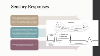
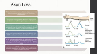
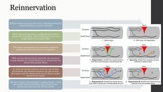
Nerve Conduction Study
(NCS)
Aditya Johan Romadhon, SST.FT, M.Fis
Introduction
The electrodiagnostic (EDX)
examination is an extension of
the clinical neurologic
examination that provides
important information about the
peripheral nervous system (PNS)
EDX testing of the PNS includes
nerve conduction studies (NCSs),
the needle electrode examination
(NEE)
The two major goals of the EDX
examination include lesion
localization (root, plexus, nerve,
neuromuscular junction [NMJ],
muscle) and lesion
characterization
(pathophysiologic features,
severity, and rate of progression)
It’s not only of diagnostic utility,
but it also contributes to patient
management and prognosis
The standard NCS assesses the larger,
more heavily myelinated nerve fibers of
the named sensory and motor
The thinly myelinated and unmyelinated
axons are not assessed by any of the
standard NCS techniques
With sensory NCSs, the recording
electrodes are positioned over the nerve
under study
Motor NCSs, they are positioned over the
muscle belly and tendon of the muscle
innervated by the nerve under study
The elicited responses are differentially
amplified and displayed on the monitor
Motor Responses
The stimulating electrodes are applied over the nerve
and the recording electrodes are applied over the
muscle
Thus, motor NCSs are orthodromically recorded (APs
traverse the nerve fibers physiological direction, from
proximal to distal)
The G1 (active) recording electrode is placed over the
muscle belly, where the motor nerve fibers enter the
muscle, whereas the G2 electrode is positioned over
the tendon
Stimulation is applied at two sites along the nerve,
yielding two separate motor responses
The response elicited by the more distal stimulation
site is termed the “distal motor response” and the one
elicited by the more proximal stimulation site is
referred to as the “proximal motor response.”
Measurements
Measurements taken from motor
responses include amplitude and
negative area under the curve
(AUC) for reflecting the number
of innervated muscle fibers
Latency, conduction velocity (CV)
for the fastest conducting nerve
fibers
And the duration of the negative
phase for the range of the CV
Sensory Responses
Sensory response represents the summation
of the individual sensory nerve fiber APs
evoked by the stimulating electrodes.
Because it is composed solely of nerve fiber
APs (i.e., no amplification effect), it is small
in size and measured in microvolts (ÎĽV)
The response is termed “antidromic” when
the recording electrodes are placed distal to
the stimulating electrodes (APs traverse the
nerve opposite to the physiologic direction)
and “orthodromic” when they are located
proximal to the stimulating electrodes
Measured parameters include latency, CV,
and negative phase duration
Fifty medical students of either sex from first year, aged between 17- 20 year, were recruited for this
nerve conduction study of radial nerve using surface electrode
Manifestation of NCS
• The resultant pathology is limited to either
demyelination (myelin disruption) or axon loss (axon
disruption-induced Wallerian degeneration)
• The resultant pathophysiologies from these two
pathologic insults include: DMCS (“demyelinating
conduction slowing”), DMCB (“demyelinating
conduction block”), and axonal conduction failure
Propagation of nerve’s action potential
Demyelination
Demyelination reduces CV in three ways
First, it increases the amount of exposed axonal
membrane, normally, only a small area of axon
membrane is exposed, it is at the nodes of Ranvier
that the AP is regenerated
Because demyelination increases the amount of
exposed axonal membrane, more charge must be
transferred to produce a given voltage change
Thus, the time required to regenerate the AP is
increased and, as a result, the CV is slowed
Second, which decreases the transmembrane
resistance to current flow, thereby increasing the
amount of current leaking across the membrane
This lessens the value of the length constant (defined
as the distance over which an AP decays by 63% of its
original value), which also decreases the CV
Third, when demyelination involves
the paranodal areas (demyelination
often begins paranodally and
becomes segmental), it exposes the
paranodally-located potassium
channels, which increases
potassium efflux, thereby favoring
nerve hyperpolarization
Since hyperpolarization opposes
depolarization, achievement of the
threshold for depolarization is
delayed and, again, CV is decreased
With larger amounts of myelin loss,
the APs are unable to traverse the
lesion, in this setting, the lesion
“blocks” the Aps from reaching their
target destinations
Axon Loss
Waller described the process of nerve degeneration that
follows axon disruption in the 1830s (Wallerian
degeneration)
The changes associated with Wallerian degeneration
involve the nerve fiber regions distant to the lesion
Proximally, there is retrograde degeneration for several
millimeters or so, as well as reactive changes in the cell
body neither of which has EDX manifestations
Unlike the proximal changes, the distal changes have
EDX manifestations (termed “conduction failure”)
in the acute axon disruption, the relationship between
the proximal and distal NCS responses is
indistinguishable from the one observed with a DMCB
lesion
As the process of Wallerian degeneration progresses,
the distal segments of the affected nerve fibers become
unable to conduct impulses
Wallerian degeneration
Thus, it is always best to perform the NCS on or after day 11
After 10-11 days, however, the sensory and motor NCS responses accurately reflect the underlying pathophysiology
After 7 days, the motor response uniformity identifies motor axon loss
Both pathophysiologies show a “conduction block” pattern on motor NCSs
During the first few days, it is not possible to differentiate an axon loss lesion from one that is due to DMCB
In general, the motor response begins to decrease around day 3 (Wallerian degeneration is becoming complete in some of the affected nerve fibers)
and reaches maximal decrement around day 6 or 7 (Wallerian degeneration is complete in all of the affected nerve fibers)
Wallerian degeneration is apparent on the motor NCS before the sensory NCSs
Reinnervation
Reinnervation occurs in two ways, collateral sprouting
and proximodistal axonal regrowth
With collateral sprouting, unaffected nerve fibers
sprout axon branches, each of which grows out to a
single denervated muscle fiber
The major requirement for successful collateral
sprouting is that the lesion be incomplete
With proximodistal axonal regrowth, the proximal
stump regrows in a distal direction to reinnervate the
denervated muscle fibers
In order for reinnervation to occur by way of this
mechanism, the distance between the site of axon
disruption and the denervated muscle fibers cannot
exceed 20-24 inches
The rate of axon advancement is approximately 1 inch
per month and muscle fibers can remain in the
denervated state for only 20-24 months
Nerve conduction study (ncs)

Nerve conduction study (ncs)

  • 1.
    Nerve Conduction Study (NCS) AdityaJohan Romadhon, SST.FT, M.Fis
  • 2.
    Introduction The electrodiagnostic (EDX) examinationis an extension of the clinical neurologic examination that provides important information about the peripheral nervous system (PNS) EDX testing of the PNS includes nerve conduction studies (NCSs), the needle electrode examination (NEE) The two major goals of the EDX examination include lesion localization (root, plexus, nerve, neuromuscular junction [NMJ], muscle) and lesion characterization (pathophysiologic features, severity, and rate of progression) It’s not only of diagnostic utility, but it also contributes to patient management and prognosis
  • 3.
    The standard NCSassesses the larger, more heavily myelinated nerve fibers of the named sensory and motor The thinly myelinated and unmyelinated axons are not assessed by any of the standard NCS techniques With sensory NCSs, the recording electrodes are positioned over the nerve under study Motor NCSs, they are positioned over the muscle belly and tendon of the muscle innervated by the nerve under study The elicited responses are differentially amplified and displayed on the monitor
  • 4.
    Motor Responses The stimulatingelectrodes are applied over the nerve and the recording electrodes are applied over the muscle Thus, motor NCSs are orthodromically recorded (APs traverse the nerve fibers physiological direction, from proximal to distal) The G1 (active) recording electrode is placed over the muscle belly, where the motor nerve fibers enter the muscle, whereas the G2 electrode is positioned over the tendon Stimulation is applied at two sites along the nerve, yielding two separate motor responses The response elicited by the more distal stimulation site is termed the “distal motor response” and the one elicited by the more proximal stimulation site is referred to as the “proximal motor response.”
  • 5.
    Measurements Measurements taken frommotor responses include amplitude and negative area under the curve (AUC) for reflecting the number of innervated muscle fibers Latency, conduction velocity (CV) for the fastest conducting nerve fibers And the duration of the negative phase for the range of the CV
  • 6.
    Sensory Responses Sensory responserepresents the summation of the individual sensory nerve fiber APs evoked by the stimulating electrodes. Because it is composed solely of nerve fiber APs (i.e., no amplification effect), it is small in size and measured in microvolts (μV) The response is termed “antidromic” when the recording electrodes are placed distal to the stimulating electrodes (APs traverse the nerve opposite to the physiologic direction) and “orthodromic” when they are located proximal to the stimulating electrodes Measured parameters include latency, CV, and negative phase duration
  • 7.
    Fifty medical studentsof either sex from first year, aged between 17- 20 year, were recruited for this nerve conduction study of radial nerve using surface electrode
  • 8.
    Manifestation of NCS •The resultant pathology is limited to either demyelination (myelin disruption) or axon loss (axon disruption-induced Wallerian degeneration) • The resultant pathophysiologies from these two pathologic insults include: DMCS (“demyelinating conduction slowing”), DMCB (“demyelinating conduction block”), and axonal conduction failure
  • 11.
  • 12.
    Demyelination Demyelination reduces CVin three ways First, it increases the amount of exposed axonal membrane, normally, only a small area of axon membrane is exposed, it is at the nodes of Ranvier that the AP is regenerated Because demyelination increases the amount of exposed axonal membrane, more charge must be transferred to produce a given voltage change Thus, the time required to regenerate the AP is increased and, as a result, the CV is slowed Second, which decreases the transmembrane resistance to current flow, thereby increasing the amount of current leaking across the membrane This lessens the value of the length constant (defined as the distance over which an AP decays by 63% of its original value), which also decreases the CV
  • 13.
    Third, when demyelinationinvolves the paranodal areas (demyelination often begins paranodally and becomes segmental), it exposes the paranodally-located potassium channels, which increases potassium efflux, thereby favoring nerve hyperpolarization Since hyperpolarization opposes depolarization, achievement of the threshold for depolarization is delayed and, again, CV is decreased With larger amounts of myelin loss, the APs are unable to traverse the lesion, in this setting, the lesion “blocks” the Aps from reaching their target destinations
  • 14.
    Axon Loss Waller describedthe process of nerve degeneration that follows axon disruption in the 1830s (Wallerian degeneration) The changes associated with Wallerian degeneration involve the nerve fiber regions distant to the lesion Proximally, there is retrograde degeneration for several millimeters or so, as well as reactive changes in the cell body neither of which has EDX manifestations Unlike the proximal changes, the distal changes have EDX manifestations (termed “conduction failure”) in the acute axon disruption, the relationship between the proximal and distal NCS responses is indistinguishable from the one observed with a DMCB lesion As the process of Wallerian degeneration progresses, the distal segments of the affected nerve fibers become unable to conduct impulses
  • 15.
    Wallerian degeneration Thus, itis always best to perform the NCS on or after day 11 After 10-11 days, however, the sensory and motor NCS responses accurately reflect the underlying pathophysiology After 7 days, the motor response uniformity identifies motor axon loss Both pathophysiologies show a “conduction block” pattern on motor NCSs During the first few days, it is not possible to differentiate an axon loss lesion from one that is due to DMCB In general, the motor response begins to decrease around day 3 (Wallerian degeneration is becoming complete in some of the affected nerve fibers) and reaches maximal decrement around day 6 or 7 (Wallerian degeneration is complete in all of the affected nerve fibers) Wallerian degeneration is apparent on the motor NCS before the sensory NCSs
  • 16.
    Reinnervation Reinnervation occurs intwo ways, collateral sprouting and proximodistal axonal regrowth With collateral sprouting, unaffected nerve fibers sprout axon branches, each of which grows out to a single denervated muscle fiber The major requirement for successful collateral sprouting is that the lesion be incomplete With proximodistal axonal regrowth, the proximal stump regrows in a distal direction to reinnervate the denervated muscle fibers In order for reinnervation to occur by way of this mechanism, the distance between the site of axon disruption and the denervated muscle fibers cannot exceed 20-24 inches The rate of axon advancement is approximately 1 inch per month and muscle fibers can remain in the denervated state for only 20-24 months