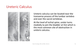
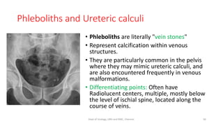
X-ray KUB
Dept of Urology
Govt Royapettah Hospital and Kilpauk Medical College
Chennai
Moderators:
Professors:
• Prof. Dr. G. Sivasankar, M.S., M.Ch.,
• Prof. Dr. A. Senthilvel, M.S., M.Ch.,
Asst Professors:
• Dr. J. Sivabalan, M.S., M.Ch.,
• Dr. R. Bhargavi, M.S., M.Ch.,
• Dr. S. Raju, M.S., M.Ch.,
• Dr. K. Muthurathinam, M.S., M.Ch.,
• Dr. D. Tamilselvan, M.S., M.Ch.,
• Dr. K. Senthilkumar, M.S., M.Ch.
Dept of Urology, GRH and KMC, Chennai. 2
History
William Roentgen, a German
Physicist, was the first to
discover X-radiation, which
earned him the Nobel Prize in
Physics in 1901.
3
Dept of Urology, GRH and KMC, Chennai.
X-Ray Word Origin
• X-ray was an unknown type of radiation when Roentgen first
observed them.
• In order to name the unknown radiation, he called it as X-radiation or
X-ray.
• Even though, later called them Roentgen rays, the name X-ray is the
one widely used.
4
Dept of Urology, GRH and KMC, Chennai.
Hand with Rings-Anna Bertha Ludwig
5
Dept of Urology, GRH and KMC, Chennai.
X-ray Spectrum
• X-rays are a type of electromagnetic waves.
• X-rays have wavelengths ranging from 0.01 to 10 nanometers, hence
their wavelengths are shorter than those of Ultraviolet rays and
typically longer than those of gamma rays.
6
Dept of Urology, GRH and KMC, Chennai.
X-ray Production
When the fast moving electrons
hit on an anode made of
Tungsten in case of medical use,
X-rays are produced.
7
Dept of Urology, GRH and KMC, Chennai.
Standard View
• The standard view is an Antero
posterior supine view.
• Patient lies supine.
• X‐ray tube is positioned overhead.
• X‐rays pass in the AP direction.
• Ideally, the patient is asked to hold
their breath (so that breathing
movement will not make the image
blurry) and the X‐ray is taken.
• Standard plate size 14”-17”
8
Dept of Urology, GRH and KMC, Chennai.
Other Views
To get spatial orientation of
opacities
• Oblique view
To look for free air and air fluid
levels
• Erect view
9
Dept of Urology, GRH and KMC, Chennai.
Systematic Reading
• Type of Radiograph-Area and View
• Patient identification and investigation timing
• Technical quality of the X-ray
• Ensure the sides with visible anatomy
• Look further into details
10
Dept of Urology, GRH and KMC, Chennai.
Assessing Technical Quality of X-ray
• Has everything been included? • The entire anatomy should be
included from level of T10 upto
2 cm below the lower margin of
symphysis pubis.
• Both the hemi‐diaphragms
should be visible, lower margin
of symphysis pubis should be
visible.
11
Dept of Urology, GRH and KMC, Chennai.
• Has the exposure been
adequate?
• Exposure is related to the
number of X‐rays that reach the
detector and make the image.
• If the exposure is adequate, the
spine should be clearly
visualized.
• In case of overexposure, soft
tissue shadows will be minimal.
Assessing Technical Quality of X-ray
12
Dept of Urology, GRH and KMC, Chennai.
Looking for Details-ABCDE of X ray
A
B
C
D
E
• Air in the Wrong Place
• Bowel
• Calcifications
• Disability of Bones
• Everything else
13
Dept of Urology, GRH and KMC, Chennai.
Looking for Details-ABCDE of X ray
A
B
C
D
E
• Air in the Wrong Place
• Bowel
• Calcifications
• Disability of Bones
• Everything else
• Pneumoperitoneum
• Pneumoretroperitoneum
• Pneumobilia
• Portal venous gas
14
Dept of Urology, GRH and KMC, Chennai.
Looking for Details-ABCDE of X ray
A
B
C
D
E
• Air in the Wrong Place
• Bowel
• Calcifications
• Disability of Bones
• Everything else
• Dilated small bowel
• Dilated large bowel
• Volvulus
• Fecal loading
15
Dept of Urology, GRH and KMC, Chennai.
Looking for Details-ABCDE of X ray
A
B
C
D
E
• Air in the Wrong Place
• Bowel
• Calcifications (Common)
• Disability of Bones
• Everything else
• Urinary tract stones
• Nephrocalcinosis
• Pancreatic calcification
• Calcified lymphnodes
• Phleboliths
16
Dept of Urology, GRH and KMC, Chennai.
Looking for Details-ABCDE of X ray
A
B
C
D
E
• Air in the Wrong Place
• Bowel
• Calcifications (Uncommon)
• Disability of Bones
• Everything else
• Calcified costal cartilage
• Adrenal calcification
• Abdominal aortic aneurysm
(AAA) calcification
• Calcified uterine fibroids
• Prostate calcification
• Abdominal aortic calcification
(normal calibre)
• Splenic artery calcification
17
Dept of Urology, GRH and KMC, Chennai.
Looking for Details-ABCDE of X ray
A
B
C
D
E
• Air in the Wrong Place
• Bowel
• Calcifications (Uncommon)
• Disability of Bones
• Everything else
• Sclerotic and lucent bone lesions
• Spine pathology
• Solid organ enlargement
• Fractures
18
Dept of Urology, GRH and KMC, Chennai.
Looking for Details-ABCDE of X ray
A
B
C
D
E
• Air in the Wrong Place
• Bowel
• Calcifications (Uncommon)
• Disability of Bones
• Everything else
• Medical and surgical objects
• Foreign bodies
• Lung bases
19
Dept of Urology, GRH and KMC, Chennai.
X-Ray KUB-Purpose
1. As a preliminary test in acute abdomen
2. To ensure the technical quality of the examination in case of
planned contrast usage.
3. To detect any calcific densities that could be urinary tract calculi.
4. To determine whether a contraindication to abdominal
compression exists.
5. To determine whether bowel preparation is adequate before
contrast study
6. To check the position of stents after urological procedures.
20
Dept of Urology, GRH and KMC, Chennai.
Abdominal Compression-Contraindications
• Spine deformity
• Abdominal distension (ileus, bowel obstruction, ascites)
• Acute abdomen findings
• Various types of urinary tract diversions and stomas
• Acute renal colic
• IVC filter (Relative contraindication)
21
Dept of Urology, GRH and KMC, Chennai.
Anatomical Landmarks
• Organ Outlines
• Major Muscles
• Properitoneal fat
• Perivesical fat plane
• Stomach and Bowel gas
22
Dept of Urology, GRH and KMC, Chennai.
Anatomical Landmarks-Organ Outline
Organ outlines are visible because
of the contrasting radiolucent
mesenteric or retroperitoneal fat
that surrounds the organs.
• Renal Outline- Bean shaped soft
tissue density, smooth outline,
extends from T12 to L2 on Lt
side & L1 to L3 on Rt side ( 3.5
vertebral bodies )
• Small kidney - if < 3 vertebrae
• Enlarged kidney - if > 4 vertebrae
23
Dept of Urology, GRH and KMC, Chennai.
Anatomical Landmarks-Major Muscles
• Psoas shadow – If absent
indicates presence of fluid in the
adjacent compartment. (In 20%
cases, normally absent on one
side).
• Quadratus lumborum- 1cm
lateral to Psoas shadow.
24
Dept of Urology, GRH and KMC, Chennai.
Anatomical Landmarks-Properitoneal fat
• It is the continuation of the
posterior paranephric space.
• It is sandwiched between
transversalis fascia and the
parietal peritoneum.
• Its medial interface marks the
lateral extent of the peritoneal
cavity
25
Dept of Urology, GRH and KMC, Chennai.
Anatomical Landmarks-Perivesical Fat line
26
Dept of Urology, GRH and KMC, Chennai.
Anatomical Landmarks- L4
• L4 vertebrae is the first lumbar
vertebrae with a transverse
process pointing upwards when
examined from above
downwards.
• Further labelling of vertebrae
should be done either above or
below L4.
27
Dept of Urology, GRH and KMC, Chennai.
Renal Calcifications-X ray KUB
Most common cause of renal calcifications is
Renal calculus disease
28
Dept of Urology, GRH and KMC, Chennai.
Renal calculus disease-
Identification with X ray
• Technical factors
• Patient factors
• Stone factors
29
Dept of Urology, GRH and KMC, Chennai.
Technical factors
• Peak kilovoltage (kVp )of xray beam of 60-70 kVp is optimal for
searching faintly opaque or small stones.
• At 80-90 kVp small stones may be invisible.
• For obese patients >70-80 kVp may be needed.
• 50 kVp is ideal for lucent uric acid stones.
30
Dept of Urology, GRH and KMC, Chennai.
Patient Factors
• Girth and obesity of the patient
• Extraurinary calcifications can obscure or mimic renal calculi.
• Calculi overlapped by bony structures
31
Dept of Urology, GRH and KMC, Chennai.
Stone Factors
• Size
• Composition
• Location
32
Dept of Urology, GRH and KMC, Chennai.
Stone Size
• Xray KUB can detect calcium containing calculi as small as 1 to 2 mm.
• Less opaque stones such as cystine are not visible until 3-4 mm in
size.
• Non opaque calculi like uric acid are generally need to be 1 cm or
larger to be faintly visible.
33
Dept of Urology, GRH and KMC, Chennai.
Stone Composition
• Two thirds of stones are composed of mixture of different crystals
• Single crystal (or Pure ) stones constitute only one third of all urinary
calculi.
• Pure stones can have characteristic radiographic appearances.
• Crystalline admixtures have varied appearances.
34
Dept of Urology, GRH and KMC, Chennai.
Stone composition and Shape
• Pure Calcium oxalate monohydrate stone- Homogeneously densely
opaque, smooth edges and may be dentate.
• Calcium oxalate monohydrate stone with >60% - Irregularly
marginated with heterogeneous, variegated radiopacity.
• Predominant Calcium oxalate dehydrate stone-Stippled and
spiculated with radial striations.
• Pure calcium phosphate stones- similar to Pure calcium oxalate stones
in appearance
• Triple phosphate stones- Laminated appearance
• Magnesium ammonium phosphate Hexahydrate(Struvite)-Staghorn
35
Dept of Urology, GRH and KMC, Chennai.
Stone composition and shape
• Pure uric acid stones- Non opaque on plain radiographs
• Uric acid stones with calcium coats- variably radiopaque on xray
• Cystine stones-moderately radiopaque, homogeneous ground glass
opacity, may be often large and multiple with staghorn configuration
• Staghorn in children or young adults should rise the suspicion of
Cystine stones.
• Matrix stones-non radiopaque on plain radiographs
• Xanthine stones- Radiolucent stones, but an admixture with calcium
may offer radiopacity
36
Dept of Urology, GRH and KMC, Chennai.
Calcium Oxalate monohydrate
37
Dept of Urology, GRH and KMC, Chennai.
Calcium Oxalate dihydrate
38
Dept of Urology, GRH and KMC, Chennai.
Cystine
39
Dept of Urology, GRH and KMC, Chennai.
Struvite
40
Dept of Urology, GRH and KMC, Chennai.
Triple phosphate
41
Dept of Urology, GRH and KMC, Chennai.
Other causes of Renal calcification
• Nephrolithiasis
• Nephrocalcinosis
• Dystrophic calcifications of vascular structures
• Neoplasms with calcifications
• Renal cysts with calcifications
• Calcified necrotic papilla
• Genitourinary tuberculosis
• Renal infarcts
• ESRD
42
Dept of Urology, GRH and KMC, Chennai.
GUTB with Renal calcifications
43
Dept of Urology, GRH and KMC, Chennai.
Medullary nephrocalcinosis
44
Dept of Urology, GRH and KMC, Chennai.
Renal artery calcification
45
Dept of Urology, GRH and KMC, Chennai.
Medullary sponge kidney
46
Dept of Urology, GRH and KMC, Chennai.
Mimicking Urinary Calcifications
• Calcified costal cartilage
• Gall stones (10% cases)
• Splenic calcification
• Splenic artery calcification
• Adrenal calcifications
• Pancreatic calcification
• Hepatic calcification
Clarification Method:
Take an Xray in Deep inspiration 47
Dept of Urology, GRH and KMC, Chennai.
Mimicking Urinary Calcifications
• Calcified costal cartilage
• Gall stones (10% cases)
• Splenic calcification
• Splenic artery calcification
• Adrenal calcifications
• Pancreatic calcification
• Hepatic calcification
Clarification Method: Number, faceted
shape, right post oblique films 48
Dept of Urology, GRH and KMC, Chennai.
Mimicking Urinary Calcifications
• Calcified costal cartilage
• Gall stones (10% cases)
• Splenic calcification
• Splenic artery calcification
• Adrenal calcifications
• Pancreatic calcification
• Hepatic calcification
49
Dept of Urology, GRH and KMC, Chennai.
Mimicking Urinary Calcifications
• Calcified costal cartilage
• Gall stones (10% cases)
• Splenic calcification
• Splenic artery calcification
• Adrenal calcifications
• Pancreatic calcification
• Hepatic calcification
50
Dept of Urology, GRH and KMC, Chennai.
Mimicking Urinary Calcifications
• Calcified costal cartilage
• Gall stones (10% cases)
• Splenic calcification
• Splenic artery calcification
• Adrenal calcifications
• Pancreatic calcification
• Hepatic calcification
51
Dept of Urology, GRH and KMC, Chennai.
Mimicking Urinary Calcifications
• Calcified costal cartilage
• Gall stones (10% cases)
• Splenic calcification
• Splenic artery calcification
• Adrenal calcifications
• Pancreatic calcification
• Hepatic calcification
52
Dept of Urology, GRH and KMC, Chennai.
Mimicking Urinary Calcifications
• Calcified costal cartilage
• Gall stones (10% cases)
• Splenic calcification
• Splenic artery calcification
• Adrenal calcifications
• Pancreatic calcification
• Hepatic calcification
53
Dept of Urology, GRH and KMC, Chennai.
Ureteric Calculus
Ureteric calculus can be located near the
transverse process of the lumbar vertebra
and over the sacral vertebrae.
At the level of ischial spine, ureter turns
medially to join the bladder at VUJ which is
the most common site of obstruction in
ureteric calculus.
54
Dept of Urology, GRH and KMC, Chennai.
Mimicking Ureteric Calculi
• Phleboliths
• Calcified costal cartilage
• Gall stones
• Enteroliths
• Calcified mesenteric lymph node
• Pancreatic calcifications
• Sacral bone islands
• Aortic or other arterial calcifications
• Fallopian tube occlusion rings
55
Dept of Urology, GRH and KMC, Chennai.
Phleboliths and Ureteric calculi
• Phleboliths are literally "vein stones"
• Represent calcification within venous
structures.
• They are particularly common in the pelvis
where they may mimic ureteric calculi, and
are also encountered frequently in venous
malformations.
• Differentiating points: Often have
Radiolucent centers, multiple, mostly below
the level of ischial spine, located along the
course of veins.
56
Dept of Urology, GRH and KMC, Chennai.
Appendicolith
57
Dept of Urology, GRH and KMC, Chennai.
Fallopian tube occlusion rings
58
Dept of Urology, GRH and KMC, Chennai.
Bladder Calculi
• Size ranges from 1-2 mm to large calculi of many cms
• May be solitary or multiple in number
• Range from densely radiopaque stones to faintly radiopaque stones
depending on the composition.
• May have variety of shapes
59
Dept of Urology, GRH and KMC, Chennai.
Bladder Calculi
60
Dept of Urology, GRH and KMC, Chennai.
Mimicking Bladder Calculi
• Feces in rectum (as low density bladder calculi)
• Retained barium in rectum after contrast study
• Opaque rectal suppositories
• Rectal foreign bodies
• Calcification in rectal tumour
• Calcification in bladder tumour
• Foreign bodies in the vagina
• Primary/secondary vaginal calculi (mostly struvite)
• Calcified fibroids or ovarian tumours
• Prostatic calcifications
61
Dept of Urology, GRH and KMC, Chennai.
Rectal Fecolith
62
Dept of Urology, GRH and KMC, Chennai.
Bladder and Vaginal Calculi
63
Dept of Urology, GRH and KMC, Chennai.
Prostatic Calcification
64
Dept of Urology, GRH and KMC, Chennai.
Bladder Wall calcification
• Schistosomiasis –Common
• Tuberculosis –Rare
• Alkaline encrysting cystitis – After radiotherapy in the presence of
necrotic or ischemic bladder tissue and alkaline urine produced by
Proteus infection.
• After cyclophosphamide or mitomycin treatment of bladder
• Primary amyloidosis –submucosal amyloid deposition
• Prune Belly Syndrome-Dystrophic calcification of dome
65
Dept of Urology, GRH and KMC, Chennai.
Bladder wall Calcification-Schistosomiasis
66
Dept of Urology, GRH and KMC, Chennai.
Alkaline Encrusting Cystitis
67
Dept of Urology, GRH and KMC, Chennai.
Prostatic Calcifications
• Primary prostatic
calcification
• Secondary prostatic
calcification
68
Dept of Urology, GRH and KMC, Chennai.
Prostatic Calcifications
• Primary prostatic
calcification
• Normally present in 30% of
subjects.
• Usually small (1-5mm) and
multiple
• Due to calcium deposition on
corpora amylacea
• Usually calcium phosphate
69
Dept of Urology, GRH and KMC, Chennai.
Prostatic Calcifications
Secondary prostatic
calcification
• In abnormal prostates with
benign hyperplasia,
obstruction and stasis,
infection or carcinoma or
after radiotherapy.
• Secondary prostatic
calcification due to
obstruction may measure
upto 10 mm or more.
70
Dept of Urology, GRH and KMC, Chennai.
Primary prostatic calculi
• Small multiple calcifications
• Due to calcification of corpora
amylacea
71
Dept of Urology, GRH and KMC, Chennai.
Secondary Prostatic Calcification
• Trilobed secondary prostatic
calcification in voiding
dysfunction due to neurogenic
bladder
72
Dept of Urology, GRH and KMC, Chennai.
Secondary Prostatic Calcification
• Dystrophic calcification of prostate
in Genitourinary tuberculosis
73
Dept of Urology, GRH and KMC, Chennai.
Secondary Prostatic Calcification
• Secondary prostatic calcification
due to urethro prostatic urinary
reflux
74
Dept of Urology, GRH and KMC, Chennai.
Urethral Calculi
• Native calculi – Rare and are formed within the urethra (Dumbell
Shaped)
• Migrant calculi- Arise in the kidney or bladder and then descend into
the urethra (Small stones struck at proximal to narrowing)
75
Dept of Urology, GRH and KMC, Chennai.
Native Calculi
• Dumbell shaped with portion of
calculus inside the bladder.
• Associated urethral stricture may
be present.
• May develop in prostate bed after
prostatectomy, particularly Open.
76
Dept of Urology, GRH and KMC, Chennai.
Migrant Calculi
• After lithotripsy, patient
underwent steinstrasse of urethra
due to urethral stricture in this
case.
77
Dept of Urology, GRH and KMC, Chennai.
Other Indicental
Findings
Vas deferens calcification
• May be due to degenerative
changes in diabetes or with
aging
• Or due to chronic infection
• Tramline calcification of ampulla
is typical
78
Dept of Urology, GRH and KMC, Chennai.
Other Indicental
Findings
Seminal vesicle calcification
• Due to chronic infection of
tuberculosis or schistosomiasis.
• Patchy, coarse and asymetrical
in TB
• Symmetrical in schistosomiasis
• Rare causes – Old age, urinary
reflux
79
Dept of Urology, GRH and KMC, Chennai.
Thank you
80
Dept of Urology, GRH and KMC, Chennai.

X RAY KUB 1

  • 1.
    X-ray KUB Dept ofUrology Govt Royapettah Hospital and Kilpauk Medical College Chennai
  • 2.
    Moderators: Professors: • Prof. Dr.G. Sivasankar, M.S., M.Ch., • Prof. Dr. A. Senthilvel, M.S., M.Ch., Asst Professors: • Dr. J. Sivabalan, M.S., M.Ch., • Dr. R. Bhargavi, M.S., M.Ch., • Dr. S. Raju, M.S., M.Ch., • Dr. K. Muthurathinam, M.S., M.Ch., • Dr. D. Tamilselvan, M.S., M.Ch., • Dr. K. Senthilkumar, M.S., M.Ch. Dept of Urology, GRH and KMC, Chennai. 2
  • 3.
    History William Roentgen, aGerman Physicist, was the first to discover X-radiation, which earned him the Nobel Prize in Physics in 1901. 3 Dept of Urology, GRH and KMC, Chennai.
  • 4.
    X-Ray Word Origin •X-ray was an unknown type of radiation when Roentgen first observed them. • In order to name the unknown radiation, he called it as X-radiation or X-ray. • Even though, later called them Roentgen rays, the name X-ray is the one widely used. 4 Dept of Urology, GRH and KMC, Chennai.
  • 5.
    Hand with Rings-AnnaBertha Ludwig 5 Dept of Urology, GRH and KMC, Chennai.
  • 6.
    X-ray Spectrum • X-raysare a type of electromagnetic waves. • X-rays have wavelengths ranging from 0.01 to 10 nanometers, hence their wavelengths are shorter than those of Ultraviolet rays and typically longer than those of gamma rays. 6 Dept of Urology, GRH and KMC, Chennai.
  • 7.
    X-ray Production When thefast moving electrons hit on an anode made of Tungsten in case of medical use, X-rays are produced. 7 Dept of Urology, GRH and KMC, Chennai.
  • 8.
    Standard View • Thestandard view is an Antero posterior supine view. • Patient lies supine. • X‐ray tube is positioned overhead. • X‐rays pass in the AP direction. • Ideally, the patient is asked to hold their breath (so that breathing movement will not make the image blurry) and the X‐ray is taken. • Standard plate size 14”-17” 8 Dept of Urology, GRH and KMC, Chennai.
  • 9.
    Other Views To getspatial orientation of opacities • Oblique view To look for free air and air fluid levels • Erect view 9 Dept of Urology, GRH and KMC, Chennai.
  • 10.
    Systematic Reading • Typeof Radiograph-Area and View • Patient identification and investigation timing • Technical quality of the X-ray • Ensure the sides with visible anatomy • Look further into details 10 Dept of Urology, GRH and KMC, Chennai.
  • 11.
    Assessing Technical Qualityof X-ray • Has everything been included? • The entire anatomy should be included from level of T10 upto 2 cm below the lower margin of symphysis pubis. • Both the hemi‐diaphragms should be visible, lower margin of symphysis pubis should be visible. 11 Dept of Urology, GRH and KMC, Chennai.
  • 12.
    • Has theexposure been adequate? • Exposure is related to the number of X‐rays that reach the detector and make the image. • If the exposure is adequate, the spine should be clearly visualized. • In case of overexposure, soft tissue shadows will be minimal. Assessing Technical Quality of X-ray 12 Dept of Urology, GRH and KMC, Chennai.
  • 13.
    Looking for Details-ABCDEof X ray A B C D E • Air in the Wrong Place • Bowel • Calcifications • Disability of Bones • Everything else 13 Dept of Urology, GRH and KMC, Chennai.
  • 14.
    Looking for Details-ABCDEof X ray A B C D E • Air in the Wrong Place • Bowel • Calcifications • Disability of Bones • Everything else • Pneumoperitoneum • Pneumoretroperitoneum • Pneumobilia • Portal venous gas 14 Dept of Urology, GRH and KMC, Chennai.
  • 15.
    Looking for Details-ABCDEof X ray A B C D E • Air in the Wrong Place • Bowel • Calcifications • Disability of Bones • Everything else • Dilated small bowel • Dilated large bowel • Volvulus • Fecal loading 15 Dept of Urology, GRH and KMC, Chennai.
  • 16.
    Looking for Details-ABCDEof X ray A B C D E • Air in the Wrong Place • Bowel • Calcifications (Common) • Disability of Bones • Everything else • Urinary tract stones • Nephrocalcinosis • Pancreatic calcification • Calcified lymphnodes • Phleboliths 16 Dept of Urology, GRH and KMC, Chennai.
  • 17.
    Looking for Details-ABCDEof X ray A B C D E • Air in the Wrong Place • Bowel • Calcifications (Uncommon) • Disability of Bones • Everything else • Calcified costal cartilage • Adrenal calcification • Abdominal aortic aneurysm (AAA) calcification • Calcified uterine fibroids • Prostate calcification • Abdominal aortic calcification (normal calibre) • Splenic artery calcification 17 Dept of Urology, GRH and KMC, Chennai.
  • 18.
    Looking for Details-ABCDEof X ray A B C D E • Air in the Wrong Place • Bowel • Calcifications (Uncommon) • Disability of Bones • Everything else • Sclerotic and lucent bone lesions • Spine pathology • Solid organ enlargement • Fractures 18 Dept of Urology, GRH and KMC, Chennai.
  • 19.
    Looking for Details-ABCDEof X ray A B C D E • Air in the Wrong Place • Bowel • Calcifications (Uncommon) • Disability of Bones • Everything else • Medical and surgical objects • Foreign bodies • Lung bases 19 Dept of Urology, GRH and KMC, Chennai.
  • 20.
    X-Ray KUB-Purpose 1. Asa preliminary test in acute abdomen 2. To ensure the technical quality of the examination in case of planned contrast usage. 3. To detect any calcific densities that could be urinary tract calculi. 4. To determine whether a contraindication to abdominal compression exists. 5. To determine whether bowel preparation is adequate before contrast study 6. To check the position of stents after urological procedures. 20 Dept of Urology, GRH and KMC, Chennai.
  • 21.
    Abdominal Compression-Contraindications • Spinedeformity • Abdominal distension (ileus, bowel obstruction, ascites) • Acute abdomen findings • Various types of urinary tract diversions and stomas • Acute renal colic • IVC filter (Relative contraindication) 21 Dept of Urology, GRH and KMC, Chennai.
  • 22.
    Anatomical Landmarks • OrganOutlines • Major Muscles • Properitoneal fat • Perivesical fat plane • Stomach and Bowel gas 22 Dept of Urology, GRH and KMC, Chennai.
  • 23.
    Anatomical Landmarks-Organ Outline Organoutlines are visible because of the contrasting radiolucent mesenteric or retroperitoneal fat that surrounds the organs. • Renal Outline- Bean shaped soft tissue density, smooth outline, extends from T12 to L2 on Lt side & L1 to L3 on Rt side ( 3.5 vertebral bodies ) • Small kidney - if < 3 vertebrae • Enlarged kidney - if > 4 vertebrae 23 Dept of Urology, GRH and KMC, Chennai.
  • 24.
    Anatomical Landmarks-Major Muscles •Psoas shadow – If absent indicates presence of fluid in the adjacent compartment. (In 20% cases, normally absent on one side). • Quadratus lumborum- 1cm lateral to Psoas shadow. 24 Dept of Urology, GRH and KMC, Chennai.
  • 25.
    Anatomical Landmarks-Properitoneal fat •It is the continuation of the posterior paranephric space. • It is sandwiched between transversalis fascia and the parietal peritoneum. • Its medial interface marks the lateral extent of the peritoneal cavity 25 Dept of Urology, GRH and KMC, Chennai.
  • 26.
    Anatomical Landmarks-Perivesical Fatline 26 Dept of Urology, GRH and KMC, Chennai.
  • 27.
    Anatomical Landmarks- L4 •L4 vertebrae is the first lumbar vertebrae with a transverse process pointing upwards when examined from above downwards. • Further labelling of vertebrae should be done either above or below L4. 27 Dept of Urology, GRH and KMC, Chennai.
  • 28.
    Renal Calcifications-X rayKUB Most common cause of renal calcifications is Renal calculus disease 28 Dept of Urology, GRH and KMC, Chennai.
  • 29.
    Renal calculus disease- Identificationwith X ray • Technical factors • Patient factors • Stone factors 29 Dept of Urology, GRH and KMC, Chennai.
  • 30.
    Technical factors • Peakkilovoltage (kVp )of xray beam of 60-70 kVp is optimal for searching faintly opaque or small stones. • At 80-90 kVp small stones may be invisible. • For obese patients >70-80 kVp may be needed. • 50 kVp is ideal for lucent uric acid stones. 30 Dept of Urology, GRH and KMC, Chennai.
  • 31.
    Patient Factors • Girthand obesity of the patient • Extraurinary calcifications can obscure or mimic renal calculi. • Calculi overlapped by bony structures 31 Dept of Urology, GRH and KMC, Chennai.
  • 32.
    Stone Factors • Size •Composition • Location 32 Dept of Urology, GRH and KMC, Chennai.
  • 33.
    Stone Size • XrayKUB can detect calcium containing calculi as small as 1 to 2 mm. • Less opaque stones such as cystine are not visible until 3-4 mm in size. • Non opaque calculi like uric acid are generally need to be 1 cm or larger to be faintly visible. 33 Dept of Urology, GRH and KMC, Chennai.
  • 34.
    Stone Composition • Twothirds of stones are composed of mixture of different crystals • Single crystal (or Pure ) stones constitute only one third of all urinary calculi. • Pure stones can have characteristic radiographic appearances. • Crystalline admixtures have varied appearances. 34 Dept of Urology, GRH and KMC, Chennai.
  • 35.
    Stone composition andShape • Pure Calcium oxalate monohydrate stone- Homogeneously densely opaque, smooth edges and may be dentate. • Calcium oxalate monohydrate stone with >60% - Irregularly marginated with heterogeneous, variegated radiopacity. • Predominant Calcium oxalate dehydrate stone-Stippled and spiculated with radial striations. • Pure calcium phosphate stones- similar to Pure calcium oxalate stones in appearance • Triple phosphate stones- Laminated appearance • Magnesium ammonium phosphate Hexahydrate(Struvite)-Staghorn 35 Dept of Urology, GRH and KMC, Chennai.
  • 36.
    Stone composition andshape • Pure uric acid stones- Non opaque on plain radiographs • Uric acid stones with calcium coats- variably radiopaque on xray • Cystine stones-moderately radiopaque, homogeneous ground glass opacity, may be often large and multiple with staghorn configuration • Staghorn in children or young adults should rise the suspicion of Cystine stones. • Matrix stones-non radiopaque on plain radiographs • Xanthine stones- Radiolucent stones, but an admixture with calcium may offer radiopacity 36 Dept of Urology, GRH and KMC, Chennai.
  • 37.
    Calcium Oxalate monohydrate 37 Deptof Urology, GRH and KMC, Chennai.
  • 38.
    Calcium Oxalate dihydrate 38 Deptof Urology, GRH and KMC, Chennai.
  • 39.
    Cystine 39 Dept of Urology,GRH and KMC, Chennai.
  • 40.
    Struvite 40 Dept of Urology,GRH and KMC, Chennai.
  • 41.
    Triple phosphate 41 Dept ofUrology, GRH and KMC, Chennai.
  • 42.
    Other causes ofRenal calcification • Nephrolithiasis • Nephrocalcinosis • Dystrophic calcifications of vascular structures • Neoplasms with calcifications • Renal cysts with calcifications • Calcified necrotic papilla • Genitourinary tuberculosis • Renal infarcts • ESRD 42 Dept of Urology, GRH and KMC, Chennai.
  • 43.
    GUTB with Renalcalcifications 43 Dept of Urology, GRH and KMC, Chennai.
  • 44.
    Medullary nephrocalcinosis 44 Dept ofUrology, GRH and KMC, Chennai.
  • 45.
    Renal artery calcification 45 Deptof Urology, GRH and KMC, Chennai.
  • 46.
    Medullary sponge kidney 46 Deptof Urology, GRH and KMC, Chennai.
  • 47.
    Mimicking Urinary Calcifications •Calcified costal cartilage • Gall stones (10% cases) • Splenic calcification • Splenic artery calcification • Adrenal calcifications • Pancreatic calcification • Hepatic calcification Clarification Method: Take an Xray in Deep inspiration 47 Dept of Urology, GRH and KMC, Chennai.
  • 48.
    Mimicking Urinary Calcifications •Calcified costal cartilage • Gall stones (10% cases) • Splenic calcification • Splenic artery calcification • Adrenal calcifications • Pancreatic calcification • Hepatic calcification Clarification Method: Number, faceted shape, right post oblique films 48 Dept of Urology, GRH and KMC, Chennai.
  • 49.
    Mimicking Urinary Calcifications •Calcified costal cartilage • Gall stones (10% cases) • Splenic calcification • Splenic artery calcification • Adrenal calcifications • Pancreatic calcification • Hepatic calcification 49 Dept of Urology, GRH and KMC, Chennai.
  • 50.
    Mimicking Urinary Calcifications •Calcified costal cartilage • Gall stones (10% cases) • Splenic calcification • Splenic artery calcification • Adrenal calcifications • Pancreatic calcification • Hepatic calcification 50 Dept of Urology, GRH and KMC, Chennai.
  • 51.
    Mimicking Urinary Calcifications •Calcified costal cartilage • Gall stones (10% cases) • Splenic calcification • Splenic artery calcification • Adrenal calcifications • Pancreatic calcification • Hepatic calcification 51 Dept of Urology, GRH and KMC, Chennai.
  • 52.
    Mimicking Urinary Calcifications •Calcified costal cartilage • Gall stones (10% cases) • Splenic calcification • Splenic artery calcification • Adrenal calcifications • Pancreatic calcification • Hepatic calcification 52 Dept of Urology, GRH and KMC, Chennai.
  • 53.
    Mimicking Urinary Calcifications •Calcified costal cartilage • Gall stones (10% cases) • Splenic calcification • Splenic artery calcification • Adrenal calcifications • Pancreatic calcification • Hepatic calcification 53 Dept of Urology, GRH and KMC, Chennai.
  • 54.
    Ureteric Calculus Ureteric calculuscan be located near the transverse process of the lumbar vertebra and over the sacral vertebrae. At the level of ischial spine, ureter turns medially to join the bladder at VUJ which is the most common site of obstruction in ureteric calculus. 54 Dept of Urology, GRH and KMC, Chennai.
  • 55.
    Mimicking Ureteric Calculi •Phleboliths • Calcified costal cartilage • Gall stones • Enteroliths • Calcified mesenteric lymph node • Pancreatic calcifications • Sacral bone islands • Aortic or other arterial calcifications • Fallopian tube occlusion rings 55 Dept of Urology, GRH and KMC, Chennai.
  • 56.
    Phleboliths and Uretericcalculi • Phleboliths are literally "vein stones" • Represent calcification within venous structures. • They are particularly common in the pelvis where they may mimic ureteric calculi, and are also encountered frequently in venous malformations. • Differentiating points: Often have Radiolucent centers, multiple, mostly below the level of ischial spine, located along the course of veins. 56 Dept of Urology, GRH and KMC, Chennai.
  • 57.
    Appendicolith 57 Dept of Urology,GRH and KMC, Chennai.
  • 58.
    Fallopian tube occlusionrings 58 Dept of Urology, GRH and KMC, Chennai.
  • 59.
    Bladder Calculi • Sizeranges from 1-2 mm to large calculi of many cms • May be solitary or multiple in number • Range from densely radiopaque stones to faintly radiopaque stones depending on the composition. • May have variety of shapes 59 Dept of Urology, GRH and KMC, Chennai.
  • 60.
    Bladder Calculi 60 Dept ofUrology, GRH and KMC, Chennai.
  • 61.
    Mimicking Bladder Calculi •Feces in rectum (as low density bladder calculi) • Retained barium in rectum after contrast study • Opaque rectal suppositories • Rectal foreign bodies • Calcification in rectal tumour • Calcification in bladder tumour • Foreign bodies in the vagina • Primary/secondary vaginal calculi (mostly struvite) • Calcified fibroids or ovarian tumours • Prostatic calcifications 61 Dept of Urology, GRH and KMC, Chennai.
  • 62.
    Rectal Fecolith 62 Dept ofUrology, GRH and KMC, Chennai.
  • 63.
    Bladder and VaginalCalculi 63 Dept of Urology, GRH and KMC, Chennai.
  • 64.
    Prostatic Calcification 64 Dept ofUrology, GRH and KMC, Chennai.
  • 65.
    Bladder Wall calcification •Schistosomiasis –Common • Tuberculosis –Rare • Alkaline encrysting cystitis – After radiotherapy in the presence of necrotic or ischemic bladder tissue and alkaline urine produced by Proteus infection. • After cyclophosphamide or mitomycin treatment of bladder • Primary amyloidosis –submucosal amyloid deposition • Prune Belly Syndrome-Dystrophic calcification of dome 65 Dept of Urology, GRH and KMC, Chennai.
  • 66.
    Bladder wall Calcification-Schistosomiasis 66 Deptof Urology, GRH and KMC, Chennai.
  • 67.
    Alkaline Encrusting Cystitis 67 Deptof Urology, GRH and KMC, Chennai.
  • 68.
    Prostatic Calcifications • Primaryprostatic calcification • Secondary prostatic calcification 68 Dept of Urology, GRH and KMC, Chennai.
  • 69.
    Prostatic Calcifications • Primaryprostatic calcification • Normally present in 30% of subjects. • Usually small (1-5mm) and multiple • Due to calcium deposition on corpora amylacea • Usually calcium phosphate 69 Dept of Urology, GRH and KMC, Chennai.
  • 70.
    Prostatic Calcifications Secondary prostatic calcification •In abnormal prostates with benign hyperplasia, obstruction and stasis, infection or carcinoma or after radiotherapy. • Secondary prostatic calcification due to obstruction may measure upto 10 mm or more. 70 Dept of Urology, GRH and KMC, Chennai.
  • 71.
    Primary prostatic calculi •Small multiple calcifications • Due to calcification of corpora amylacea 71 Dept of Urology, GRH and KMC, Chennai.
  • 72.
    Secondary Prostatic Calcification •Trilobed secondary prostatic calcification in voiding dysfunction due to neurogenic bladder 72 Dept of Urology, GRH and KMC, Chennai.
  • 73.
    Secondary Prostatic Calcification •Dystrophic calcification of prostate in Genitourinary tuberculosis 73 Dept of Urology, GRH and KMC, Chennai.
  • 74.
    Secondary Prostatic Calcification •Secondary prostatic calcification due to urethro prostatic urinary reflux 74 Dept of Urology, GRH and KMC, Chennai.
  • 75.
    Urethral Calculi • Nativecalculi – Rare and are formed within the urethra (Dumbell Shaped) • Migrant calculi- Arise in the kidney or bladder and then descend into the urethra (Small stones struck at proximal to narrowing) 75 Dept of Urology, GRH and KMC, Chennai.
  • 76.
    Native Calculi • Dumbellshaped with portion of calculus inside the bladder. • Associated urethral stricture may be present. • May develop in prostate bed after prostatectomy, particularly Open. 76 Dept of Urology, GRH and KMC, Chennai.
  • 77.
    Migrant Calculi • Afterlithotripsy, patient underwent steinstrasse of urethra due to urethral stricture in this case. 77 Dept of Urology, GRH and KMC, Chennai.
  • 78.
    Other Indicental Findings Vas deferenscalcification • May be due to degenerative changes in diabetes or with aging • Or due to chronic infection • Tramline calcification of ampulla is typical 78 Dept of Urology, GRH and KMC, Chennai.
  • 79.
    Other Indicental Findings Seminal vesiclecalcification • Due to chronic infection of tuberculosis or schistosomiasis. • Patchy, coarse and asymetrical in TB • Symmetrical in schistosomiasis • Rare causes – Old age, urinary reflux 79 Dept of Urology, GRH and KMC, Chennai.
  • 80.
    Thank you 80 Dept ofUrology, GRH and KMC, Chennai.