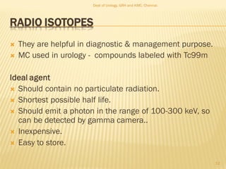
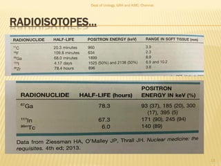
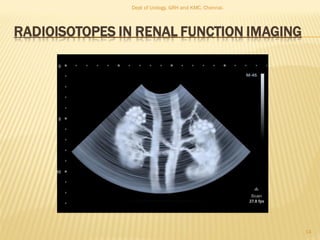
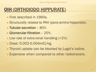
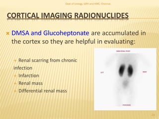
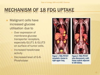
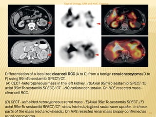
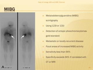
This document discusses the role of radioisotopes in urological diagnosis and management. It provides background on the history of radioisotopes and cyclotrons. It describes common radioisotopes used in urology like technetium-99m, iodine-131, gallium-67 and indium-111. The document discusses how different radioisotopes can be used to image renal function and structure, renal infections, and urological cancers like kidney cancer, bladder cancer and prostate cancer. Key applications of different radioisotopes are summarized.
Management of renal cell carcinoma - presented at Asian Oncology Summit 2013Siewhong Ho
Dr Ho lectured at the Asian Oncology Summit 2013 in Bangkok on the surgical opinion on management of renal cell carcinoma. He presented to a varied audience of medical oncologist, radiation oncologist, urologists, researchers, para clinical staff and nurses. The most interesting aspect of the lecture was on the role of urologists in management of Stage 4 kidney cancer in the era of 'targeted therapy'. The role of cytoreductive nephrectomy was reviewed potential future developments in this area was discussed
A prostate biopsy is a procedure used to obtain tissue samples from the prostate gland in order to detect cancer. The biopsy is best performed with a spring-driven needle core biopsy device (or biopsy gun)
No prostate-specific antigen (PSA) value can establish with absolute certainty whether a patient has prostate cancer. Thus, the decision to proceed with prostate biopsy must be individualized. Urinary biomarkers have been shown to be useful in identifying patients at risk for prostate cancer prior to the initial biopsy. [1]
Even more difficult is the decision to perform a repeat biopsy. Patients with atypical small acinar neoplasia have an absolute indication for repeat biopsy soon after the initial biopsy. However, patients with focal high-grade prostatic intraepithelial neoplasia (HGPIN) do not need to undergo automatic biopsy, because they are not at significantly higher risk for prostate cancer. By contrast, patients with multifocal HGPIN are at significant risk for prostate cancer and should undergo delayed interval biopsy every 3 years as long as they remain healthy. Patients who have persistently abnormal or rising PSA levels or very low percentages of free PSA (< 13%) are at some risk for harboring unrecognized prostate cancer and thus should be considered for repeat biopsy.
Contraindications for prostate biopsy include the surgical absence of a rectum or the presence of a rectal fistula.
The complications encountered after TRUS biopsy are commonly minor and self-limited, including mild hematuria, hematospermia, and transient rectal bleeding. Urinary tract infection is another frequently noted complication of prostate biopsy.
Trimodal Management of Locally Invasive Urinary Bladder CancerNainaAnon
To evaluate the response of the modern bladder-preservation treatment modality; Trimodal Therapy (TMT) in Muscle-Invasive Bladder Cancer (MIBC). Aiming at bladder preservation in MIBC, TMT was to offer a quality- of-life advantage and avoid potential morbidity and mortality of Radical Cystectomy (RC) without compromising oncologic outcomes.
New Directions in Targeted Therapeutic Approaches for Older Adults With Mantl...i3 Health
i3 Health is pleased to make the speaker slides from this activity available for use as a non-accredited self-study or teaching resource.
This slide deck presented by Dr. Kami Maddocks, Professor-Clinical in the Division of Hematology and
Associate Division Director for Ambulatory Operations
The Ohio State University Comprehensive Cancer Center, will provide insight into new directions in targeted therapeutic approaches for older adults with mantle cell lymphoma.
STATEMENT OF NEED
Mantle cell lymphoma (MCL) is a rare, aggressive B-cell non-Hodgkin lymphoma (NHL) accounting for 5% to 7% of all lymphomas. Its prognosis ranges from indolent disease that does not require treatment for years to very aggressive disease, which is associated with poor survival (Silkenstedt et al, 2021). Typically, MCL is diagnosed at advanced stage and in older patients who cannot tolerate intensive therapy (NCCN, 2022). Although recent advances have slightly increased remission rates, recurrence and relapse remain very common, leading to a median overall survival between 3 and 6 years (LLS, 2021). Though there are several effective options, progress is still needed towards establishing an accepted frontline approach for MCL (Castellino et al, 2022). Treatment selection and management of MCL are complicated by the heterogeneity of prognosis, advanced age and comorbidities of patients, and lack of an established standard approach for treatment, making it vital that clinicians be familiar with the latest research and advances in this area. In this activity chaired by Michael Wang, MD, Professor in the Department of Lymphoma & Myeloma at MD Anderson Cancer Center, expert faculty will discuss prognostic factors informing treatment, the promising results of recent trials in new therapeutic approaches, and the implications of treatment resistance in therapeutic selection for MCL.
Target Audience
Hematology/oncology fellows, attending faculty, and other health care professionals involved in the treatment of patients with mantle cell lymphoma (MCL).
Learning Objectives
1.) Identify clinical and biological prognostic factors that can guide treatment decision making for older adults with MCL
2.) Evaluate emerging data on targeted therapeutic approaches for treatment-naive and relapsed/refractory MCL and their applicability to older adults
3.) Assess mechanisms of resistance to targeted therapies for MCL and their implications for treatment selection
Acute scrotum is a general term referring to an emergency condition affecting the contents or the wall of the scrotum.
There are a number of conditions that present acutely, predominantly with pain and/or swelling
A careful and detailed history and examination, and in some cases, investigations allow differentiation between these diagnoses. A prompt diagnosis is essential as the patient may require urgent surgical intervention
Testicular torsion refers to twisting of the spermatic cord, causing ischaemia of the testicle.
Testicular torsion results from inadequate fixation of the testis to the tunica vaginalis producing ischemia from reduced arterial inflow and venous outflow obstruction.
The prevalence of testicular torsion in adult patients hospitalized with acute scrotal pain is approximately 25 to 50 percent
Ethanol (CH3CH2OH), or beverage alcohol, is a two-carbon alcohol
that is rapidly distributed in the body and brain. Ethanol alters many
neurochemical systems and has rewarding and addictive properties. It
is the oldest recreational drug and likely contributes to more morbidity,
mortality, and public health costs than all illicit drugs combined. The
5th edition of the Diagnostic and Statistical Manual of Mental Disorders
(DSM-5) integrates alcohol abuse and alcohol dependence into a single
disorder called alcohol use disorder (AUD), with mild, moderate,
and severe subclassifications (American Psychiatric Association, 2013).
In the DSM-5, all types of substance abuse and dependence have been
combined into a single substance use disorder (SUD) on a continuum
from mild to severe. A diagnosis of AUD requires that at least two of
the 11 DSM-5 behaviors be present within a 12-month period (mild
AUD: 2–3 criteria; moderate AUD: 4–5 criteria; severe AUD: 6–11 criteria).
The four main behavioral effects of AUD are impaired control over
drinking, negative social consequences, risky use, and altered physiological
effects (tolerance, withdrawal). This chapter presents an overview
of the prevalence and harmful consequences of AUD in the U.S.,
the systemic nature of the disease, neurocircuitry and stages of AUD,
comorbidities, fetal alcohol spectrum disorders, genetic risk factors, and
pharmacotherapies for AUD.
Title: Sense of Smell
Presenter: Dr. Faiza, Assistant Professor of Physiology
Qualifications:
MBBS (Best Graduate, AIMC Lahore)
FCPS Physiology
ICMT, CHPE, DHPE (STMU)
MPH (GC University, Faisalabad)
MBA (Virtual University of Pakistan)
Learning Objectives:
Describe the primary categories of smells and the concept of odor blindness.
Explain the structure and location of the olfactory membrane and mucosa, including the types and roles of cells involved in olfaction.
Describe the pathway and mechanisms of olfactory signal transmission from the olfactory receptors to the brain.
Illustrate the biochemical cascade triggered by odorant binding to olfactory receptors, including the role of G-proteins and second messengers in generating an action potential.
Identify different types of olfactory disorders such as anosmia, hyposmia, hyperosmia, and dysosmia, including their potential causes.
Key Topics:
Olfactory Genes:
3% of the human genome accounts for olfactory genes.
400 genes for odorant receptors.
Olfactory Membrane:
Located in the superior part of the nasal cavity.
Medially: Folds downward along the superior septum.
Laterally: Folds over the superior turbinate and upper surface of the middle turbinate.
Total surface area: 5-10 square centimeters.
Olfactory Mucosa:
Olfactory Cells: Bipolar nerve cells derived from the CNS (100 million), with 4-25 olfactory cilia per cell.
Sustentacular Cells: Produce mucus and maintain ionic and molecular environment.
Basal Cells: Replace worn-out olfactory cells with an average lifespan of 1-2 months.
Bowman’s Gland: Secretes mucus.
Stimulation of Olfactory Cells:
Odorant dissolves in mucus and attaches to receptors on olfactory cilia.
Involves a cascade effect through G-proteins and second messengers, leading to depolarization and action potential generation in the olfactory nerve.
Quality of a Good Odorant:
Small (3-20 Carbon atoms), volatile, water-soluble, and lipid-soluble.
Facilitated by odorant-binding proteins in mucus.
Membrane Potential and Action Potential:
Resting membrane potential: -55mV.
Action potential frequency in the olfactory nerve increases with odorant strength.
Adaptation Towards the Sense of Smell:
Rapid adaptation within the first second, with further slow adaptation.
Psychological adaptation greater than receptor adaptation, involving feedback inhibition from the central nervous system.
Primary Sensations of Smell:
Camphoraceous, Musky, Floral, Pepperminty, Ethereal, Pungent, Putrid.
Odor Detection Threshold:
Examples: Hydrogen sulfide (0.0005 ppm), Methyl-mercaptan (0.002 ppm).
Some toxic substances are odorless at lethal concentrations.
Characteristics of Smell:
Odor blindness for single substances due to lack of appropriate receptor protein.
Behavioral and emotional influences of smell.
Transmission of Olfactory Signals:
From olfactory cells to glomeruli in the olfactory bulb, involving lateral inhibition.
Primitive, less old, and new olfactory systems with different path
Tom Selleck Health: A Comprehensive Look at the Iconic Actor’s Wellness Journeygreendigital
Tom Selleck, an enduring figure in Hollywood. has captivated audiences for decades with his rugged charm, iconic moustache. and memorable roles in television and film. From his breakout role as Thomas Magnum in Magnum P.I. to his current portrayal of Frank Reagan in Blue Bloods. Selleck's career has spanned over 50 years. But beyond his professional achievements. fans have often been curious about Tom Selleck Health. especially as he has aged in the public eye.
Follow us on: Pinterest
Introduction
Many have been interested in Tom Selleck health. not only because of his enduring presence on screen but also because of the challenges. and lifestyle choices he has faced and made over the years. This article delves into the various aspects of Tom Selleck health. exploring his fitness regimen, diet, mental health. and the challenges he has encountered as he ages. We'll look at how he maintains his well-being. the health issues he has faced, and his approach to ageing .
Early Life and Career
Childhood and Athletic Beginnings
Tom Selleck was born on January 29, 1945, in Detroit, Michigan, and grew up in Sherman Oaks, California. From an early age, he was involved in sports, particularly basketball. which played a significant role in his physical development. His athletic pursuits continued into college. where he attended the University of Southern California (USC) on a basketball scholarship. This early involvement in sports laid a strong foundation for his physical health and disciplined lifestyle.
Transition to Acting
Selleck's transition from an athlete to an actor came with its physical demands. His first significant role in "Magnum P.I." required him to perform various stunts and maintain a fit appearance. This role, which he played from 1980 to 1988. necessitated a rigorous fitness routine to meet the show's demands. setting the stage for his long-term commitment to health and wellness.
Fitness Regimen
Workout Routine
Tom Selleck health and fitness regimen has evolved. adapting to his changing roles and age. During his "Magnum, P.I." days. Selleck's workouts were intense and focused on building and maintaining muscle mass. His routine included weightlifting, cardiovascular exercises. and specific training for the stunts he performed on the show.
Selleck adjusted his fitness routine as he aged to suit his body's needs. Today, his workouts focus on maintaining flexibility, strength, and cardiovascular health. He incorporates low-impact exercises such as swimming, walking, and light weightlifting. This balanced approach helps him stay fit without putting undue strain on his joints and muscles.
Importance of Flexibility and Mobility
In recent years, Selleck has emphasized the importance of flexibility and mobility in his fitness regimen. Understanding the natural decline in muscle mass and joint flexibility with age. he includes stretching and yoga in his routine. These practices help prevent injuries, improve posture, and maintain mobilit
Knee anatomy and clinical tests 2024.pdfvimalpl1234
This includes all relevant anatomy and clinical tests compiled from standard textbooks, Campbell,netter etc..It is comprehensive and best suited for orthopaedicians and orthopaedic residents.
Pulmonary Thromboembolism - etilogy, types, medical- Surgical and nursing man...VarunMahajani
Disruption of blood supply to lung alveoli due to blockage of one or more pulmonary blood vessels is called as Pulmonary thromboembolism. In this presentation we will discuss its causes, types and its management in depth.
New Drug Discovery and Development .....NEHA GUPTA
The "New Drug Discovery and Development" process involves the identification, design, testing, and manufacturing of novel pharmaceutical compounds with the aim of introducing new and improved treatments for various medical conditions. This comprehensive endeavor encompasses various stages, including target identification, preclinical studies, clinical trials, regulatory approval, and post-market surveillance. It involves multidisciplinary collaboration among scientists, researchers, clinicians, regulatory experts, and pharmaceutical companies to bring innovative therapies to market and address unmet medical needs.
ARTIFICIAL INTELLIGENCE IN HEALTHCARE.pdfAnujkumaranit
Artificial intelligence (AI) refers to the simulation of human intelligence processes by machines, especially computer systems. It encompasses tasks such as learning, reasoning, problem-solving, perception, and language understanding. AI technologies are revolutionizing various fields, from healthcare to finance, by enabling machines to perform tasks that typically require human intelligence.
HOT NEW PRODUCT! BIG SALES FAST SHIPPING NOW FROM CHINA!! EU KU DB BK substit...GL Anaacs
Contact us if you are interested:
Email / Skype : kefaya1771@gmail.com
Threema: PXHY5PDH
New BATCH Ku !!! MUCH IN DEMAND FAST SALE EVERY BATCH HAPPY GOOD EFFECT BIG BATCH !
Contact me on Threema or skype to start big business!!
Hot-sale products:
NEW HOT EUTYLONE WHITE CRYSTAL!!
5cl-adba precursor (semi finished )
5cl-adba raw materials
ADBB precursor (semi finished )
ADBB raw materials
APVP powder
5fadb/4f-adb
Jwh018 / Jwh210
Eutylone crystal
Protonitazene (hydrochloride) CAS: 119276-01-6
Flubrotizolam CAS: 57801-95-3
Metonitazene CAS: 14680-51-4
Payment terms: Western Union,MoneyGram,Bitcoin or USDT.
Deliver Time: Usually 7-15days
Shipping method: FedEx, TNT, DHL,UPS etc.Our deliveries are 100% safe, fast, reliable and discreet.
Samples will be sent for your evaluation!If you are interested in, please contact me, let's talk details.
We specializes in exporting high quality Research chemical, medical intermediate, Pharmaceutical chemicals and so on. Products are exported to USA, Canada, France, Korea, Japan,Russia, Southeast Asia and other countries.
For Better Surat #ℂall #Girl Service ❤85270-49040❤ Surat #ℂall #Girls
Radioisotopes in urology
1. ROLE OF RADIOISOTOPES IN DIAGNOSIS &
MANAGEMENT OF UROLOGICAL PROCEDURE
Dept of Urology
Govt Royapettah Hospital and Kilpauk Medical College
Chennai
2. MODERATORS:
Professors:
Prof. Dr. G. Sivasankar, M.S., M.Ch.,
Prof. Dr. A. Senthilvel, M.S., M.Ch.,
Asst Professors:
Dr. J. Sivabalan, M.S., M.Ch.,
Dr. R. Bhargavi, M.S., M.Ch.,
Dr. S. Raju, M.S., M.Ch.,
Dr. K. Muthurathinam, M.S., M.Ch.,
Dr. D. Tamilselvan, M.S., M.Ch.,
Dr. K. Senthilkumar, M.S., M.Ch.
Dept of Urology, GRH and KMC, Chennai.
2
3. RADIOISOTOPE
The terms Radionuclide, Radioisotope, Radio Tracer or
Nuclear Imaging agent all refer to the radioactive
material that is used to make the nuclear medicine
images.
These substances are molecules that contain
radioactive atoms.
When those atoms decay, they emit energy in the form
of gamma rays or alpha or beta particles & they are
detected by the nuclear medicine camera.
3
Dept of Urology, GRH and KMC, Chennai.
4. HISTORY
Cyclotron invention in
1931 by Lawrence and
Sloan.
It accelerates charged
particles outwards from
the center of a flat
cylindrical vacuum
chamber along a spiral
path.
4
Dept of Urology, GRH and KMC, Chennai.
6. HISTORY
Discovery of fission reaction in 1938 with
production of Iodine-131.
First used for evaluation of thyroid function with
I131.
Diodrast studied forGU tract in 1950s but
disadvantage was significant hepatic uptake.
In 1960 131I-ortho-iodo-hippurate (OIH) was
introduced.
6
Dept of Urology, GRH and KMC, Chennai.
7. HISTORY
Development of various radiotracers such as
99mTc labeled to DTPA, DMSA, GHA, MAG3
have further developed the field.
Development of SPECT and PET scans are
latest in the field.
7
Dept of Urology, GRH and KMC, Chennai.
8. SCINTIGRAPHY
Scintigraphy (Latin scintilla, spark) is a
diagnostic test used in nuclear medicine,
wherein radioisotopes are taken internally and
the emitted radiation.
This raditaion activity is captured by external
detector gamma camera to form two-
dimensional images.
It is not like a X-ray/CT scan where external
radiation is passed through the body to form an
image.
8
Dept of Urology, GRH and KMC, Chennai.
10. WHY RADIOTRACER STUDIES
Provides useful and often valuable
functional information that is not easily
available from other methods.
Associated with lower absorbed radiation
dose and significantly lower morbidity than
with contrast assisted procedures.
10
Dept of Urology, GRH and KMC, Chennai.
11. Scintigraphy does not damage the kidney, has
no lingering toxicity and is free from allergic
reactions.
It is noninvasive, has minimal risk, minimal
discomfort, and allows determination of the
function of the kidney.
11
Dept of Urology, GRH and KMC, Chennai.
12. RADIO ISOTOPES
They are helpful in diagnostic & management purpose.
MC used in urology - compounds labeled with Tc99m
Ideal agent
Should contain no particulate radiation.
Shortest possible half life.
Should emit a photon in the range of 100-300 keV, so
can be detected by gamma camera..
Inexpensive.
Easy to store.
12
Dept of Urology, GRH and KMC, Chennai.
15. OIH (ORTHOIODO HIPPURATE)
First described in 1960s.
Structurally related to PAH (para-amino-hippurate).
Tubular secretion – 80%
Glomerular filtration – 20%.
Low rate of extra-renal handling (<2%)
Dose: 0.002-0.004mCi/kg.
Thyroid uptake can be blocked by Lugol’s iodine.
Expensive when compared to other radiotracers.
15
Dept of Urology, GRH and KMC, Chennai.
16. 99MTC-DTPA
Technetium 99m-diethylene triamine pentaacetic acid
(99mTc-DTPA) is primarily a glomerular filtration agent .
It is most useful for evaluation of obstruction and renal
function because it is excreted through the kidney and
dependent on glomerular filtration rate (GFR).
It is less useful in patients with renal failure because
impaired GFR may limit adequate evaluation of the
collecting system and ureters.
T1/2 = <12 min
It is readily available and relatively inexpensive.
16
Dept of Urology, GRH and KMC, Chennai.
17. 99MTC-MAG3
Technetium 99m-mercaptoacetyl triglycine (99mTc-
MAG3) cleared mainly by tubular secretion.
A small amount, approximately 10%, of MAG3 is
excreted by extrarenal , mostly by hepatobiliary
excretion.
T1/2 = 6 hrs
Because it is extensively bound to protein in plasma, it
is limited in its ability to measure GFR but is an
excellent choice for patients with renal insufficiency
and urinary obstruction.
17
Dept of Urology, GRH and KMC, Chennai.
18. 99MTC-ETHYLENE DICYSTEINE
Similar to MAG property (tubular secretion)
Can be labeled at room temp
18
Dept of Urology, GRH and KMC, Chennai.
19. 99MTC-DMSA
Technetium 99m-dimercaptosuccinic acid (99mTc-
DMSA) is cleared by both filtration and secretion.
99mTc-DMSA localizes to the renal cortex with little
accumulation in the renal papilla and medulla.
Therefore it is most useful for identifying cortical
defects and ectopic or aberrant kidneys.
99mTc-DMSA can distinguish a benign functioning
abnormality in the kidney from a space-occupying
malignant lesion, which would not have normal renal
function.
19
Dept of Urology, GRH and KMC, Chennai.
20. RADIONUCLIDES
Three basic classes of radionuclide for kidney
function & structure:
Filtered agents : DTPA and MAG3
Excreted agents : MAG3 and Hippuran,EC2
Cortical imaging agents : DMSA and
Glucoheptonate
20
Dept of Urology, GRH and KMC, Chennai.
21. FILTERED RADIONUCLIDES
DTPA ,MAG3 are filtered through the glomerulus. This is
useful in evaluating:
Perfusion
Vascular supply
Filtration
Measuring renal function (GFR)
Drainage
Detects obstruction
21
Dept of Urology, GRH and KMC, Chennai.
22. EXCRETED RADIONUCLIDES
MAG3 and Hippuran, EC2 are excreted by the
renal tubules. These radionuclides are helpful in
evaluating patients with:
Diminished renal function
Kidney transplants
22
Dept of Urology, GRH and KMC, Chennai.
23. CORTICAL IMAGING RADIONUCLIDES
DMSA and Glucoheptonate are accumulated in
the cortex so they are helpful in evaluating:
Renal scarring from chronic
infection
Infarction
Renal mass
Differential renal mass
23
Dept of Urology, GRH and KMC, Chennai.
24. RADIO ISOTOPES IN RENAL INFECTION IMAGING
Infection suspected–
99mTc- DMSA,
Ga 67- citrate bind to transferin
Leucocyte labeled with 111 In- oxine
Leucocyte labeled with 99mTc-HMPAO(Hexa-methyl-
prpylenemine –oxime)
95-100% sensitivity for DMSA in acute pyelonephritis
Cannot distinguish infection from sterile inflammation
24
Dept of Urology, GRH and KMC, Chennai.
26. 67 GA - CITRATE
It bind to transferin and transported to area of
inflammation in the body.
67Ga T1/2 = 78.3 hours ,
68Ga t1/2 = 68 min
Single photon emission tomography (SPECT) fused
with CT- markedly improved lesion localization
Gallium in the kidneys can persist up to 24 hours - can
obscure renal infection.
So Imaging is taken after 48 hours.
67Ga- citrate lacks specificity, uptake can be taken by
many neoplastic process. 26
Dept of Urology, GRH and KMC, Chennai.
27. 111 INDIUM- OXINE
For leucocyte labeled 111Indium-oxine imaging:
1. 50 mL of blood
2. labeling with tracer
3. reinjection
Normally accumulate in liver, spleen & bone
marrow
Other side uptake indicate presence of active
infection
T1/2 = 67.3 hours
Imaging with 111indium WBC - 24 hours
27
Dept of Urology, GRH and KMC, Chennai.
28. 99MTC-DTPA
Most widely used radiopharmaceutical agent
for imaging of kidney infections.
Decreased uptake in areas of active
pyelonephritis.
28
Dept of Urology, GRH and KMC, Chennai.
29. 99M TC DMSA
29
99mTc-DMSA scan
shows a focal wedge-shaped abnormality in the
upper pole of the left kidney consistent with Acute
Pyelonephritis
Dept of Urology, GRH and KMC, Chennai.
30. IMAGING OF GU MALIGNANCIES
30
Dept of Urology, GRH and KMC, Chennai.
31. 18FDG
Most commonly performed using the PET
radiotracer 18 FDG.
18FDG – uptake by metabolic active organs
including brain , kidney.
Additionally taken up by malignant cells by
aerobic glycolysis (Warburg Effect)
31
Dept of Urology, GRH and KMC, Chennai.
32. MECHANISM OF 18 FDG UPTAKE
Malignant cells have
increased glucose
utilisation due to
Over expression of
membrane glucose
transporter receptors,
especially GLUT1 & GLUT3
on surface of tumor cells.
Increased hexokinase
activity
Decreased level of G-6-
Phoshotase
32
Dept of Urology, GRH and KMC, Chennai.
33. KIDNEY CANCER
18F- FDG PET
Highest sensitivity for papillary RCC type II with
metastasis.
124I – girentuximab
target Carbonic anhydrase IX (CAIX)
CAIX universally expressed by clear cell RCC
99mTc – sestamibi
Bind with high mitochondrial content
Bening oncocytoma, parathyroid adenoma
33
Dept of Urology, GRH and KMC, Chennai.
34. 34
Differentiation of a localized clear cell RCC (A to C) from a benign renal oncocytoma (D to
F) using 99mTc-sestamibi SPECT/CT.
(A) CECT -heterogeneous mass in the left kidney . (B)Axial 99mTc-sestamibi SPECT (C)
axial 99mTc-sestamibi SPECT/ CT - NO radiotracer uptake. On HPE resected mass -
clear cell RCC.
(D) CECT - left-sided heterogeneousrenal mass (E)Axial 99mTc-sestamibi SPECT .(F)
axial 99mTc-sestamibi SPECT/CT - show intrinsic/highest radiotracer uptake, in those
parts of the mass (red arrowheads).On HPE resectedrenal mass biopsy confirmed as
Dept of Urology, GRH and KMC, Chennai.
35. BLADDER CANCER
FDG-PET
Limitation- because excreted in urine
Not detect micromets
Indication:
1. equivocal conventional imaging
2. metastatic lesions , those recurring in bed after
cystectomy
C11 choline- not excreted in urine
35
Dept of Urology, GRH and KMC, Chennai.
36. PROSTATE CANCER
HISTORY:
The first radiotracer to be approved by the FDA specifically for
prostate cancer imaging was 11C-choline .
This agent has been shown to provide added value over
conventional imaging in the detection of -occult pelvic lymph
nodes in patients undergoing radical prostatectomy.
11C-choline that have been studied for prostate cancer
imaging, including 18F-fluorocholine, 18F-fluoromethylcholine,
and 11C-acetate but giving false positive result.
36
Dept of Urology, GRH and KMC, Chennai.
37. Current and new agents undergoing clinical study for radionuclide
imaging of PCainclude:
2-18F-fluoro-2-deoxy-D-glucose (FDG)
111In-7E11 antibody(ProstaScint™)
18F-fluorodihydrotestosterone (18F-FDHT)
radioacetateanalogs
radiocholine analogs
anti-1-amino-3-18Ffluorocyclobutane-1-carboxylic acid(anti-
[18F]FACBC)
37
Dept of Urology, GRH and KMC, Chennai.
39. FDG PET
FDGwas not useful in:
Detection of primary organ confined /early stage prostate cancer
To differentiate post operative scar and local recurrence
However, FDGis usefulin:
detecting bone and soft-tissue PCametastases, although it is less sensitive
than bonescan
It has also been shown that FDGuptake correlates with elevated
prostate-specific antigen (PSA) levels and the rate of increase in PSAasa
measure of metastatic diseaseprogression
From these results it has been suggested that FDGis useful for imaging PCa
in selected populations of patients with aggressive disease
39
Dept of Urology, GRH and KMC, Chennai.
40. 18F-FACBC
(ANTI-1-AMINO-3-18F-CARBOXYLICACID)
FDA approved radiotracer.
Also known as 18F-fluciclovine
18F-FACBC functions as a substrate for the amino acid
transporters LAT1(L-type AA transporter) and ASCT2( alanine-
serine-cysteine transporter 2), which are overexpressed by
multiple malignancies including prostate cancer.
Useful in suspected prostate cancer recurrence on elevated
PSA level.
40
Dept of Urology, GRH and KMC, Chennai.
41. PSMA TARGETED RADIOTRACERS
Early - 68Ga-labeled
agents (e.g., 68Ga-PSMA-
11 and 68Ga-PSMA-I&T),
Now -18F-labeled
compounds (e.g., 18F-
DCFPyL and 18F-PSMA-
1007) that take advantage
of this radionuclide’s
longer half-life and
superior imaging
characteristics.
41
Dept of Urology, GRH and KMC, Chennai.
42. 18F- DCFPYL
42
Imaging studies of a patient with metastatic prostate cancer. (A) 99mTc-MDP
(methylene diphosphonate) bone scan demonstrates intense radiotraceruptake
at a focus near the left sacroiliac joint (red arrowhead).(B) Axial, CE-CTimage- a
sclerotic lesion in the left iliac that corresponds to the site of uptake on the bone
scan (red arrowhead). (C) Axial 18F-DCFPyLPSMA-targeted PET and (D) 18F-
DCFPyL PET/CT fusion images demonstrate intense radiotracer uptake at the
same location (red arrowheads),corroborating the findings on bone scan and
CT.
Dept of Urology, GRH and KMC, Chennai.
43. 68GA-RM2
Gastrin-releasing peptide receptor (GRPR), which
is overexpressed in many human malignancies
including prostate cancer.
68Ga-RM2 is a synthetic bombesin receptor
antagonist targeting gastrin-releasing peptide
receptors (GRPr) .Peptide derivatives of bombesin
bind to GRPR with high affinity.
Radionuclides have been used to label bombesin
derivatives, including 18F, 68Ga, and 64Cu.
43
Dept of Urology, GRH and KMC, Chennai.
44. INDIUM (111IN) CAPROMAB PENDETIDE
(PROSTASCINT)
Capromab is a mouse monoclonal
antibody which recognizes a protein found
on both prostate cancer cells and normal
prostate tissue. It is linked to pendetide, a
derivative of DTPA.
Pendetide acts as a chelating agent for
the radionuclide indium-111.
Following an intravenous injection of
Prostascint, imaging is performed
using single photon emission computed
tomography (SPECT).
44
Dept of Urology, GRH and KMC, Chennai.
45. 18F- FDG
Squamous cell carcinoma of the penis &
inguinal LN staging
Recurrence of Seminoma & Non seminomatous
GCT
45
Dept of Urology, GRH and KMC, Chennai.
46. MIBG
Metaiodobenzylguanidine (MIBG)
scintigraphy
Using 123I or 131I
Detection of ectopic pheochromocytomas-
gold standard
Metastatic or locally recurrent disease
Focal areas of increased MIBG activity
Sensitivity less than 90%
Specificity exceeds 90% if correlated with
CT or MRI
46
Dept of Urology, GRH and KMC, Chennai.
47. MALE INFERTILITY
Penile scintigraphy
By using RBC labeled Tc99m.
To evaluation of penile blood flow for erectile
dysfunction
Radiological evaluation of priapism
47
Dept of Urology, GRH and KMC, Chennai.
48. RADIONEUCLIDE CYSTOGRAM
Sensitive in detecting reflux
Significant lower absorbed
radiation
Use
1. Follow up of documented
reflux
2. Screening siblings of children
with reflux
Agents–DTPA, sulfur colloid,
pertechnate [ preferred],
MAG3
48
Dept of Urology, GRH and KMC, Chennai.
50. HISTORY
Prostate cancer
Pasteau (1913) first described radium containing needles in
to prostate through urethra
Huge Hampton Young (1922)- prostate brachytherapy by
using intracavitary radium sources in bladder, rectum &
urethra.
Benjamin Barrington pioneered for – implanting radon gas
containing needles in to the prostate by transperineal
approach.
50
Dept of Urology, GRH and KMC, Chennai.
51. Brachytherapy is a method of treatment in
which sealed radioactive source are used to
deliver radiation at a short distance by
various methods.
It developed largely through the use of
sealed radium and radon sources.
In the 1950s, alternative artificially
produced nuclides became available.
Gradually radium and radon were
replaced with 137Cs, 192Ir, 60Co, 198Au,
and 125I sources
51
Dept of Urology, GRH and KMC, Chennai.
53. DIFFERENT DOSE OF BRACHYTHERAPY
Ultra LDR : 001 to 0.3 Gy/hr : dose rate used in
permanent implants with I125 and Pd103
LDR : 0.4 to 2 Gy/hr, compatible with conventional
manual or automatic after loading technique
MDR : 2 – 12 Gy/hr, can also be delivered by manual or
automatic after loading, although the latter is far more
frequent
HDR : >12 Gy/hr and only automatic after loading can be
used because of the high source activity
PDR : pulses of 1 to 3 Gy/hr, delivers the dose in a
large number of small fractions with short intervals
Permanent Implants : deliver a high total dose
53
Dept of Urology, GRH and KMC, Chennai.
54. LDR Interstitial Brachytherapy
1.Classical LDR permanent brachytherapy
Radon 222
Au 198 seeds
2.ULDR brachytherapy
low energy emitters –Pd103 and I125
LDR intracavitary brachytherapy
Average photon energy : 60 to 100 keV
Sources : Radium226 and Cesium137
54
Dept of Urology, GRH and KMC, Chennai.
55. PULSE DOSE RATE
Combined physical advantage of HDR BT and
radiobiological advantage of LDR BT
advantages of PDR brachytherapy compared with LDR
full radiation protection for caregivers
no source preparation necessary
no extensive source inventory, that is, only one iridium-
192 source per after loader to be replaced every 2 or 3
months
55
Dept of Urology, GRH and KMC, Chennai.
56. 1) Low dose rate: permanent implant
2)High dose rate : Temporary catheter
56
Radio isotopes Half life Energy emission
103 pallidium 21 d 21 kev
125 Iodine 59.6 d 28 kev
131 Cesium 9.7 d 30 kev
198 Gold 2.7 d 412 kev
222 Radon 3.83 d 83 kev
Radioisotpes Halflife Emissionenergy
192 Iridium 74 d 400kev
Dept of Urology, GRH and KMC, Chennai.
57. PALLIATION OF BONE PAIN IN CANCER PATIENTS
Radiopharmaceutics - available for
reduction of metastatic bone pain
Duration of response - few weeks to a few
months
Radiopharmaceutics are commercially
available (beta emitters):
Sodium phosphate (32P)
Strontium-89 chloride (89sr)
Samarium-153 (153sm).
Now alpha emitters – radium 223 improve
bone pain in mCRPC with improve overall
survival
57
Dept of Urology, GRH and KMC, Chennai.
59. SODIUM PHOSPHATE (32P)
Since the 1950s
Radioactive phosphate is incorporated into
hydroxyapatite
Good response rates, 60% to 85%
Advantages of 32P
1. Oral
2. Does not have to be sterile or completely free of
pyrogens
3. Low expense
59
Dept of Urology, GRH and KMC, Chennai.
60. STRONTIUM-89 CHLORIDE
Silberstein and colleagues
Overall response rate of 25-65%
Half life 4-5 days
Beta emitter
Retention is longer , lead to myelotoxicity
Amount of response directly proportional to the
administered dose
Use: castration resistent prostate cancer bone
pain
60
Dept of Urology, GRH and KMC, Chennai.
61. SAMARIUM-153-ETHYLENE DIAMINE
TETRAMETHYLENE PHOSPHONATE
Binds hydroxyapatite
Response rates of 55% to 80%
Half life 2 days
No additional effects demonstrated at higher doses
Primary adverse reaction - myelotoxicity
Fatalities if using both 89sr and 153sm
Result of severe thrombocytopenia
Initial 48 to 72 hour increase in pain- ‘‘flare phenomenon,’’
Associated with a therapeutic palliative response.
61
Dept of Urology, GRH and KMC, Chennai.