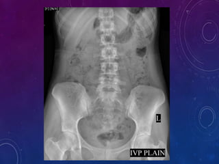
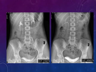
GOOD MORNING TO EVERYONE…
INTRAVENOUS UROGRAM (IVU)
MR.JAI KUMAR,
DRDT 2ND YEAR,
KILPAUK MEDICAL COLLEGE,
CHENNAI-10
INTRODUCTION
 IVU is the radiographic examination of urinary tract including renal
parenchyma, calyces and pelvis after intravenous injection of contrast
media.
 Intravenous pyelogram (IVP) is a misnomer as it implies visualization of
the pelvis and calyces without parenchyma.
 The term pyelogram is reserved for retrograde studies visualizing only the
collecting system.
 There has been a decline in the intravenous urograms done over the last
10 years.
This is because of
1. Development of newer imaging modalities like CT Scan, Ultrasound etc.
2. Cost-containment
3. Adverse effects of contrast media
 Introduction of excretory Urography was done in 1929, by American urologist
Moses Swick.
 He injected an organically-bound iodide compound into a vein, taking X-rays
as the material cleared the body through the urinary tract.
In 1937 Berger made several recommendations
 Routine tomography
 High dose of contrast agents
 Urethral compression
INDICATIONS
 Screening of entire urinary tract especially in cases of heamaturia or pyuria
 Diseases of renal collecting system and renal pelvis
 Differentiate function of both kidneys
 Abnormalities of the ureter
 Obstructive uropathy tract
 TB of the urinary tract
 Calculus diseases
 Potential of the renal doners
 Surgery of urinary tract
 Suspected renal injury
 Renal colic or flunk pain
IN CHILDERN
 VATER anomalies. Renal anomalies are seen in the 90% of patients.
 Malformation of urinary tract, e.g. polycystic disease, PUJ obstruction etc.
 Neurological disorders affecting urinary tract.
 Malformation of genitalia like bilateral cryptochidism.
 Anorectal anomalies.
 Enuresis in the presence of bacteriuria.
 Abnormal urinary sediment.
 History of recurrent urinary tract infection.
No absolute contraindication
Relative contraindications
 Severe history of anaphylaxis previously carries 30% risk.
 Renal failure (raised serum creatinine level >1.5 mg/dL).
 Hepatorenal syndrome.
 Previous allergy to the contrast agent/iodine.
 Generalized allergic conditions.
 Multiple myeloma.
 Pregnancy.
 Infancy.
 Hyperthyroidism.
 Diabetes.
CONTRAINDICATIONS
OVERVIEW OF URINARY SYSTEM
• Consist of 2 kidneys, 2 ureter, 1
urinary bladder and 1 urethra.
• After kidney filter the blood, they
return most of the water and other
soluter to the blood stream.
• The remaining water (urine),
passes through the ureters and is
stored in the urinary bladder.
GROSS ANATOMY OF THE KIDNEY
INTERNAL STRUCTURE OF THE KIDNEY
 The parenchyma of the kidney is divided into two major structures:
1) Superficial is the renal cortex
2) Deep is the renal medulla.
 Grossly, these structures take the shape of 8 to 18 cone-shaped renal lobes, each
containing renal cortex surrounding a portion of medulla called as renal pyramid.
 Between the renal pyramids are projections of cortex called renal columns.
 Nephrons, the urine-producing functional structures of the kidney, span the cortex
and medulla.
 The tip or papilla, of each pyramid empties urine into a minor calyx
 minor calyces empty into major calyces, and major calyces empty into the renal
pelvis, which becomes the ureter.
FUNCTION OF THE URINARY SYSTEM
 KIDNEY – Regulate blood volume and
composition, Regulate pH, Produce two
hormones and Excrete waste products
 URETERS- transport urine from kidney to
urinary bladder
 URINARY BLADDER- store urine and
expels through urethra
 URETHRA- discharge urine from the body
 Ask for any history of Diabetes mellitus,Phechrocytoma,Renal Disease,
Allergy to drugs and any specific foods.
 Fasting for 4 hours
 Do not dehydrate the patient
 Bowel preparation:
1.Dulcolax is given 2-4 tablets at bedtime for 2 days prior to the I.V.U
2.Because colon should be empty for I.V.U
 Take informed consent.
PREPERATION
PROCEDURE REQUIREMENTS
 600mA fluoroscopy guided x-ray unit
 Abdominal compression equipment.
 Medium / Regular film screen combination in a variety of sizes.
 Pads and immobilization aids.
 Intravenous administration equipment:
50 ml syringe, filling needle, skin prep, sticky tape,
 Selection of needles, venflon 19 gause.
 Tourniquet or blood pressure cuff.
 Emergency drugs and equipment.
EQUIPMENTS
In adults In children
Non ionic contrast
media( iohexol -
omnipaque)
300 mg I/ml 40-80 ml
350 mg I/ml 40-80 ml
240 mg I/ml
Below 7 Kg 4ml/ kg
Above 7 kg 3ml/kg
300 mg I/ml
Below 7 Kg 3ml/ kg
Above 7 kg 2ml/kg
Ionic contrast media 300-600 mg iodine
equivalent/kg body
weight.
Maximum of 40 gm of
iodine.
Meglumine iothalamate or
diatrizoate 60 % containing
equivalent of 280 mg I/ml of
iodine. Dose is 1-2ml/kg body
weight.
Below 6 months : 10 ml
6 months – 2 years : 20 ml
2-10 years : 20-40 ml
CONTRAST MEDIA DOSES
 Contrast Media is usually given as a I.V. bolus injection with in 30-60
seconds.
 The density of the nephrogram is directly proportional to the plasma
concentration of contrast media.
 More iodine increases the density of the nephrogram.
 Large Doses of Contrast Media increase diuresis which distends the
collecting system thus Increasing the diagnostic information from the
Urogram
MODE OF INJECTION
MODE OF INJECTION
 Patient is placed in supine position with pelvis at cathode side of the tube.
 A support is placed under patient’s knees to reduce lordotic curvature of
lumbosacral spine and provide comfort.
 A scout film is taken including the Kidneys,Ureters,Bladder and Urethral
Regions on a large size film.
 Contrast media is injected intravenously into a prominent vein in the arm.
 Test injection of 1ml of contrast is given and patient is observed for 1 min
to look for any contrast reactions.
 Then the rest of the contrast is rapidly injected within 30-60 seconds.
PROCEDURE
Low KV(65-75) high mA (600-1000) and short exposure should be used to get
optimum image contrast.
Standard films taken
 Plain X-Ray KUB/Scout film -14x17
 1 minute film - 10x12
 5 minute film - 10x12
 10 minute film - 15x12
 15 minute film - 15x12
 35 minute film - 14x17
 Post Void film - 10x8
FILMING TECHNIQUES
Plain X-ray KUB /Scout film provides valuable information and sometimes
indicates provable diagnosis.
Useful in assessing :
1) Calculus
2) Intestinal abnormalities
3) Intestinal gas pattern
4) Calcification
5) Abdominal mass
6) Foreign body
PLAIN XRAY KUB / SCOUT FILM
 1 minute film shows Nephrogram. This radiograph is often omitted has the
renal outlines are usually adequately visualized on 5 minute film
 5 minute film shows nephrogram, Renal pelvis, upper part of ureter.
 Compression band is now applied on Patients abdomen and the balloon is
positioned on anterior, superior iliac spine where cross the pelvic brim.
 This is to produce better pelviccalyceal distension.
1) Renal trauma
2) Large abdominal mass
3) Abdominal aneurysm
4) After abdominal surgery
 If 5 minute film shows dilated calyces or if calyces and pelvis are not
adequately opacified, obstruction exists and compression band not be
applied.
 If compression band is applied a film is taken after 10 minutes , the film
should shows centered kidneys to demonstrate distended collecting system
and proximal ureter.
Compression contraindicated in:
15 minutes film :
1) Visualization of ureter is better in prone position as the fill better.
2) This position reverses curve of the inferior course of the ureters making it
anti-dependent is to gravity.
3) Another method to see ureter is modified trendelenberg technique with 15-20
degrees head low tilt with the patient supine.
30 minute film:
1) It gives complete over view of the urinary tract ; Kidneys,Ureters,Bladder.
Bladder distension can be evaluated
Post Void film:
Taken immediately after voiding it is used to assess for :
1) Residual urine
2) Bladder mucosal lesions
3) Diverticula
4) Bladder tumour
5) Outlet obstruction VUR
Note: All films are taken expiratory phase only
Oblique view:
1. To project the ureter away to supine and to separate overlying radio opaque
shadows mimicking calculi.
2. Oblique views are also used for visualization of posterolateral aspects of
bladder and for doubtful urethral masses.
Erect film:
1. Provoke emptying of urinary tract.
2. Demonstrate layering of calculi in cysts and abscesses.
3. Detect urinary tract gas not seen in other films.
4. Have optimum demonstration of renal ptosis, bladder hernia,cystole and
areas of obstruction in ureter.
Prone film:
1. Viewing of urethral areas not seen in supine films.
2. Demonstration of renal ptosis and bladder hernia.
SPECIAL FILMS IN IVU
Delayed films in IVU are taken 1-24 hours after injection. Patients should
always be instructed to void prior to delayed films so that a calculus in the
distal ureter seen in the well.
Usual sequence of delayed films is after 1 hr, 3hrs, 6hrs, 12hrs and 24hrs.
Delayed films are used in :
 Cases of obstruction where early nephrogram is seen but collecting
system is not seen
 Long standing hydronephrosis in which renal parenchyma is seen but
collecting system is not visualized until many hours later
 Congenital lesions like non-visualized upper calyceal system with
obstructed ureter
Delayed films:
1.Diuretic urograms
 It is useful when intermittent obstruction is suspected but cannot be confirmed
by standard urogram.therefore the use of diuretic shows an acutely developing
hydronephrosis if true intermittent hydronephrosis is present.
 I.V.frusemide is used to induce dieresis which distends the renal pelvis.
 The film is taken 5-10 minutes after administrating the diuretic.
MODIFICATIONS OF UROGRAM
2.Tailored Urogram:
 It modifies the urograms to provide the information needed to include or exclude
the clinical problem and tailor the Urogram for that.
 The study is terminated as soon as the desired information is available.
3.Hypertensive Urogram :
 It is also called minute sequence urograms.
 The films are taken 1,2,3,5 minutes after injection of contrast media.
 Although the findings are of value, IVU cannot be used for screening of
hypertensives as there are any many positive and false negative results
Contrast is given in 500 ml normal saline. Now this procedure is not widely used.
Advantages:
 Nephrogram persists for longer time
 Enhanced dieresis from the additional contrast media and water volume will distend the
collecting system
 Collecting system is visualized for long times
 No significant increase the contrast reactions
 Administration is easy
Disadvantages:
 Overloads the patient with more iodine than necessary
 Calyceal blunting may be produced, Suggesting abnormal dilation
 May lead to pyelosinus extravasations and pain in patients with partial obstruction
 An initial vascular nephrogram is not obtained
4.Drip infusion Urography:
5.Limited Urography:
The procedure is useful for follow up to earlier pathology.
Film taken:
1) KUB
2) 15 mins AP
3) Post void
6.Emergency Urography:
It is done in cases of urinary colic.
Film taken :
1) KUB
2) 15mins
COMPLICATIONS:
Due to contrast
Minor reactions(5%):
1) Nausea
2) Vomiting
3) Mild rash
4) Light head ache
5) Mild dyspnoea
Intermediate reactions (1%):
1) Extensive urticaria
2) Facial oedema
3) Bronchospasm
4) Laryngeal oedema
5) Dyspnoea
6) Hypotension
Severe reactions (0.05%):
1) Circulatory collapse
2) pulmonary oedema
3) Severe angina
4) Myocardial infraction
5) Convulsions
6) Coma
7) Cardiac or respiratory arrest
Due to technique:
1) Upper arm or shoulder pain
2) Extravasation of contrast at the injection site.
AFTER CARE
1) Observation for 6 hours
2) Watch for late contrast reactions
3) Prevention of dehydration
4) In high risk patients-renal function test should be done to
watch for deterioration
 Clear outline of the entire urinary system so can see even mild
hydronephrosis.
 Easier to pick out obstructing stone when there are multiple pelvic
calcifications.
 Can show non-opaque stones as filling defects.
 Demonstrate renal function and allow for verification that the opposite
kidney is functioning normally.
ADVANTAGES
 Need for IV contrast material
 May provoke an allergic response
 Multiple delayed films (Can take hours as contrast passes quite slowly into
the blocked renal unit and ureter.)
 May not have sufficient Opacification to define the anatomy and point of
obstruction.
 Requires a significant amount of radiation exposure and may not be ideal for
young children or pregnant women
DISADVANTAGES
THANK YOU GUYZZ….

INTRAVENOUS UROGRAM (IVU)

  • 1.
    GOOD MORNING TOEVERYONE…
  • 2.
    INTRAVENOUS UROGRAM (IVU) MR.JAIKUMAR, DRDT 2ND YEAR, KILPAUK MEDICAL COLLEGE, CHENNAI-10
  • 3.
    INTRODUCTION  IVU isthe radiographic examination of urinary tract including renal parenchyma, calyces and pelvis after intravenous injection of contrast media.  Intravenous pyelogram (IVP) is a misnomer as it implies visualization of the pelvis and calyces without parenchyma.  The term pyelogram is reserved for retrograde studies visualizing only the collecting system.  There has been a decline in the intravenous urograms done over the last 10 years. This is because of 1. Development of newer imaging modalities like CT Scan, Ultrasound etc. 2. Cost-containment 3. Adverse effects of contrast media
  • 4.
     Introduction ofexcretory Urography was done in 1929, by American urologist Moses Swick.  He injected an organically-bound iodide compound into a vein, taking X-rays as the material cleared the body through the urinary tract. In 1937 Berger made several recommendations  Routine tomography  High dose of contrast agents  Urethral compression
  • 5.
    INDICATIONS  Screening ofentire urinary tract especially in cases of heamaturia or pyuria  Diseases of renal collecting system and renal pelvis  Differentiate function of both kidneys  Abnormalities of the ureter  Obstructive uropathy tract  TB of the urinary tract  Calculus diseases  Potential of the renal doners  Surgery of urinary tract  Suspected renal injury  Renal colic or flunk pain
  • 6.
    IN CHILDERN  VATERanomalies. Renal anomalies are seen in the 90% of patients.  Malformation of urinary tract, e.g. polycystic disease, PUJ obstruction etc.  Neurological disorders affecting urinary tract.  Malformation of genitalia like bilateral cryptochidism.  Anorectal anomalies.  Enuresis in the presence of bacteriuria.  Abnormal urinary sediment.  History of recurrent urinary tract infection.
  • 7.
    No absolute contraindication Relativecontraindications  Severe history of anaphylaxis previously carries 30% risk.  Renal failure (raised serum creatinine level >1.5 mg/dL).  Hepatorenal syndrome.  Previous allergy to the contrast agent/iodine.  Generalized allergic conditions.  Multiple myeloma.  Pregnancy.  Infancy.  Hyperthyroidism.  Diabetes. CONTRAINDICATIONS
  • 8.
    OVERVIEW OF URINARYSYSTEM • Consist of 2 kidneys, 2 ureter, 1 urinary bladder and 1 urethra. • After kidney filter the blood, they return most of the water and other soluter to the blood stream. • The remaining water (urine), passes through the ureters and is stored in the urinary bladder.
  • 9.
    GROSS ANATOMY OFTHE KIDNEY
  • 10.
    INTERNAL STRUCTURE OFTHE KIDNEY  The parenchyma of the kidney is divided into two major structures: 1) Superficial is the renal cortex 2) Deep is the renal medulla.  Grossly, these structures take the shape of 8 to 18 cone-shaped renal lobes, each containing renal cortex surrounding a portion of medulla called as renal pyramid.  Between the renal pyramids are projections of cortex called renal columns.  Nephrons, the urine-producing functional structures of the kidney, span the cortex and medulla.  The tip or papilla, of each pyramid empties urine into a minor calyx  minor calyces empty into major calyces, and major calyces empty into the renal pelvis, which becomes the ureter.
  • 11.
    FUNCTION OF THEURINARY SYSTEM  KIDNEY – Regulate blood volume and composition, Regulate pH, Produce two hormones and Excrete waste products  URETERS- transport urine from kidney to urinary bladder  URINARY BLADDER- store urine and expels through urethra  URETHRA- discharge urine from the body
  • 12.
     Ask forany history of Diabetes mellitus,Phechrocytoma,Renal Disease, Allergy to drugs and any specific foods.  Fasting for 4 hours  Do not dehydrate the patient  Bowel preparation: 1.Dulcolax is given 2-4 tablets at bedtime for 2 days prior to the I.V.U 2.Because colon should be empty for I.V.U  Take informed consent. PREPERATION
  • 13.
  • 14.
     600mA fluoroscopyguided x-ray unit  Abdominal compression equipment.  Medium / Regular film screen combination in a variety of sizes.  Pads and immobilization aids.  Intravenous administration equipment: 50 ml syringe, filling needle, skin prep, sticky tape,  Selection of needles, venflon 19 gause.  Tourniquet or blood pressure cuff.  Emergency drugs and equipment. EQUIPMENTS
  • 15.
    In adults Inchildren Non ionic contrast media( iohexol - omnipaque) 300 mg I/ml 40-80 ml 350 mg I/ml 40-80 ml 240 mg I/ml Below 7 Kg 4ml/ kg Above 7 kg 3ml/kg 300 mg I/ml Below 7 Kg 3ml/ kg Above 7 kg 2ml/kg Ionic contrast media 300-600 mg iodine equivalent/kg body weight. Maximum of 40 gm of iodine. Meglumine iothalamate or diatrizoate 60 % containing equivalent of 280 mg I/ml of iodine. Dose is 1-2ml/kg body weight. Below 6 months : 10 ml 6 months – 2 years : 20 ml 2-10 years : 20-40 ml CONTRAST MEDIA DOSES
  • 16.
     Contrast Mediais usually given as a I.V. bolus injection with in 30-60 seconds.  The density of the nephrogram is directly proportional to the plasma concentration of contrast media.  More iodine increases the density of the nephrogram.  Large Doses of Contrast Media increase diuresis which distends the collecting system thus Increasing the diagnostic information from the Urogram MODE OF INJECTION
  • 17.
  • 18.
     Patient isplaced in supine position with pelvis at cathode side of the tube.  A support is placed under patient’s knees to reduce lordotic curvature of lumbosacral spine and provide comfort.  A scout film is taken including the Kidneys,Ureters,Bladder and Urethral Regions on a large size film.  Contrast media is injected intravenously into a prominent vein in the arm.  Test injection of 1ml of contrast is given and patient is observed for 1 min to look for any contrast reactions.  Then the rest of the contrast is rapidly injected within 30-60 seconds. PROCEDURE
  • 19.
    Low KV(65-75) highmA (600-1000) and short exposure should be used to get optimum image contrast. Standard films taken  Plain X-Ray KUB/Scout film -14x17  1 minute film - 10x12  5 minute film - 10x12  10 minute film - 15x12  15 minute film - 15x12  35 minute film - 14x17  Post Void film - 10x8 FILMING TECHNIQUES
  • 20.
    Plain X-ray KUB/Scout film provides valuable information and sometimes indicates provable diagnosis. Useful in assessing : 1) Calculus 2) Intestinal abnormalities 3) Intestinal gas pattern 4) Calcification 5) Abdominal mass 6) Foreign body PLAIN XRAY KUB / SCOUT FILM
  • 22.
     1 minutefilm shows Nephrogram. This radiograph is often omitted has the renal outlines are usually adequately visualized on 5 minute film  5 minute film shows nephrogram, Renal pelvis, upper part of ureter.  Compression band is now applied on Patients abdomen and the balloon is positioned on anterior, superior iliac spine where cross the pelvic brim.  This is to produce better pelviccalyceal distension.
  • 24.
    1) Renal trauma 2)Large abdominal mass 3) Abdominal aneurysm 4) After abdominal surgery  If 5 minute film shows dilated calyces or if calyces and pelvis are not adequately opacified, obstruction exists and compression band not be applied.  If compression band is applied a film is taken after 10 minutes , the film should shows centered kidneys to demonstrate distended collecting system and proximal ureter. Compression contraindicated in:
  • 25.
    15 minutes film: 1) Visualization of ureter is better in prone position as the fill better. 2) This position reverses curve of the inferior course of the ureters making it anti-dependent is to gravity. 3) Another method to see ureter is modified trendelenberg technique with 15-20 degrees head low tilt with the patient supine. 30 minute film: 1) It gives complete over view of the urinary tract ; Kidneys,Ureters,Bladder. Bladder distension can be evaluated
  • 27.
    Post Void film: Takenimmediately after voiding it is used to assess for : 1) Residual urine 2) Bladder mucosal lesions 3) Diverticula 4) Bladder tumour 5) Outlet obstruction VUR Note: All films are taken expiratory phase only
  • 29.
    Oblique view: 1. Toproject the ureter away to supine and to separate overlying radio opaque shadows mimicking calculi. 2. Oblique views are also used for visualization of posterolateral aspects of bladder and for doubtful urethral masses. Erect film: 1. Provoke emptying of urinary tract. 2. Demonstrate layering of calculi in cysts and abscesses. 3. Detect urinary tract gas not seen in other films. 4. Have optimum demonstration of renal ptosis, bladder hernia,cystole and areas of obstruction in ureter. Prone film: 1. Viewing of urethral areas not seen in supine films. 2. Demonstration of renal ptosis and bladder hernia. SPECIAL FILMS IN IVU
  • 31.
    Delayed films inIVU are taken 1-24 hours after injection. Patients should always be instructed to void prior to delayed films so that a calculus in the distal ureter seen in the well. Usual sequence of delayed films is after 1 hr, 3hrs, 6hrs, 12hrs and 24hrs. Delayed films are used in :  Cases of obstruction where early nephrogram is seen but collecting system is not seen  Long standing hydronephrosis in which renal parenchyma is seen but collecting system is not visualized until many hours later  Congenital lesions like non-visualized upper calyceal system with obstructed ureter Delayed films:
  • 33.
    1.Diuretic urograms  Itis useful when intermittent obstruction is suspected but cannot be confirmed by standard urogram.therefore the use of diuretic shows an acutely developing hydronephrosis if true intermittent hydronephrosis is present.  I.V.frusemide is used to induce dieresis which distends the renal pelvis.  The film is taken 5-10 minutes after administrating the diuretic. MODIFICATIONS OF UROGRAM
  • 34.
    2.Tailored Urogram:  Itmodifies the urograms to provide the information needed to include or exclude the clinical problem and tailor the Urogram for that.  The study is terminated as soon as the desired information is available. 3.Hypertensive Urogram :  It is also called minute sequence urograms.  The films are taken 1,2,3,5 minutes after injection of contrast media.  Although the findings are of value, IVU cannot be used for screening of hypertensives as there are any many positive and false negative results
  • 35.
    Contrast is givenin 500 ml normal saline. Now this procedure is not widely used. Advantages:  Nephrogram persists for longer time  Enhanced dieresis from the additional contrast media and water volume will distend the collecting system  Collecting system is visualized for long times  No significant increase the contrast reactions  Administration is easy Disadvantages:  Overloads the patient with more iodine than necessary  Calyceal blunting may be produced, Suggesting abnormal dilation  May lead to pyelosinus extravasations and pain in patients with partial obstruction  An initial vascular nephrogram is not obtained 4.Drip infusion Urography:
  • 36.
    5.Limited Urography: The procedureis useful for follow up to earlier pathology. Film taken: 1) KUB 2) 15 mins AP 3) Post void 6.Emergency Urography: It is done in cases of urinary colic. Film taken : 1) KUB 2) 15mins
  • 37.
    COMPLICATIONS: Due to contrast Minorreactions(5%): 1) Nausea 2) Vomiting 3) Mild rash 4) Light head ache 5) Mild dyspnoea Intermediate reactions (1%): 1) Extensive urticaria 2) Facial oedema 3) Bronchospasm 4) Laryngeal oedema 5) Dyspnoea 6) Hypotension
  • 38.
    Severe reactions (0.05%): 1)Circulatory collapse 2) pulmonary oedema 3) Severe angina 4) Myocardial infraction 5) Convulsions 6) Coma 7) Cardiac or respiratory arrest Due to technique: 1) Upper arm or shoulder pain 2) Extravasation of contrast at the injection site.
  • 39.
    AFTER CARE 1) Observationfor 6 hours 2) Watch for late contrast reactions 3) Prevention of dehydration 4) In high risk patients-renal function test should be done to watch for deterioration
  • 40.
     Clear outlineof the entire urinary system so can see even mild hydronephrosis.  Easier to pick out obstructing stone when there are multiple pelvic calcifications.  Can show non-opaque stones as filling defects.  Demonstrate renal function and allow for verification that the opposite kidney is functioning normally. ADVANTAGES
  • 41.
     Need forIV contrast material  May provoke an allergic response  Multiple delayed films (Can take hours as contrast passes quite slowly into the blocked renal unit and ureter.)  May not have sufficient Opacification to define the anatomy and point of obstruction.  Requires a significant amount of radiation exposure and may not be ideal for young children or pregnant women DISADVANTAGES
  • 42.

Editor's Notes

  • #7 these patients have vertebral, anal, tracheo-oesphageal, and renal anomalies