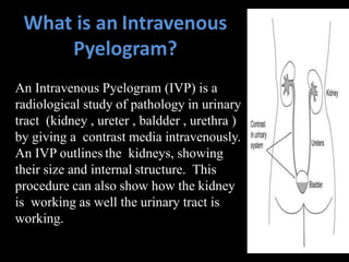
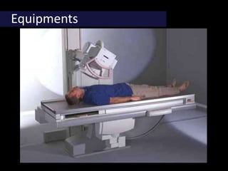
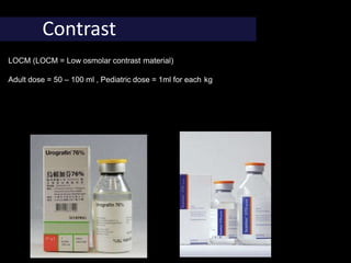
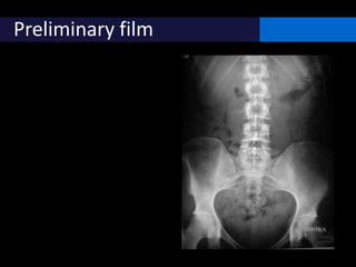
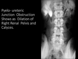
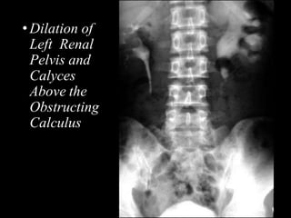
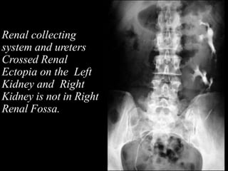
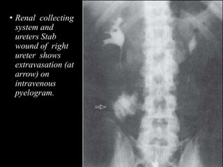
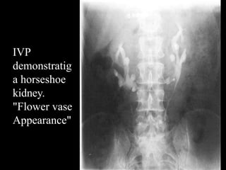
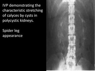
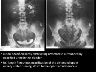
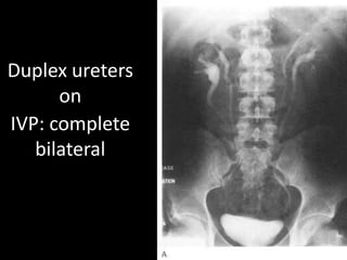
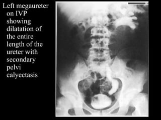
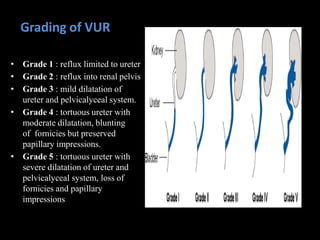
IVP, RGU & MCU
DR. NISHIT VIRADIA
IVP
• It is a radiographic examination of urinary
tract including renal parenchyma, calyces and
pelvis after iv injection of contrast.
• Intravenous pyelogram is a misnomer as it
implies visualization of pelvis and calyces
without parenchyma.
• The term pyelogram is reserved for retrograde
studies visualizing only collecting system
What is an Intravenous
Pyelogram?
An Intravenous Pyelogram (IVP) is a
radiological study of pathology in urinary
tract (kidney , ureter , baldder , urethra )
by giving a contrast media intravenously.
An IVP outlines the kidneys, showing
their size and internal structure. This
procedure can also show how the kidney
is working as well the urinary tract is
working.
Benefits
• Imaging of the urinary tract with IVP is a minimally invasive
procedure.
• IVP images provide valuable, detailed information to assist
physicians in diagnosing and treating urinary tract
conditions from kidney stones to cancer.
• An IVP can often provide enough information about kidney
stones and urinary tract obstructions to direct treatment with
medication and avoid more invasive surgical procedures.
Indications
1. Haematuria
2. Calculus in urinary tract
3. Tumour of kidney,Ureter,Bladder
4. Infection in urinary tract
e.g.Tuberculosis, Nephritis, Cystitis
5. Congenital anamolies of Urinary tract
e.g.Unilateral kidney, Bifid ureter, Ectopic kidney, polycystic kidney
6. Suspected urinary tract pathology
7. To study kidney function
8. Post urinary tract surgery
Contra-Indications:
1. History Of allergy to iodinated contrast media.
2. Pregnancy
3. Cardiac and Renal failure.
4. Multiple Myeloma
5. Hepatorenal syndrome
6. Raised serum creatinin level
7. Thyrotoxicosis
8. Metformin
The procedure
Equipments
Contrast
LOCM (LOCM = Low osmolar contrast material)
Adult dose = 50 – 100 ml , Pediatric dose = 1ml for each kg
Compound Name type Osmolality
Ionic Diatrizoate Monomer 1550 High
Ionic Metrizoate Monomer 2100 High
Ionic Iothalamate 600-2100 High
Ionic Ioxaglate Dimer 580 Low
Non-ionic Iopamidol Monomer 796 Low
Non-ionic Iohexol Monomer 884 Low
Non-ionic Ioxilan Monomer 695 Low
Non-ionic Iopromide Monomer 774 Low
Non-ionic Ioversol Monomer 350 Low
Non-ionic Iodixanol Dimer 290 Low
Adverse effects of contrast
• Chemotoxic
• Nephrotoxicity
• Nonoliguric
• Creatinine peaks in 3-5 days
• Risk 1 in 1000-5000 if no risk factors
• Risk factors: renal insufficiency, DM, CHF,
hyperuricemia, proteinuria, multiple doses of
contrast
• Metformin (Glucophage) overdose causes
lactic acidosis  withhold for 48 hours after
contrast
• Anaphylactoid / Idiosyncratic
Adverse effects of contrast
• Anaphylactoid / Idiosyncratic
• Mild: metallic taste, warmth, sneezing, coughing,
mild hives  notreatment
• Moderate: vomiting, severe hives, HA, palpitations,
facial edema
• Severe: hypotension, bronchospasm, laryngeal
edema, pulmonary edema, LOC
• Idiosyncratic rxns for ionic contrast = as high as
12%, most mild.
• Tx with Antihistamine = mild, Epi or beta agonist =
severe.
• Non-ionic = 3%
Preparation
1.Ask patient for history.
2.Bowel Preparation
3.Do not dehydrate the patient.
Preliminary film
Mode of injection
• Contrast media usually given iv bolus injection
within 30-60 seconds
• The density of nephrogram is directly
proportional to the plasma concentration.
Immediate film (Nephrogram phase)
• It shows nephrogram.
• This radiograph often omitted as the renal
outline are usually adequately visualized in 5
min film.
5 minute film(Nephrogram)
• It shows nephrogram, renal pelvis, upper part
of ureter.
Compression Band
• Compression band is now applied.
• It is to produce better pelvicalyceal distention
Contraindication
Renal trauma
Large abdominal mass
After abdominal Sx
If in 5 min film pelvis & calyces not adequately
opacified
15 minutes film
• Visualization of ureter is
better in prone position
• This position reverse
the curve of the inferior
course of ureter making
it anti-dependent to
gravity.
35 minutes film (Cystogram phase)
inspect :
- Bladder size and shape
- Contrast is filling the bladder or
not
- Bladder surface is smooth or
rough
- Is there any diverticlula, filling
defect or prostate indentation?
Postvoiding film
look for:
- Residual urine
- Bladder mucosal lesion
- Diverticulitis
- Bladder tumor
- Outlet obstruction
- VUR
Pyelo- ureteric
Junction Obstruction
Shows as Dilation of
Right Renal Pelvis and
Calyces.
•Dilation of
Left Renal
Pelvis and
Calyces
Above the
Obstructing
Calculus
Renal collecting
system and ureters
Crossed Renal
Ectopia on the Left
Kidney and Right
Kidney is not in Right
Renal Fossa.
• Renal collecting
system and
ureters Stab
wound of right
ureter shows
extravasation (at
arrow) on
intravenous
pyelogram.
IVP
demonstratig
a horseshoe
kidney.
"Flower vase
Appearance"
IVP demonstrating the
characteristic stretching
of calyces by cysts in
polycystic kidneys.
Spider leg
appearance
• a Non-opacified partly obstructing ureterocele surrounded by
opacified urine in the bladder
• full length film shows opacification of the distended upper
moiety ureter running down to the opacified ureterocele
Duplex ureters
on
IVP: complete
bilateral
Left megaureter
on IVP
showing
dilatation of
the entire
length of the
ureter with
secondary
pelvi
calyectasis
Benign prostatic hyperplasia. White = bladder. Dark = benign
enlargement of prostate, pushing down on inferior bladder
Intravenous pyelogram
showed no obstructive
uropathy, but symmetric
diverticula could be seen
near both ureteral orifices
(arrows). These lesions,
known as Hutch
diverticula, are usually
congenital rather than
occurring as a result of a
neurogenic bladder or an
infection or obstruction
Nodular squamous
cell carcinoma of
the urinary bladder.
Dilated left lower
ureter probably
secondary to
obstruction by
tumor.
Nonvisualization of
the right ureter
caused by complete
occlusion
rgu
ANATOMY OF URETHRA
• MALE URETHRA IS 18-20 CMS LONG
• EXTENDS FROM BLADDER NECK TILL THE MEATAL OPENING AT PENIS
• It has four named regions:
 Prostatic urethra:
 Is approximately 3 cm in length.
 Passes through the prostate gland.
 Membranous urethra:
 Is approximately 1 cm in length.
 Passes through the urogenital diaphragm.
 Bulbar urethra
 From inferior aspect of urogenital diaphragm to penoscrotal junction.
 Spongy (penile) urethra:
 Passes through the length of the penis.
PARTS OF URETHRA
• ANTERIOR URETHRA
-PENILE URETHRA
-BULBAR URETHRA
• POSTERIOR URETHRA
- MEMBRANOUS URETHRA
- PROSTATIC URETHRA
PROSTATIC URETHRA
• BEGINS ATINTERNAL URETHRAL ORIFICE &
RUNS VERTICALLY DOWNWARDS
• WIDEST & MOST DILATABLE PART,
NARROWEST WHERE IT JOINS MEMBRANOUS
URETHRA
• VERU MONTANUM IS A MEDIAN
LONGITUDINAL RIDGE OF MUCOUS
MEMBRANE
MEMBRANOUS URETHRA
• SHORTEST , NARROWEST & LEAST
DISTENSIBLE PART OF THE URETHRA
• 2-2.5 CMS
• SURROUNDED BY EXTERNAL URETHRAL
SPHINCTER
• THE BULBOURETHRAL GLANDS OF COWPER
ARE PLACED ONE ON EACH SIDE OF
MEMBRANOUS URETHRA
• URETHRAL GLANDS ALSO OPEN HERE
PENILE (SPONGY) URETHRA
• LENGTH 14-15 CMS & 6MM DIAMETER
• IT IS DILATED WITHIN THE GLANS PENIS TO
FORM FOSSA NAVICULARIS
• URETHRAL GLANDS OF LITTRE OPEN HERE
Female urethra
• Widest at bladder
neck.
• 4-5cms in length
• Narrowest & least
distensible at meatus.
• This forms the
Spinning top
configuration of
urethra on normal
MCU.
Definition
 A retrograde urethrogram is a routine radiologicprocedure
(most typically in males) used to image the integrity of
the urethra by using iodinated contrast media.
Indications
• Strictures
• Pelvic trauma/ Urethral tears
• Urethral obstruction
• Urethral foreign body.
• Urethral diverticulum.
• Periurethral / prostatic abscess.
• Fistula / false passages.
• Urethral mucosal tumours
• Post operative evaluation
Contraindications
Acute urinarytract
infection
Technique
 Patient lies supineon thex-ray table
 Retract the foreskin and clean the tipof penis with betadineor
antisepticsolution.
 Inject a small amountof local anesthetic into the urethrawith a 8-
F Foley catheter or syringe and balloon is inflated with 1-3 ml of
water.
 Local anesthesia helps to relax thesphincteras the patient may
contract it during the procedure thus leading to a diagnosis of a
stricture
 Contrast medium is injected under fluoroscopy control andfilms
are taken
 The male urethra is best seen in theobliqueposition. Female
urethra is best seen in lateral oranteroposterior position .
Image interpretation
• Normal retrograde urethrogram (RGU):
• If the radiopaque contrast is injected properly,
the entire anterior and posterior urethra should
be filled with contrast and seen to jet into the
bladder neck. The verumontanum is seen as an
ovoid filling defect in the posterior urethra
• The distal end of the verumontanum marks the
proximal boundary of the membranous urethra
and constitutes the urethra that passes through
the urogenital diaphragm.
Affect of patient positioning on the appearance of the urethra during retrograde
urethrography. (a) Retrograde urethrogram obtained with the patient supine shows
the bulbous urethra as a diverticulum-like outpouching. (b) On a retrograde
urethrogram obtained after the patient was placed in a steep oblique position with
the penis stretched, the penoscrotal junction and bulbous urethra have a normal
appearance.
COMPLICATIONS
Contrast reaction ( due to absorption through
bladder mucosa )
UTI
Urethral trauma or rupture.
Extravasation of contrast – due to use of
excessive pressure in stricture.
Role of urethrography in stricture
• Accurately delineates the anatomy of urethra.
• Location, number and extent of the strictures are
very well displayed
• Delineation of the bladder neck and urethra is best
achieved on the MCU in the oblique projection.
• Secondary changes in the bladder.
• Todemonstrate the VUR
• Visualisation of any associated fistulas.
Penile urethra stricture
a)Urethral stricture, b)periurethral
abscess
Urethral calculi
 Mostly expelled from bladder into the urethra during voiding- migrant calculi.
 Primary calculi may be seen in association with urethral stricture or urethral
diverticulum.
 Symptoms include weak stream, dysuria, and hematuria.
 RGU usually depicts a rounded filling defect in the urethra.
Tuberculous urethritis
 Descending infection and renal tuberculosis is
evident.
 In the acute phase, there is urethral discharge
with associated involvement of the epididymis,
prostate, and other parts of the urinary system.
 In chronic phase patients present with
obstructive symptoms secondary to urethral
strictures.
 May lead to periurethral abscesses, which,
unless treated, produce numerous perineal and
scrotal fistulas- Watering can perineum.
 Retrograde urethrography typically
demonstrates an anterior urethral stricture
associated with multiple prostatocutaneous and
urethrocutaneous fistulas.
Gonococcal urethral stricture. Retrograde
urethrogram reveals a segment of irregular,
beaded narrowing in the distal bulbous urethra
with opacification of the left Cowper duct.
Blunt Urethral Trauma
Classified Anatomically as - Anterior
- Posterior
Anterior urethral injury
MC iatrogenic (due to instrumentation)
May occur if pt falls on a blunt object or direct injury to
perineum
Straddle Injury - compression of urethra against
anterior pelvic ring
Posterior urethral injury results from
A crushing force to the pelvis
Is associated with pelvic fractures.
Goldman & Sander classification (Based on findings at retrograde urethrography)
• Type I injury
 Rupture of the puboprostatic ligaments which stretches the prostatic
urethra
 Continuity of the urethra is maintained
 Type II injury (15%)
The membranous urethra is torn above an intact
urogenital diaphragm, which prevents contrast material
extravasation from extending into the perineum
 Type III injury (MC)
The membranous urethra is ruptured but the injury extends into the
proximal bulbous urethra because of laceration of the urogenital
diaphragm
Extravasation not only into the pelvic extraperitoneal space but also
into the perineum.
• Type IV
Bladder neck injury with extension to the urethra.
Type V injury
Injury to the Anterior urethra - partial or complete.
Extravasation seen to penile soft tissue.
MCU
MICTURATING CYSTOURETHROGRAM
• Voiding cystourethrogram demonstrates the
lower urinary tract & helps to detect VUR ,
bladder pathology, congenital or aquired
anamolies of bladder
• It is performed by passing a catheter through
the urethra into the bladder, filling the
bladder with contrast material and then taking
radiographs while the patient voids.
ANTEROGRADE URETHROGRAPHY/
MICTURATING CYSTOURETHROGRAPHY
• INDICATIONS
 CHILDREN
- UTI
-Voiding difficulties.
-Vesico ureteric reflux.
-Baseline study prior to urinary tract surgery.
-Post operative evaluation of ureteric abnormalities.
-Trauma.
-Suspected anatomic abnormalities of bladder neck &
urethra. ( posterior urethral valve )
ADULTS
- Functional disorders of bladder & urethra.
-Suspected vesicovaginal / vesicocolic fistula.
-Suspected bladder / urethral trauma.
-Urethral diverticula
Procedure
• Using a sterile technique , a catheter is introduced into
the bladder.
• A 5F feeding tube with side holes are used for children
and in older children or adults 8F or 10F catheters are
used .
• In girls after initial inspection of perineum to identify
any local genitilia abnormalities (cystoceles or labial
fusion ) the catheter is introduced..
• When it enters the bladder a varying amount of urine
will flow through it. If no flow a catheter is introduced
till urine is obtained.
 Suprapubic pressure Is sometimes helpful.
 In males , foreskin is retracted and catheter is introduced .
The catheter should be lubricated with anaesthetic jelly
and inserted slowly and gently into the urthera holding the
penis is vertical position .
 The normal bladder capacity in children is estimated to be
1 ounce ie 29 cc .
 For newborns -30 to 35 cc can be instilled.
• For upto 3 yrs – 200 to 250 cc
 Adequate capacity is reached when the child becomes
uncomfortable and begins voiding around the catheter.
 Bladder capacity (in milliliters) is variable but can often be
predicted with the previous mentioned formula
Filming
 In children : upto 2 yrs of age bladder is filled by hand
injection. For older children contrast medium is instilled from
a bottle elevated one metre above the examination table.
 During filming, fluroscopic screening is performed at short
intervals to see any vu reflux,diverticuli.
 The child is turned oblique on both sides to ensure that
minimal reflux is not overlooked.
 In infants : voiding starts the moment catheter is removed. At
the end of voiding ,frontal film is taken which includes entire
abdomen including the kidney region to prevent overlooking
the vu reflux which is apparent only on termination of voiding
and may reach upper collecting system.
In adult male : bladder is filled in the usual
way as in older child and voiding filming is
done in both oblique projection views.
The voiding study in male adults can be
modified by getting the patient to void against
resistance i.e. by compression of distal part of
penis thus enhancing the visualization of
urethra by artificial distention .
• ALTERNATE TECHNIQUES
1) SUPRAPUBIC BLADDER PUNCTURE.
 Sometimes in PUV & pelvic trauma – not possible to catheterize.
2) URETHROCYSTOGRAPHY
 Contrast medium introduced into the bladder during RGU.
3)EXCRETION MCU ( MCU followed by IVU )
Advantage – avoid catheterization and related risk of infection.
Disadvantage - VUR can not be visualized properly .
takes longer time.
Posterior urethral valve in newborn and in a 7 yr. Old boy
Posterior urethral valve -image shows a
dilated posterior urethra with an abrupt
transition to a normal-calibre anteriorurethra
with bladder neck hypertrophy, the irregular
trabeculated bladder wall, and the left-sided
grade III vesicoureteric reflux.
Grading of VUR
• Grade 1 : reflux limited to ureter
• Grade 2 : reflux into renal pelvis
• Grade 3 : mild dilatation of
ureter and pelvicalyceal system.
• Grade 4 : tortuous ureter with
moderate dilatation, blunting
of fornicies but preserved
papillary impressions.
• Grade 5 : tortuous ureter with
severe dilatation of ureter and
pelvicalyceal system, loss of
fornicies and papillary
impressions
• Congenital megalourethra
• This is a rare congenital anomaly resulting
from the faulty development of the corpora
cavernosa and corpus spongiosum.
megalourethra in an infant. Lateral mcu image
reveals an extensively dilated anterior and
posterior urethra
Urachal diverticulum. Posteroanterior mcu
image shows a gross urachal diverticulum
bladder.
MCU image shows a diverticulum resulting from spontaneous
opacification of a prostatic utricle
(a) Early anteroposterior voiding
cystourethrogram demonstrates a
ureterocele
Tuberculous urethritis
 Descending infection and renal tuberculosis is
evident.
 In the acute phase, there is urethral discharge
with associated involvement of the
epididymis, prostate, and other parts of the
urinary system.
 In chronic phase patients present with
obstructive symptoms secondary to urethral
strictures.
 May lead to periurethral
abscesses, which, unless treated, produce
numerous perineal and scrotal fistulas-
Watering can perineum.
 Retrograde urethrography typically
demonstrates an anterior urethral stricture
associated with multiple prostatocutaneous and
urethrocutaneous fistulas.
IVP, RGU & MCU

IVP, RGU & MCU

  • 1.
    IVP, RGU &MCU DR. NISHIT VIRADIA
  • 2.
    IVP • It isa radiographic examination of urinary tract including renal parenchyma, calyces and pelvis after iv injection of contrast. • Intravenous pyelogram is a misnomer as it implies visualization of pelvis and calyces without parenchyma. • The term pyelogram is reserved for retrograde studies visualizing only collecting system
  • 3.
    What is anIntravenous Pyelogram? An Intravenous Pyelogram (IVP) is a radiological study of pathology in urinary tract (kidney , ureter , baldder , urethra ) by giving a contrast media intravenously. An IVP outlines the kidneys, showing their size and internal structure. This procedure can also show how the kidney is working as well the urinary tract is working.
  • 4.
    Benefits • Imaging ofthe urinary tract with IVP is a minimally invasive procedure. • IVP images provide valuable, detailed information to assist physicians in diagnosing and treating urinary tract conditions from kidney stones to cancer. • An IVP can often provide enough information about kidney stones and urinary tract obstructions to direct treatment with medication and avoid more invasive surgical procedures.
  • 5.
    Indications 1. Haematuria 2. Calculusin urinary tract 3. Tumour of kidney,Ureter,Bladder 4. Infection in urinary tract e.g.Tuberculosis, Nephritis, Cystitis 5. Congenital anamolies of Urinary tract e.g.Unilateral kidney, Bifid ureter, Ectopic kidney, polycystic kidney 6. Suspected urinary tract pathology 7. To study kidney function 8. Post urinary tract surgery
  • 6.
    Contra-Indications: 1. History Ofallergy to iodinated contrast media. 2. Pregnancy 3. Cardiac and Renal failure. 4. Multiple Myeloma 5. Hepatorenal syndrome 6. Raised serum creatinin level 7. Thyrotoxicosis 8. Metformin
  • 7.
  • 8.
  • 9.
    Contrast LOCM (LOCM =Low osmolar contrast material) Adult dose = 50 – 100 ml , Pediatric dose = 1ml for each kg
  • 10.
    Compound Name typeOsmolality Ionic Diatrizoate Monomer 1550 High Ionic Metrizoate Monomer 2100 High Ionic Iothalamate 600-2100 High Ionic Ioxaglate Dimer 580 Low Non-ionic Iopamidol Monomer 796 Low Non-ionic Iohexol Monomer 884 Low Non-ionic Ioxilan Monomer 695 Low Non-ionic Iopromide Monomer 774 Low Non-ionic Ioversol Monomer 350 Low Non-ionic Iodixanol Dimer 290 Low
  • 11.
    Adverse effects ofcontrast • Chemotoxic • Nephrotoxicity • Nonoliguric • Creatinine peaks in 3-5 days • Risk 1 in 1000-5000 if no risk factors • Risk factors: renal insufficiency, DM, CHF, hyperuricemia, proteinuria, multiple doses of contrast • Metformin (Glucophage) overdose causes lactic acidosis  withhold for 48 hours after contrast • Anaphylactoid / Idiosyncratic
  • 12.
    Adverse effects ofcontrast • Anaphylactoid / Idiosyncratic • Mild: metallic taste, warmth, sneezing, coughing, mild hives  notreatment • Moderate: vomiting, severe hives, HA, palpitations, facial edema • Severe: hypotension, bronchospasm, laryngeal edema, pulmonary edema, LOC • Idiosyncratic rxns for ionic contrast = as high as 12%, most mild. • Tx with Antihistamine = mild, Epi or beta agonist = severe. • Non-ionic = 3%
  • 13.
    Preparation 1.Ask patient forhistory. 2.Bowel Preparation 3.Do not dehydrate the patient.
  • 14.
  • 15.
    Mode of injection •Contrast media usually given iv bolus injection within 30-60 seconds • The density of nephrogram is directly proportional to the plasma concentration.
  • 16.
    Immediate film (Nephrogramphase) • It shows nephrogram. • This radiograph often omitted as the renal outline are usually adequately visualized in 5 min film.
  • 17.
    5 minute film(Nephrogram) •It shows nephrogram, renal pelvis, upper part of ureter.
  • 18.
    Compression Band • Compressionband is now applied. • It is to produce better pelvicalyceal distention Contraindication Renal trauma Large abdominal mass After abdominal Sx If in 5 min film pelvis & calyces not adequately opacified
  • 19.
    15 minutes film •Visualization of ureter is better in prone position • This position reverse the curve of the inferior course of ureter making it anti-dependent to gravity.
  • 20.
    35 minutes film(Cystogram phase) inspect : - Bladder size and shape - Contrast is filling the bladder or not - Bladder surface is smooth or rough - Is there any diverticlula, filling defect or prostate indentation?
  • 21.
    Postvoiding film look for: -Residual urine - Bladder mucosal lesion - Diverticulitis - Bladder tumor - Outlet obstruction - VUR
  • 22.
    Pyelo- ureteric Junction Obstruction Showsas Dilation of Right Renal Pelvis and Calyces.
  • 23.
    •Dilation of Left Renal Pelvisand Calyces Above the Obstructing Calculus
  • 24.
    Renal collecting system andureters Crossed Renal Ectopia on the Left Kidney and Right Kidney is not in Right Renal Fossa.
  • 25.
    • Renal collecting systemand ureters Stab wound of right ureter shows extravasation (at arrow) on intravenous pyelogram.
  • 26.
  • 27.
    IVP demonstrating the characteristicstretching of calyces by cysts in polycystic kidneys. Spider leg appearance
  • 28.
    • a Non-opacifiedpartly obstructing ureterocele surrounded by opacified urine in the bladder • full length film shows opacification of the distended upper moiety ureter running down to the opacified ureterocele
  • 29.
  • 30.
    Left megaureter on IVP showing dilatationof the entire length of the ureter with secondary pelvi calyectasis
  • 31.
    Benign prostatic hyperplasia.White = bladder. Dark = benign enlargement of prostate, pushing down on inferior bladder
  • 32.
    Intravenous pyelogram showed noobstructive uropathy, but symmetric diverticula could be seen near both ureteral orifices (arrows). These lesions, known as Hutch diverticula, are usually congenital rather than occurring as a result of a neurogenic bladder or an infection or obstruction
  • 33.
    Nodular squamous cell carcinomaof the urinary bladder. Dilated left lower ureter probably secondary to obstruction by tumor. Nonvisualization of the right ureter caused by complete occlusion
  • 34.
  • 35.
    ANATOMY OF URETHRA •MALE URETHRA IS 18-20 CMS LONG • EXTENDS FROM BLADDER NECK TILL THE MEATAL OPENING AT PENIS • It has four named regions:  Prostatic urethra:  Is approximately 3 cm in length.  Passes through the prostate gland.  Membranous urethra:  Is approximately 1 cm in length.  Passes through the urogenital diaphragm.  Bulbar urethra  From inferior aspect of urogenital diaphragm to penoscrotal junction.  Spongy (penile) urethra:  Passes through the length of the penis.
  • 37.
    PARTS OF URETHRA •ANTERIOR URETHRA -PENILE URETHRA -BULBAR URETHRA • POSTERIOR URETHRA - MEMBRANOUS URETHRA - PROSTATIC URETHRA
  • 38.
    PROSTATIC URETHRA • BEGINSATINTERNAL URETHRAL ORIFICE & RUNS VERTICALLY DOWNWARDS • WIDEST & MOST DILATABLE PART, NARROWEST WHERE IT JOINS MEMBRANOUS URETHRA • VERU MONTANUM IS A MEDIAN LONGITUDINAL RIDGE OF MUCOUS MEMBRANE
  • 39.
    MEMBRANOUS URETHRA • SHORTEST, NARROWEST & LEAST DISTENSIBLE PART OF THE URETHRA • 2-2.5 CMS • SURROUNDED BY EXTERNAL URETHRAL SPHINCTER • THE BULBOURETHRAL GLANDS OF COWPER ARE PLACED ONE ON EACH SIDE OF MEMBRANOUS URETHRA • URETHRAL GLANDS ALSO OPEN HERE
  • 40.
    PENILE (SPONGY) URETHRA •LENGTH 14-15 CMS & 6MM DIAMETER • IT IS DILATED WITHIN THE GLANS PENIS TO FORM FOSSA NAVICULARIS • URETHRAL GLANDS OF LITTRE OPEN HERE
  • 41.
    Female urethra • Widestat bladder neck. • 4-5cms in length • Narrowest & least distensible at meatus. • This forms the Spinning top configuration of urethra on normal MCU.
  • 42.
    Definition  A retrogradeurethrogram is a routine radiologicprocedure (most typically in males) used to image the integrity of the urethra by using iodinated contrast media.
  • 43.
    Indications • Strictures • Pelvictrauma/ Urethral tears • Urethral obstruction • Urethral foreign body. • Urethral diverticulum. • Periurethral / prostatic abscess. • Fistula / false passages. • Urethral mucosal tumours • Post operative evaluation
  • 44.
  • 45.
    Technique  Patient liessupineon thex-ray table  Retract the foreskin and clean the tipof penis with betadineor antisepticsolution.  Inject a small amountof local anesthetic into the urethrawith a 8- F Foley catheter or syringe and balloon is inflated with 1-3 ml of water.  Local anesthesia helps to relax thesphincteras the patient may contract it during the procedure thus leading to a diagnosis of a stricture  Contrast medium is injected under fluoroscopy control andfilms are taken  The male urethra is best seen in theobliqueposition. Female urethra is best seen in lateral oranteroposterior position .
  • 46.
    Image interpretation • Normalretrograde urethrogram (RGU): • If the radiopaque contrast is injected properly, the entire anterior and posterior urethra should be filled with contrast and seen to jet into the bladder neck. The verumontanum is seen as an ovoid filling defect in the posterior urethra • The distal end of the verumontanum marks the proximal boundary of the membranous urethra and constitutes the urethra that passes through the urogenital diaphragm.
  • 48.
    Affect of patientpositioning on the appearance of the urethra during retrograde urethrography. (a) Retrograde urethrogram obtained with the patient supine shows the bulbous urethra as a diverticulum-like outpouching. (b) On a retrograde urethrogram obtained after the patient was placed in a steep oblique position with the penis stretched, the penoscrotal junction and bulbous urethra have a normal appearance.
  • 49.
    COMPLICATIONS Contrast reaction (due to absorption through bladder mucosa ) UTI Urethral trauma or rupture. Extravasation of contrast – due to use of excessive pressure in stricture.
  • 52.
    Role of urethrographyin stricture • Accurately delineates the anatomy of urethra. • Location, number and extent of the strictures are very well displayed • Delineation of the bladder neck and urethra is best achieved on the MCU in the oblique projection. • Secondary changes in the bladder. • Todemonstrate the VUR • Visualisation of any associated fistulas.
  • 53.
  • 54.
  • 55.
    Urethral calculi  Mostlyexpelled from bladder into the urethra during voiding- migrant calculi.  Primary calculi may be seen in association with urethral stricture or urethral diverticulum.  Symptoms include weak stream, dysuria, and hematuria.  RGU usually depicts a rounded filling defect in the urethra.
  • 56.
    Tuberculous urethritis  Descendinginfection and renal tuberculosis is evident.  In the acute phase, there is urethral discharge with associated involvement of the epididymis, prostate, and other parts of the urinary system.  In chronic phase patients present with obstructive symptoms secondary to urethral strictures.  May lead to periurethral abscesses, which, unless treated, produce numerous perineal and scrotal fistulas- Watering can perineum.  Retrograde urethrography typically demonstrates an anterior urethral stricture associated with multiple prostatocutaneous and urethrocutaneous fistulas.
  • 57.
    Gonococcal urethral stricture.Retrograde urethrogram reveals a segment of irregular, beaded narrowing in the distal bulbous urethra with opacification of the left Cowper duct.
  • 58.
    Blunt Urethral Trauma ClassifiedAnatomically as - Anterior - Posterior Anterior urethral injury MC iatrogenic (due to instrumentation) May occur if pt falls on a blunt object or direct injury to perineum Straddle Injury - compression of urethra against anterior pelvic ring Posterior urethral injury results from A crushing force to the pelvis Is associated with pelvic fractures.
  • 59.
    Goldman & Sanderclassification (Based on findings at retrograde urethrography) • Type I injury  Rupture of the puboprostatic ligaments which stretches the prostatic urethra  Continuity of the urethra is maintained
  • 60.
     Type IIinjury (15%) The membranous urethra is torn above an intact urogenital diaphragm, which prevents contrast material extravasation from extending into the perineum
  • 61.
     Type IIIinjury (MC) The membranous urethra is ruptured but the injury extends into the proximal bulbous urethra because of laceration of the urogenital diaphragm Extravasation not only into the pelvic extraperitoneal space but also into the perineum.
  • 62.
    • Type IV Bladderneck injury with extension to the urethra.
  • 63.
    Type V injury Injuryto the Anterior urethra - partial or complete. Extravasation seen to penile soft tissue.
  • 64.
  • 65.
    MICTURATING CYSTOURETHROGRAM • Voidingcystourethrogram demonstrates the lower urinary tract & helps to detect VUR , bladder pathology, congenital or aquired anamolies of bladder • It is performed by passing a catheter through the urethra into the bladder, filling the bladder with contrast material and then taking radiographs while the patient voids.
  • 66.
    ANTEROGRADE URETHROGRAPHY/ MICTURATING CYSTOURETHROGRAPHY •INDICATIONS  CHILDREN - UTI -Voiding difficulties. -Vesico ureteric reflux. -Baseline study prior to urinary tract surgery. -Post operative evaluation of ureteric abnormalities. -Trauma. -Suspected anatomic abnormalities of bladder neck & urethra. ( posterior urethral valve ) ADULTS - Functional disorders of bladder & urethra. -Suspected vesicovaginal / vesicocolic fistula. -Suspected bladder / urethral trauma. -Urethral diverticula
  • 67.
    Procedure • Using asterile technique , a catheter is introduced into the bladder. • A 5F feeding tube with side holes are used for children and in older children or adults 8F or 10F catheters are used . • In girls after initial inspection of perineum to identify any local genitilia abnormalities (cystoceles or labial fusion ) the catheter is introduced.. • When it enters the bladder a varying amount of urine will flow through it. If no flow a catheter is introduced till urine is obtained.
  • 68.
     Suprapubic pressureIs sometimes helpful.  In males , foreskin is retracted and catheter is introduced . The catheter should be lubricated with anaesthetic jelly and inserted slowly and gently into the urthera holding the penis is vertical position .  The normal bladder capacity in children is estimated to be 1 ounce ie 29 cc .  For newborns -30 to 35 cc can be instilled. • For upto 3 yrs – 200 to 250 cc  Adequate capacity is reached when the child becomes uncomfortable and begins voiding around the catheter.  Bladder capacity (in milliliters) is variable but can often be predicted with the previous mentioned formula
  • 69.
    Filming  In children: upto 2 yrs of age bladder is filled by hand injection. For older children contrast medium is instilled from a bottle elevated one metre above the examination table.  During filming, fluroscopic screening is performed at short intervals to see any vu reflux,diverticuli.  The child is turned oblique on both sides to ensure that minimal reflux is not overlooked.  In infants : voiding starts the moment catheter is removed. At the end of voiding ,frontal film is taken which includes entire abdomen including the kidney region to prevent overlooking the vu reflux which is apparent only on termination of voiding and may reach upper collecting system.
  • 70.
    In adult male: bladder is filled in the usual way as in older child and voiding filming is done in both oblique projection views. The voiding study in male adults can be modified by getting the patient to void against resistance i.e. by compression of distal part of penis thus enhancing the visualization of urethra by artificial distention .
  • 71.
    • ALTERNATE TECHNIQUES 1)SUPRAPUBIC BLADDER PUNCTURE.  Sometimes in PUV & pelvic trauma – not possible to catheterize. 2) URETHROCYSTOGRAPHY  Contrast medium introduced into the bladder during RGU. 3)EXCRETION MCU ( MCU followed by IVU ) Advantage – avoid catheterization and related risk of infection. Disadvantage - VUR can not be visualized properly . takes longer time.
  • 73.
    Posterior urethral valvein newborn and in a 7 yr. Old boy
  • 74.
    Posterior urethral valve-image shows a dilated posterior urethra with an abrupt transition to a normal-calibre anteriorurethra with bladder neck hypertrophy, the irregular trabeculated bladder wall, and the left-sided grade III vesicoureteric reflux.
  • 75.
    Grading of VUR •Grade 1 : reflux limited to ureter • Grade 2 : reflux into renal pelvis • Grade 3 : mild dilatation of ureter and pelvicalyceal system. • Grade 4 : tortuous ureter with moderate dilatation, blunting of fornicies but preserved papillary impressions. • Grade 5 : tortuous ureter with severe dilatation of ureter and pelvicalyceal system, loss of fornicies and papillary impressions
  • 77.
    • Congenital megalourethra •This is a rare congenital anomaly resulting from the faulty development of the corpora cavernosa and corpus spongiosum.
  • 78.
    megalourethra in aninfant. Lateral mcu image reveals an extensively dilated anterior and posterior urethra
  • 79.
    Urachal diverticulum. Posteroanteriormcu image shows a gross urachal diverticulum bladder.
  • 80.
    MCU image showsa diverticulum resulting from spontaneous opacification of a prostatic utricle
  • 81.
    (a) Early anteroposteriorvoiding cystourethrogram demonstrates a ureterocele
  • 82.
    Tuberculous urethritis  Descendinginfection and renal tuberculosis is evident.  In the acute phase, there is urethral discharge with associated involvement of the epididymis, prostate, and other parts of the urinary system.  In chronic phase patients present with obstructive symptoms secondary to urethral strictures.  May lead to periurethral abscesses, which, unless treated, produce numerous perineal and scrotal fistulas- Watering can perineum.  Retrograde urethrography typically demonstrates an anterior urethral stricture associated with multiple prostatocutaneous and urethrocutaneous fistulas.