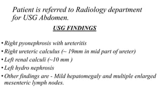
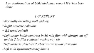
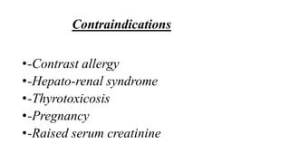
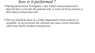
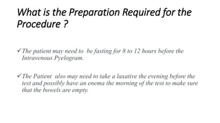
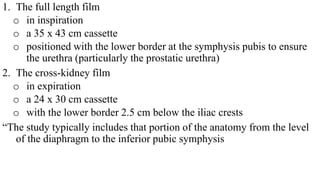
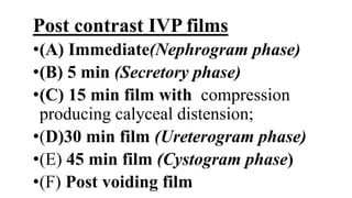
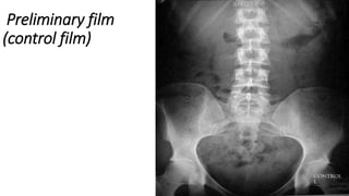
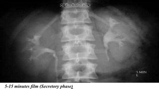
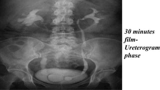
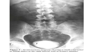
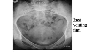
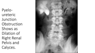
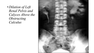
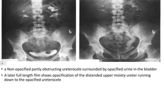
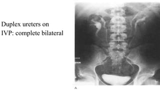
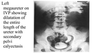
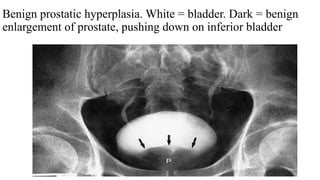
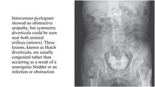
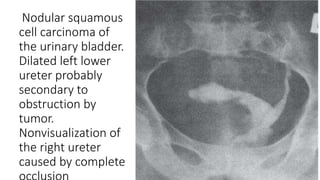
The document describes a case presentation of a 10-year-old male child who presented with abdominal pain. An ultrasound and intravenous pyelogram (IVP) were performed. The ultrasound found right pyonephrosis, right and left renal calculi, and left hydroureteronephrosis. The IVP confirmed these findings and also found a right ureteric calculus and question of a left ureteric stricture. The document then provides details on IVP, including how it is performed, indications, advantages, limitations, normal findings, and examples of various abnormalities that can be seen on an IVP.
Learn Barium Meal & Follow Through for the beginners from a Radiology Resident.For some image description please go through the text book "David Sutton" because i have described these image during my presentation Verbally..There are many animations used inside this presentation so to see all the pictures which are placed layer by layer with the help of animations you simple need to download this presentation first.... Thanx.
Barium meal ppt presentation is very important for radiology resident , radiologist and radiographers. this slide contents lots of barium image and technique, position, indication and modification and lots of information. this presentation help alot thanks .
A presentation about Imaging the urinary tract using contrast.
contains 45 slides, and covers the following methods :
1 - Antegrade urography
2 - Retrograde urography
3 - Retrograde cystography
4 - Voiding cystography
5 - Retrograde Urethrography
Intravenous urography is covered in a separate presentation, that you can read and download from here :
http://www.slideshare.net/abdallamutwakil/intravenous-urography-ivu-35107052
This presentation was prepared and presented by me in the tutorials of the Radiology Department of Sebha Medical Center.
Excretionurography
Also known as intravenous urography (IVU).
Most frequently employed radiologic investigation of renal rainage.
The contrast material is administered intravenously.
Best method for adults unless use of other methods is specified and is used in examinations of upper urinary tracts of infants and children.
The Indian economy is classified into different sectors to simplify the analysis and understanding of economic activities. For Class 10, it's essential to grasp the sectors of the Indian economy, understand their characteristics, and recognize their importance. This guide will provide detailed notes on the Sectors of the Indian Economy Class 10, using specific long-tail keywords to enhance comprehension.
For more information, visit-www.vavaclasses.com
How to Create Map Views in the Odoo 17 ERPCeline George
The map views are useful for providing a geographical representation of data. They allow users to visualize and analyze the data in a more intuitive manner.
How to Split Bills in the Odoo 17 POS ModuleCeline George
Bills have a main role in point of sale procedure. It will help to track sales, handling payments and giving receipts to customers. Bill splitting also has an important role in POS. For example, If some friends come together for dinner and if they want to divide the bill then it is possible by POS bill splitting. This slide will show how to split bills in odoo 17 POS.
Synthetic Fiber Construction in lab .pptxPavel ( NSTU)
Synthetic fiber production is a fascinating and complex field that blends chemistry, engineering, and environmental science. By understanding these aspects, students can gain a comprehensive view of synthetic fiber production, its impact on society and the environment, and the potential for future innovations. Synthetic fibers play a crucial role in modern society, impacting various aspects of daily life, industry, and the environment. ynthetic fibers are integral to modern life, offering a range of benefits from cost-effectiveness and versatility to innovative applications and performance characteristics. While they pose environmental challenges, ongoing research and development aim to create more sustainable and eco-friendly alternatives. Understanding the importance of synthetic fibers helps in appreciating their role in the economy, industry, and daily life, while also emphasizing the need for sustainable practices and innovation.
Ethnobotany and Ethnopharmacology:
Ethnobotany in herbal drug evaluation,
Impact of Ethnobotany in traditional medicine,
New development in herbals,
Bio-prospecting tools for drug discovery,
Role of Ethnopharmacology in drug evaluation,
Reverse Pharmacology.
Read| The latest issue of The Challenger is here! We are thrilled to announce that our school paper has qualified for the NATIONAL SCHOOLS PRESS CONFERENCE (NSPC) 2024. Thank you for your unwavering support and trust. Dive into the stories that made us stand out!
Welcome to TechSoup New Member Orientation and Q&A (May 2024).pdfTechSoup
In this webinar you will learn how your organization can access TechSoup's wide variety of product discount and donation programs. From hardware to software, we'll give you a tour of the tools available to help your nonprofit with productivity, collaboration, financial management, donor tracking, security, and more.
Welcome to TechSoup New Member Orientation and Q&A (May 2024).pdf
IVP Best presnetation
1. A case presentation on IVP
Speaker - Dr.Sachin
Guide- Dr.Kirti Rana Chaturvedi
2. •A patient Bheraram 10 year male child
presented to pediatric emergency with c/o
Abdominal pain since 9 days which increases
day by day.
•No h/o fever and vomiting.
•No significant past history and family history.
3. Patient is referred to Radiology department
for USG Abdomen.
USG FINDINGS
•Right pyonephrosis with ureteritis
•Right ureteric calculus (~ 19mm in mid part of ureter)
•Left renal calculi (~10 mm )
•Left hydro nephrosis
•Other findings are - Mild hepatomegaly and multiple enlarged
mesenteric lymph nodes.
4. For confirmation of USG abdomen report IVP has been
done.
IVP REPORT
•Normally excreting both kidney.
• Right ureteric calculus
• B/l renal calculi
• Left ureter holds contrast in 30 min film with abrupt cut off
and in 2 hr film contrast wash away s/o
?left ureteric stricture ? Aberrant vascular structure
-Left mild hydroureteronephrosis.
6. An intravenous pyelogram (IVP) is a special x-ray
examination of the kidneys, bladder, and ureters.
It is also called Intravenous Urogram.
An Intravenous Pyelogram can show the size, shape, and
position of the urinary tract, and it can evaluate the
collecting system inside the kidneys.
Intravenous Pyelogram can be done as a emergency
Procedure without any preparation.
7. Indications of IVP
An abdominal injury.
ureteric fistulas and strictures
Bladder and kidney infections.
Blood in the urine.
Flank pain (possibly due to kidney stones).
Tumors.
8. •Advantages of IVP
•-Detailed anatomy of the collecting systems
•-Demonstration of major calcification
•-Sensitive for acute obstruction
•-Low cost
9. Limitations of IVP
• -It depends on kidney function
• -Do not differentiate solid or cystic lesion
• -Requires contrast medium and radiation.
• -Missing small stones.
• -Quality of study may be limited by inadequate bowel
preparation, bowel ileus, swallowed air and technician
variability.
• -Inconvenience of a long filming sequence.
11. how is it performed ?
During Intravenous Pyelogram, a dye called contrast material is
injected into a vein into the patients arm. A series of X-ray pictures is
then taken at timed intervals.
The test should be done in a X Ray Department where a doctor is
available as on occasions the contrast can cause severe reactions
which may lead to medical emergencies.
12. What is the Preparation Required for the
Procedure ?
The patient may need to be fasting for 8 to 12 hours before the
Intravenous Pyelogram.
The Patient also may need to take a laxative the evening before the
test and possibly have an enema the morning of the test to make sure
that the bowels are empty.
13. The standard plain radiographic imaging of the
urinary tract is the
KUB (kidneys, ureters and bladder), which
consists of
I. a full length abdominal film and
II. an upper abdominal (cross-kidney) film
•taken with the patient supine using a low
voltage technique (60-65 kV) to maximise
soft-tissue contrast.
14. 1. The full length film
o in inspiration
o a 35 x 43 cm cassette
o positioned with the lower border at the symphysis pubis to ensure
the urethra (particularly the prostatic urethra)
2. The cross-kidney film
o in expiration
o a 24 x 30 cm cassette
o with the lower border 2.5 cm below the iliac crests
“The study typically includes that portion of the anatomy from the level
of the diaphragm to the inferior pubic symphysis
16. The outline of several anatomical structures can be seen
on the KUB
orenal,
opsoas and
obladder outlines,
1. the KUB is a relatively unreliable diagnostic tool
2. principal use is in the assessment of urinary tract
calculi.
3. It is, however, extremely unreliable in the diagnosis
of ureteric calculi, with an accuracy of only around
50%.
18. Contrast
LOCM 370 (LOCM = Low osmolar contrast material)
Adult dose = 50 – 100 ml
Pediatric dose = 1ml for each kg
19. Post contrast IVP films
•(A) Immediate(Nephrogram phase)
•(B) 5 min (Secretory phase)
•(C) 15 min film with compression
producing calyceal distension;
•(D)30 min film (Ureterogram phase)
•(E) 45 min film (Cystogram phase)
•(F) Post voiding film
21. Immediate film (Nephrogram phase)
•A.P. view of the renal areas to show the nephrogram, i.e.
the renal parenchyma opacified by the contrast medium
in the renal tubules.
• (taking it after injection equals about 10 to 14 seconds
which is the approximate arm-to-kidney time).
25. 30 minutes film (Ureterogram
phase) :
Is there any collecting systems and ureters dilatation or
filling defect? (normal ureter filling is rarely demonstrate
the whole ureter from proximal to distal as there is a
peristaltic wave )
27. •45 minutes film (Cystogram phase) :
•-Bladder size and shape
•-Contrast is filling the bladder or not
•-Bladder surface is smooth or rough
•-Is there any diverticlula, filling defect or prostate
indentation?
28.
29. •Post voiding film :
•-Residual urine
•-Contrast left on upper tract? (normally there is
no contrast left on upper urinary tract on post
voiding film)
31. IVP REPORTING FORMAT
• Indication
• Technique : an IVP was performed following the intravenous administration
of contrast.
• Comparison
• The scout films demonstrates—
• Bony shadowing
• Bowel gas pattern-adequates or inadequates bowel preparation.
• Renal shadowing
• Psoas shadowing
• Any evidences of masses or organomegaly.
32. Conti….
• Any radiopaque calculi visible in the kidney or along the ureteral tracts.
• Following the uneventful administration of …….cc”s of omnipaque 300
intravenously, the nephrograms and pyelograms are prompt and
bilateral. Kidneys are normal in size, shape and axis. There are no
masses evident.
• There is no dilatation or filling defects of the renal calyces or pelvis
bilaterally. The ureter are normal in course or calibre, without any
filling defects.
• The bladder is normal in size, shape and contour. No filling defects are
evident.
• There is no significant post void residual.
• Impression- normal IVP.
39. • a Non-opacified partly obstructing ureterocele surrounded by opacified urine in the bladder
• A later full length film shows opacification of the distended upper moiety ureter running
down to the opacified ureterocele
42. Benign prostatic hyperplasia. White = bladder. Dark = benign
enlargement of prostate, pushing down on inferior bladder
43. Intravenous pyelogram
showed no obstructive
uropathy, but symmetric
diverticula could be seen
near both ureteral
orifices (arrows). These
lesions, known as Hutch
diverticula, are usually
congenital rather than
occurring as a result of a
neurogenic bladder or an
infection or obstruction
44. Nodular squamous
cell carcinoma of
the urinary bladder.
Dilated left lower
ureter probably
secondary to
obstruction by
tumor.
Nonvisualization of
the right ureter
caused by complete