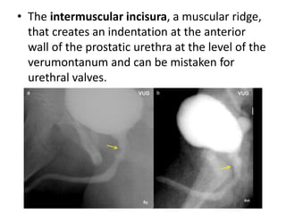
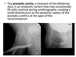
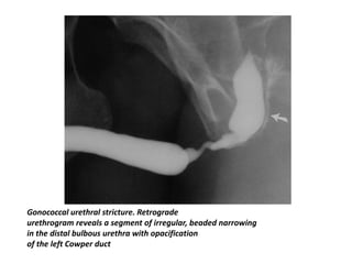
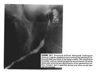
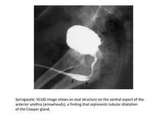
IMAGING OF URETHRAL
PATHOLOGIES
DR.D.SUNIL KUMAR
Normal Anatomy of the Urethra
• Male Urethra:
• The male urethra varies from 17.5 to 20 cm in length and consists of
anterior and posterior portions, each of which is subdivided into two
parts.
• The anterior urethra extends from the external meatus to the inferior
edge of the urogenital diaphragm, coursing through the corpus
spongiosum.
• The anterior urethra is conventionally divided into the penile (or
pendulous) and bulbous parts at the penoscrotal junction on the basis of
clinical and imaging findings.
• The proximal portion of the bulbous urethra is dilated and
termed the “sump” of the bulbous urethra; just proximal to
the sump, the bulbous urethra assumes a conical shape at
the bulbomembranous junction. This portion of the bulb is
known as the “cone.”
• The penoscrotal junction can be identified on
urethrograms by a mild angulation causedby the
suspensory ligament.
• The posterior urethra is divided into the prostatic and
membranous urethras.
• The prostatic urethra is approximately 3.5 cm long and
passes through the prostate slightly anterior to midline.
• The prostatic urethra tapers distally into the
membranous urethra, which is approximately 1–1.5 cm
long and ends at the inferior aspect of the urogenital
diaphragm.
• Verumontanum is a
1-cm-long ovoid
mound that lies in
the posterior wall
of the prostatic
urethra.
• The distal end of
the verumontanum
marks the proximal
boundary of the
membranous
urethra. This is also
the region ofthe
external sphincter
of the urethra.
• The distal
boundary of the
membranous
urethra is the
conical tip of the
bulbar urethra.
The verumontanum produces an oval filling
defect at the middle of the posterior
prostatic wall.
• The landmarks of the membranous urethra must be
recognized, so that it can be accurately located on
retrograde or voiding studies.
• If the membranous urethra can be identified, it will not
be confused for a stricture. Narrowing elsewhere in the
urethra will be defined clearly as separate from the
membranous urethra, and therefore, most likely
representative of a pathologic stricture.
• On voiding studies, the bladder neck opens widely and
becomes funnel-shaped. While the verumontanum
usually still can be seen, the proximal bulbar urethra
has less of a conical appearance. However, the
membranous urethra remains the narrowest segment
between these parts of the urethra, although it may
dilate up to 6 or 7 mm in diameter during voiding.
The membranous urethra lies specifically between two radiologic landmarks:
the distal end of the verumontanum (proximally-yellow arrow) and the conical end of
the bulbar urethra (distally-red arrow).
• The identification of the bulbomembranous junction
on a retrograde urethrogram is very important for
assessing patients with urethral disease as well as for
planning urologic procedures.
• When the posterior urethra is optimally opacified and
the verumontanum visible, the bulbomembranous
junction can be identified 1–1.5 cm distal to the
inferior margin of the verumontanum.
• When the posterior urethra is suboptimally opacified,
the bulbomembranous junction can be arbitrarily
localized where an imaginary line connecting the
inferior margins of the obturator foramina intersects
the urethra.
Urethrographic Techniques
• Conventional retrograde and voiding
urethrography still remain the best initial imaging
methods for the evaluation of urethral anatomy
and pathology.
• Anterior urethra is visualized on retrograde
urethrography and a voiding study is more
appropriate for the posterior urethra.
• In most cases though, it is necessary to perform
both studies in order to ensure that a significant
abnormality is not missed out or a normal variant
is not misunderstood as pathology, since the two
techniques have complementary roles.
MIMICS OF URETHRAL PATHOLOGY IN
RCUG
• Contraction or spasm of the
constrictor nudae muscle, a
deep musculotendinous
sling of the bulbocavernous
muscle, may cause anterior
or, less frequently,
circumferential indentation
of the proximal bulbous
urethra at retrograde
urethrography.
• This bulbous urethral
indentation should not be
confused with urethral
stricture
• Filling of the Cowper
ducts should not be
misinterpreted as
extravasation.
• However opacification
of the prostatic ducts,
Cowper ducts, and
periurethral Littre´
glands is often, but not
necessarily, associated
with urethral
inflammatory and
stricture disease.
• The intermuscular incisura, a muscular ridge,
that creates an indentation at the anterior
wall of the prostatic urethra at the level of the
verumontanum and can be mistaken for
urethral valves.
• The prostatic utricle, a remnant of the Müllerian
duct, is an anatomic variant that may occasionally
fill with contrast during urethrography, creating a
small diverticulum at the posterior aspect of the
prostatic urethra at the apex of the
verumontanum
Acquired Inflammatory Diseases
• Gonococcal and Nongonococcal Urethritis
• Complications associated with gonococcal urethritis are more
common and more serious than those associated with
nongonococcal urethritis and include urethral stricture, periurethral
abscess, and periurethral fistula.
• An estimated 15% of men with gonococcal urethritis go on to
develop stricture, with an interval of 2–30 years between infection
and the onset of obstructive symptoms.
• The typical urethrographic finding in gonococcal urethral stricture is
an irregular urethral narrowing several centimeters long.
• While the bulbar urethra is the most common area of occurrence,
gonorrheal strictures may occur anywhere in the anterior urethra or
may even involve the entire anterior urethra
Gonococcal urethral stricture. Retrograde
urethrogram reveals a segment of irregular, beaded narrowing
in the distal bulbous urethra with opacification
of the left Cowper duct
• If the disease has spread proximaly to the
membranous urethra, the normal cone shape of
the proximal bulbous urethra becomes
asymmetric and narrowed, giving an elongated
appearance to the membranous urethra.
• Abnormality of the normal convex cone shape of
the proximal bulbous urethra indicates scarring
extending into the membranous urethra.
• This radiologic finding is of prime importance to
the urologist, because surgical treatment may
involve cutting the scar tissue and consequently
the distal sphincter, which can result in iatrogenic
incontinence.
• Periurethral abscess is a life-threatening infection of the male urethra and
periurethral tissue and frequently a sequela of gonococcal infection,
urethral stricture disease, or urethral catheterization.
• Periurethral abscess arises initially when a Littre´ gland( mucus glands that
branch off the wall of the urethra of male) becomes obstructed by
inspissated pus or fibrosis.
• Pseudodiverticulum formation results from urethral communication with a
periurethral abscess.
• Because the tunica albuginea of the penis prevents the dorsal spread of
infection, the abscess tends to track ventrally along the corpus
spongiosum, where it is confined by the Buck fascia.
• However, when the Buck fascia is perforated, there can be extensive
necrosis of the subcutaneous tissue and fascia.
• An abscess that drains into the urethra may be demonstrated at
urethrography
• Ultrasonography (US) can demonstrate the presence of periurethral
abscess, and CT and MR imaging are helpful for assessing the extent of the
periurethral abscess and complications such as fasciitis and Fournier
gangrene.
Gonococcal urethral stricture
with periurethral
abscess. Retrograde
urethrogram shows a long
segment of irregular, beaded
narrowing in the bulbous
urethra with opacification of the
Littre´ glands (arrow).
Note the irregular periurethral
cavity originating from
the ventral aspect of the
bulbous urethra.
• Urethroperineal fistulas are most often the
consequence of a periurethral abscess. In
general, the initial abscess cavity contracts by
means of healing fibrosis, which leaves only
the narrow fistulous tract from the urethra to
the perineum.
• Consequently, urination usually occurs
through the perineal fistulas, which results in
the so-called “watering can perineum”.
• They are usually the result of tuberculosis and
schistosomiasis infections.
• High intraurethral pressure proximal to a stricture
not only results in dilation of the urethra, but also
can cause reflux of urine into the prostatic ducts.
• Ostia for these ducts, 30 to 40 in number, are
found in the floor of the prostatic urethra around
the verumontanum.
• This reflux may be massive and may allow
infection to enter the prostate, potentially
resulting in a prostatic abscess or formation of
multiple prostatic calculi.
Condyloma Acuminata
• Condyloma acuminata are caused by viral infection and
produce soft, sessile, squamous papillomas on the
penile glans and shaft and the prepuce.
• Urethral involvement occurs in 0.5%–5% of male
patients.
• The use of catheterization, instrumentation, and
retrograde urethrography is not recommended
because of the possibility of retrograde seeding.
• The diagnostic procedure of choice is voiding
cystourethrography.
• The typical urethrographic findings are multiple
papillary filling defects in the anterior urethra.
Condyloma acuminata. Retrograde
urethrogram
demonstrates multiple small filling defects in
the anterior urethra
Strictures of the Urethra
• In general, the term urethral stricture refers to a fibrous
scarring of the urethra caused by collagen and fibroblast
proliferation.
• The causes of anterior urethral strictures may be
inflammatory (eg, infectious urethritis, balanitis xerotica
obliterans), traumatic (straddle injury, iatrogenic
instrumentation) or congenital.
• The most common external cause of traumatic stricture is
straddle injury.
• Iatrogenic trauma to the urethra may result from pressure
necrosis at fixed points in the urethra from indwelling
catheters.
• Instrumentation-related strictures usually occur in the
bulbomembranous region and, less commonly, at the
penoscrotal junction.
• Alternatively, posterior urethral stricture is
often an obliterative process that occurs as a
result of urethral distraction or disruption
caused by either trauma or surgery.
• Iatrogenic stricture of the prostatic posterior
urethra (“bladder neck contracture”) usually
occurs after transurethral resection of the
prostate or open radical prostatectomy.
• Retrograde urethrography is the primary method
used to image anterior urethral stricture
• The length of the stricture will be underestimated
if the patient is not placed in a steep oblique
position for retrograde urethrography.
• Sonourethrography is best used adjunctively to
guide treatment planning in patients with known
bulbous urethral strictures and has been reported
to be more accurate than retrograde
urethrography for estimating the length of
urethral strictures
Urethral Calculi
• Most urethral calculi consist of small stones
expelled from the bladder into the urethra during
voiding; these are referred to as migrant calculi.
• Occasionally, however, a stone may be large
enough to become lodged at a point of urethral
narrowing such as the membranous urethra.
• Retrograde urethrography will usually depict a
rounded filling defect in the urethra. On a
preprocedural low abdominal radiograph, the
stone may be identified before contrast material
is injected.
Calculi associated with urethral stricture. (a)
Conventional radiograph reveals faintly
opaque stones
projected over the penis (arrows). (b)
Retrograde urethrogram demonstrates the
stones (arrowhead) lying in a segment
of anterior urethral stricture.
Sonourethrography in Evaluation of
Abnormalities of Anterior Male
Urethra.
• The technique involved is the use of a 5 MHz linear
array transducer applied to the dorsal surface of the
penis.
• Images are obtained during retrograde instillation of
normal saline.
• As a dynamic, three-dimensional study, which can be
repeated without radiation exposure,
sonourethrography offers important advantages over
conventional techniques.
• Panoramic reconstruction of the sonographic images is
done for better understanding of urethral pathologies.
Normal sonourethrography appearance of
anterior urethra longitudinal view.
Long penile urethral stricture with
periurethral abscess and false tract on
sonourethrography.
Patient with short segment bulbar urethral
stricture.
Patient with penile urethral mucosal
irregularity and bulbar stricture on
Sonourethrography . A case of chronic
urethritis
Female Urethra
• The female urethra is 4 cm long and extends
from the bladder neck at the urethrovesical
junction to the vestibule, where it forms the
external meatus between the labia minora.
• Many small periurethral glands open into the
urethra. Distally, these glands group together
on either side of the urethra (Skene glands)
and empty through two small ducts to either
side of the external meatus.
Acquired Female
Urethral Diverticula
• The diagnosis of female urethral diverticula is being made with greater
frequency owing to awareness of the condition and of its coexistence with
stress urinary incontinence and urinary infection.
• Urethral diverticulum has been reported in 1.4% of women with stress
urinary incontinence.
• Female urethral diverticulum is currently thought to be acquired and is
attributed to the rupture of dilated and infected periurethral glands, which
results in pseudodiverticulum formation.
• Urethral diverticula are usually located posterolateral to the urethra.
• When a diverticulum originates from the proximal urethra, there may be a
mass effect on the bladder base similar to that seen in elderly men with
an enlarged prostate, a finding that is referred to as the “female prostate”
sign.
• The classic manifestation of urethral diverticulum hasbeen described as
the three Ds (dysuria, postvoid dribbling, and dyspareunia).
• The diagnosis is usually made with voiding
cystourethrography or cross-sectional imaging.
• A wide-neck communicating diverticulum can
also be demonstrated on a postvoiding image
obtained during excretory urography.
Female urethral diverticulum.
Postvoiding
image obtained during excretory
urography demonstrates
a contrast material–filled urethral
diverticulum
• Double balloon
(positive pressure)
urethrography is more
sensitive than voiding
cystourethrography
and may allow
contrast material to be
forced into a
diverticular ostium by
creating a relatively
closed urethral system
in which the contrast
material passes into
the defect by means of
concentric pressure
rather than
opportunistic stream
diversion.
• Transvaginal US has also been reported to be
helpful for identifying urethral diverticula in
women.
• US can demonstrate a relatively echo-free cavity
adjacent to the urethra and may also
demonstrate intracavitary debris or surrounding
inflammatory edema.
• MR imaging is more sensitive than voiding
cystourethrography and double balloon
urethrography in the detection of urethral
diverticula, particularly in the detection of
narrow-neck noncommunicating urethral
diverticula.
• The complex appearance of urethral diverticula is
best demonstrated at MR imaging performed
with pelvic phased array coils.
• The use of endovaginal or endorectal coils at MR
imaging can provide high-resolution details of
urethral diverticula
Urethral diverticulum (female prostate sign). Sagittal fast spin-echo T2-weighted MR image
demonstrates a large diverticulum surrounding the urethra (arrow), with a septum that
results in an impression at the bladder base. B bladder, S pubic symphysis.
Anomalies of the Urethra
• Posterior Urethral Valves
• The most common congenital obstructive
lesion of the urethra, occurring only in
phenotypic boys.
• Posterior urethral valves result from the
formation of a thick, valvelike membrane from
tissue of wolffian duct origin that courses
obliquely from the verumontanum to the
most distal portion of the prostatic urethra
• VCUG is the best imaging technique for the
diagnosis of posterior urethral valves.
• Radiologic findings include dilatation and
elongation of the posterior urethra and,
occasionally, a linear radiolucent band
corresponding to the valve.
• The bladder neck becomes hypertrophic and
appears narrow in relation to the dilated
posterior urethra.
• VUR occurs in 50% of patients.
• Bladder trabeculation, hypertrophy, and
diverticula are also demonstrated at VCUG
Posterior urethral valve. (a) VCUG image shows the typical distention of the posterior
urethra and abrupt change in caliber in the region of the external sphincter (arrow) at the
junction of the posterior and anterior urethra. Note also the bladder wall thickening and
trabeculation. (b) Transverse US image through the bladder shows significant thickening of
the bladder wall
(arrows).
Posterior urethral valves with
bilateral vesicoureteral reflux.
Posterior urethral valves. An image from a VCUG demonstrates a thick-walled trabeculated
bladder and dilatation of the posterior urethra (long arrow), and the location of the valve
(small arrow)
• Ultrasound
• Antenatal ultrasound
• On antenatal ultrasound the appearance is that of
marked distention and hypertrophy of the bladder,
hydronephrosis and hydroureter may or may not be
present
• in severe cases oligohydramnios and renal dysplasia.
• keyhole sign may be seen on ultrasound due to the
distention of both the bladder and the urethra
immediately proximal to the valve
• Unfortunately such findings are generally not seen
before 26 weeks of gestation, and as such are not
frequently identified on routine morphology screening,
usually carried out around 18 weeks gestation
Typical key-hole appearance of urinary bladder
when seen in long axis.
Two ' stomach bubbles' are posterior to
kidney and abutting them. They should be
urinoma.
• Postnatal ultrasound
• Following birth, findings are the same as those on
antenatal ultrasound, although as patients who present
after birth usually have less severe obstruction, the
features may be less evident:
• examination of the posterior urethra can be performed
longitudinally through the perineum. Ideally this is
performed during micturition (which may take some
patience) at which time the proximal urethra can be
seen to dilate;diameter of more than 6mm is
considered abnormal and is highly specific and
sensitive to the diagnosis (sensitivity 100%, specificity
89%, positive predictive value 88%)
• additionally the valve may actually be seen as an
echogenic line
Urethral sonogram showing
a linear echogenic line (arrow) within the
urethral lumen at the transition zone
between the proximal urethra and the more
distal urethra with widening of the
proximal urethra and bladder neck, consistent
with a PUV
Anterior Urethral Valves
• Anterior urethral valves are rare congenital anomalies
that cause lower urinary tract obstruction in children.
• They can occur as an isolated entity or in association
with a proximal diverticulum
• Anterior urethral valves may be found anywhere in the
anterior urethra. Forty percent of the valves are
located in the bulbar urethra, 30% at the penoscrotal
junction, and 30% in the pendulous urethra.
• VCUG is the diagnostic modality of choice for anterior
urethral valves. Typically, the urethra appears dilated
proximal to the valve and narrowed distal to it
• In addition to demonstrating a lesion in the
urethra, VCUG may also reveal an associated
anomaly. VUR has been reported in one-third of
cases and upper tract deterioration in one-half.
Endoscopic examination of the urethra usually
helps confirm the diagnosis.
Anterior urethral valve. VCUG
image shows urethral dilatation proximal to
an anterior urethral valve (arrow) and
narrowing
distal to it. Note the abrupt change in the
caliber of the urethra below the valve.
Congenital urethral stricture
• Although most urethral strictures in males are
posttraumatic, there are rare reports of congenital urethral
strictures of the bulbous urethra in neonates and older
children
• It is secondary to a failure of canalization of the cloacal
membrane during fetal development
• Diagnosis is by VCUG or retrograde urethrography. VCUG
will demonstrate focal narrowing of the bulbous urethra,
while retrograde urethrography will confirm a normal
penile urethra. Other causes of urethral stricture, including
trauma, must be excluded.
• The site of urethral narrowing in congenital urethral
stricture is distal to the external urethral sphincter, which
differentiates this entity from PUV
Congenital urethral stricture.
A markedly dilated urethra is
seen proximal to a
congenital stricture in the
bulbous urethra.
Retrograde urethrography in
this patient (not shown)
demonstrated a
focal narrowing at the
bulbous urethra with a
normal penile urethra.
The site of obstruction is
more distal than that seen
with PUV
Congenital urethral polyps
• Congenital urethral polyps are benign and arise from
the prostatic urethra near the verumontanum.
• Because they have a stalk, these polyps are mobile and
can move proximally into the bladder or distally into
the bulbous urethra.
• They can be a cause of urethral obstruction or
bleeding.
• VCUG is diagnostic and demonstrates a mobile filling
defect in the bladder neck or below the
verumontanum .
• Endoscopic resection is the treatment of choice
Urethral polyp. a Image from a VCUG
demonstrates a
polypoid filling defect arising from the
prostatic urethra (arrow). b
Note change of position of the filling defect
(arrow) during the
examination
Urethral Duplication
• Urethral duplication is a rare anomaly frequently seen
in association with other anomalies including
hypospadias, epispadias, cleft lip and palate, congenital
heart disease, tracheoesophageal fistula, imperforate
anus and musculoskeletal anomalies
• Duplication commonly occurs along the sagittal plane.
• The ventral urethra is the more functional urethra and
contains the verumontanum and sphincters.
• When urethral duplication is present along the coronal
plane, bladder duplication is always present
• Urethral duplication can be classified into
three types using Effmann’s classification.
– In type I, there is partial duplication of the
urethra.
– In type II, there is complete duplication of the
urethra.
– Type III urethral duplication consists of complete
duplication of the urethra and bladder
A perineal or rectal fistula (Y-type fistula) associated with a stenotic, normally located
penile urethra is placed in the IIa category.
Type IIB
VCUG image shows two different
urethral channels
(1, 2) arising from two different
bladder orifices
(arrows). In the midurethra, the two
channels
join to form a single anterior urethra.
Complete urethral duplication. (a) VCUG image obtained in a 6-month-old boy shows two
complete urethral channels. The duplicate urethra (arrowheads) is located in the dorsal
surface of the penis. The ventral urethra () is normal.
Congenital urethroperineal fistula
• Although congenital urethroperineal fistula (CUF)
resembles Y-type urethral duplication, it should be
considered a separate entity.
• In Y-type urethral duplication the ventral urethra opens
to the perineum and, as in all urethral duplications, is
the functional urethra.
• In contrast, in CUF, the dorsal urethra is the functional
urethra and the ventral urethra (fistula) is hypoplastic.
• The differentiation between Y-type duplication and
CUF is particularly important in the surgical
management of these patients.
• In Y-duplication the functional ventral channel
should not be resected, while in the CUF
resection of the ventral channel is curative
• Diagnosis is made by retrograde
urethrography or VCUG to determine the
dominant urethra. In the case of congenital
urethroperineal fistula, voiding will be
predominantly through the dorsal urethra.
Megalourethra
• Megalourethra is caused by defective formation
of the corpus spongiosum and corpora cavernosa
secondary to a mesodermal defect.
• Megalourethra is often associated with other
congenital abnormalities including
cryptorchidism, renal agenesis etc.
• These patients have a functional rather than
anatomic urethral obstruction, causing stasis and
back pressure into the upper urinary tracts.
Diagnosis is by VCUG.
• Reconstructive surgery is required.
Megalourethra. Single view
from a VCUG in a boy with
partial sacral agenesis
demonstrates focal dilatation
of the urethra
• There are two
types of
megalourethra:
– scaphoid, in
which there is
ventral
urethral
dilatation and
hypoplasia of
the corpus
spongiosum
oblique VCUG
image reveals a huge scaphoid, contrast-filled
structure at the distal
penile urethra (*); it is more prominent
ventrally. The posterior urethra
and bulbar urethra are normal
– fusiform, in which
there is
circumferential
urethral dilatation
and hypoplasia of the
corpus spongiosa
and corpora
cavernosa.
Fusiform megalourethra in an infant. Lateral
VCUG image
reveals an extensively dilated anterior and
posterior urethra
Urethral Diverticula
• Anterior urethral diverticulum, although uncommon, is
the second most common cause of congenital urethral
obstruction in boys.
• A diverticulum of the anterior urethra develops on the
ventral surface of the penile urethra as a result of
either incomplete development of the corpus
spongiosum focally or incomplete fusion of a segment
of the urethral plate.
• A lip of tissue may be seen around the diverticulum. As
the diverticulum distends, the lip of tissue is pressed
against the urethral wall and results in a valve like
obstruction .
Anterior urethral diverticulum. Arrow points
to a diverticulum
arising from and communicating with the
ventral aspect of the
urethra
• Most children are diagnosed in infancy with
dribbling-type micturation or infection.
• The dribbling may be due to emptying of the
diverticulum or to overflow incontinence. If the
obstruction is distal, ballooning of the urethra
may occur with voiding. VCUG is the key to
diagnosis. During VCUG, the typical saccular
diverticulum of the anterior urethra fills with
contrast material and appears as an oval
structure on the ventral aspect of the anterior
urethra
A huge anterior urethral diverticulum arising from the bulbar
urethra in a 10-year-old male child. The boy had a history of a swelling
at the penoscrotal region during micturition. An oblique VCUG image
reveals a large ventral diverticulum (*) with a narrow neck
Cowper’s syringocele
• Cowper’s glands are
small paired glands
located dorsal to and on
either side of the
membranous urethra.
They secrete a mucous
substance during
ejaculation that acts as a
lubricant. The main duct
draining Cowper’s
glands drain below the
urogenital diaphragm
into the ventral aspect
of the bulbous urethra
• During VCUG, the
main duct and
Cowper’s glands can
fill with contrast
material and appear
as a tubular channel
paralleling the ventral
aspect of the
undersurface of the
bulbous membranous
urethra and ending at
the urogenital
diaphragm.
• This finding is usually
of no clinical
significance.
• Cowper’s syringocele, a rare anomaly, occurs when
there is dilatation of the main draining duct.
• It is usually congenital but can be seen in adults too.
• Cowper’s syringocele can also be classified as open or
closed, open if it communicates with the urethra and
closed if it does not.
• Open syringoceles present with post-void dribbling,
fever, urethral discharge, perineal pain, hematuria and
urinary incontinence.
• Closed syringoceles is usually asymptomatic until it is
not infected. In this case, obstructive voiding
symptoms, dysuria, urinary retention and perineal pain
can be present
• Diagnosis is made by VCUG, retrograde urethrography
or urethrocystoscopy.
Syringocele. VCUG image shows an oval structure on the ventral aspect of the
anterior urethra (arrowheads), a finding that represents tubular dilatation
of the Cowper gland.
• The main differential diagnoses are with:
• - urethral fistulae (fistulae are irregular in
contours with a tendency to enlarge)
• - contrast-media spillage and extravasation;
• - urethral duplications;
• - diverticula;
• - overlapping images.
Imaging of urethral pathologies

Imaging of urethral pathologies

  • 1.
  • 2.
    Normal Anatomy ofthe Urethra • Male Urethra: • The male urethra varies from 17.5 to 20 cm in length and consists of anterior and posterior portions, each of which is subdivided into two parts. • The anterior urethra extends from the external meatus to the inferior edge of the urogenital diaphragm, coursing through the corpus spongiosum. • The anterior urethra is conventionally divided into the penile (or pendulous) and bulbous parts at the penoscrotal junction on the basis of clinical and imaging findings.
  • 3.
    • The proximalportion of the bulbous urethra is dilated and termed the “sump” of the bulbous urethra; just proximal to the sump, the bulbous urethra assumes a conical shape at the bulbomembranous junction. This portion of the bulb is known as the “cone.” • The penoscrotal junction can be identified on urethrograms by a mild angulation causedby the suspensory ligament.
  • 5.
    • The posteriorurethra is divided into the prostatic and membranous urethras. • The prostatic urethra is approximately 3.5 cm long and passes through the prostate slightly anterior to midline. • The prostatic urethra tapers distally into the membranous urethra, which is approximately 1–1.5 cm long and ends at the inferior aspect of the urogenital diaphragm.
  • 7.
    • Verumontanum isa 1-cm-long ovoid mound that lies in the posterior wall of the prostatic urethra. • The distal end of the verumontanum marks the proximal boundary of the membranous urethra. This is also the region ofthe external sphincter of the urethra. • The distal boundary of the membranous urethra is the conical tip of the bulbar urethra.
  • 8.
    The verumontanum producesan oval filling defect at the middle of the posterior prostatic wall.
  • 9.
    • The landmarksof the membranous urethra must be recognized, so that it can be accurately located on retrograde or voiding studies. • If the membranous urethra can be identified, it will not be confused for a stricture. Narrowing elsewhere in the urethra will be defined clearly as separate from the membranous urethra, and therefore, most likely representative of a pathologic stricture. • On voiding studies, the bladder neck opens widely and becomes funnel-shaped. While the verumontanum usually still can be seen, the proximal bulbar urethra has less of a conical appearance. However, the membranous urethra remains the narrowest segment between these parts of the urethra, although it may dilate up to 6 or 7 mm in diameter during voiding.
  • 10.
    The membranous urethralies specifically between two radiologic landmarks: the distal end of the verumontanum (proximally-yellow arrow) and the conical end of the bulbar urethra (distally-red arrow).
  • 11.
    • The identificationof the bulbomembranous junction on a retrograde urethrogram is very important for assessing patients with urethral disease as well as for planning urologic procedures. • When the posterior urethra is optimally opacified and the verumontanum visible, the bulbomembranous junction can be identified 1–1.5 cm distal to the inferior margin of the verumontanum. • When the posterior urethra is suboptimally opacified, the bulbomembranous junction can be arbitrarily localized where an imaginary line connecting the inferior margins of the obturator foramina intersects the urethra.
  • 12.
    Urethrographic Techniques • Conventionalretrograde and voiding urethrography still remain the best initial imaging methods for the evaluation of urethral anatomy and pathology. • Anterior urethra is visualized on retrograde urethrography and a voiding study is more appropriate for the posterior urethra. • In most cases though, it is necessary to perform both studies in order to ensure that a significant abnormality is not missed out or a normal variant is not misunderstood as pathology, since the two techniques have complementary roles.
  • 13.
    MIMICS OF URETHRALPATHOLOGY IN RCUG • Contraction or spasm of the constrictor nudae muscle, a deep musculotendinous sling of the bulbocavernous muscle, may cause anterior or, less frequently, circumferential indentation of the proximal bulbous urethra at retrograde urethrography. • This bulbous urethral indentation should not be confused with urethral stricture
  • 14.
    • Filling ofthe Cowper ducts should not be misinterpreted as extravasation. • However opacification of the prostatic ducts, Cowper ducts, and periurethral Littre´ glands is often, but not necessarily, associated with urethral inflammatory and stricture disease.
  • 15.
    • The intermuscularincisura, a muscular ridge, that creates an indentation at the anterior wall of the prostatic urethra at the level of the verumontanum and can be mistaken for urethral valves.
  • 16.
    • The prostaticutricle, a remnant of the Müllerian duct, is an anatomic variant that may occasionally fill with contrast during urethrography, creating a small diverticulum at the posterior aspect of the prostatic urethra at the apex of the verumontanum
  • 17.
    Acquired Inflammatory Diseases •Gonococcal and Nongonococcal Urethritis • Complications associated with gonococcal urethritis are more common and more serious than those associated with nongonococcal urethritis and include urethral stricture, periurethral abscess, and periurethral fistula. • An estimated 15% of men with gonococcal urethritis go on to develop stricture, with an interval of 2–30 years between infection and the onset of obstructive symptoms. • The typical urethrographic finding in gonococcal urethral stricture is an irregular urethral narrowing several centimeters long. • While the bulbar urethra is the most common area of occurrence, gonorrheal strictures may occur anywhere in the anterior urethra or may even involve the entire anterior urethra
  • 18.
    Gonococcal urethral stricture.Retrograde urethrogram reveals a segment of irregular, beaded narrowing in the distal bulbous urethra with opacification of the left Cowper duct
  • 20.
    • If thedisease has spread proximaly to the membranous urethra, the normal cone shape of the proximal bulbous urethra becomes asymmetric and narrowed, giving an elongated appearance to the membranous urethra. • Abnormality of the normal convex cone shape of the proximal bulbous urethra indicates scarring extending into the membranous urethra. • This radiologic finding is of prime importance to the urologist, because surgical treatment may involve cutting the scar tissue and consequently the distal sphincter, which can result in iatrogenic incontinence.
  • 22.
    • Periurethral abscessis a life-threatening infection of the male urethra and periurethral tissue and frequently a sequela of gonococcal infection, urethral stricture disease, or urethral catheterization. • Periurethral abscess arises initially when a Littre´ gland( mucus glands that branch off the wall of the urethra of male) becomes obstructed by inspissated pus or fibrosis. • Pseudodiverticulum formation results from urethral communication with a periurethral abscess. • Because the tunica albuginea of the penis prevents the dorsal spread of infection, the abscess tends to track ventrally along the corpus spongiosum, where it is confined by the Buck fascia. • However, when the Buck fascia is perforated, there can be extensive necrosis of the subcutaneous tissue and fascia.
  • 23.
    • An abscessthat drains into the urethra may be demonstrated at urethrography • Ultrasonography (US) can demonstrate the presence of periurethral abscess, and CT and MR imaging are helpful for assessing the extent of the periurethral abscess and complications such as fasciitis and Fournier gangrene. Gonococcal urethral stricture with periurethral abscess. Retrograde urethrogram shows a long segment of irregular, beaded narrowing in the bulbous urethra with opacification of the Littre´ glands (arrow). Note the irregular periurethral cavity originating from the ventral aspect of the bulbous urethra.
  • 24.
    • Urethroperineal fistulasare most often the consequence of a periurethral abscess. In general, the initial abscess cavity contracts by means of healing fibrosis, which leaves only the narrow fistulous tract from the urethra to the perineum. • Consequently, urination usually occurs through the perineal fistulas, which results in the so-called “watering can perineum”. • They are usually the result of tuberculosis and schistosomiasis infections.
  • 26.
    • High intraurethralpressure proximal to a stricture not only results in dilation of the urethra, but also can cause reflux of urine into the prostatic ducts. • Ostia for these ducts, 30 to 40 in number, are found in the floor of the prostatic urethra around the verumontanum. • This reflux may be massive and may allow infection to enter the prostate, potentially resulting in a prostatic abscess or formation of multiple prostatic calculi.
  • 28.
    Condyloma Acuminata • Condylomaacuminata are caused by viral infection and produce soft, sessile, squamous papillomas on the penile glans and shaft and the prepuce. • Urethral involvement occurs in 0.5%–5% of male patients. • The use of catheterization, instrumentation, and retrograde urethrography is not recommended because of the possibility of retrograde seeding. • The diagnostic procedure of choice is voiding cystourethrography. • The typical urethrographic findings are multiple papillary filling defects in the anterior urethra.
  • 29.
    Condyloma acuminata. Retrograde urethrogram demonstratesmultiple small filling defects in the anterior urethra
  • 30.
    Strictures of theUrethra • In general, the term urethral stricture refers to a fibrous scarring of the urethra caused by collagen and fibroblast proliferation. • The causes of anterior urethral strictures may be inflammatory (eg, infectious urethritis, balanitis xerotica obliterans), traumatic (straddle injury, iatrogenic instrumentation) or congenital. • The most common external cause of traumatic stricture is straddle injury. • Iatrogenic trauma to the urethra may result from pressure necrosis at fixed points in the urethra from indwelling catheters. • Instrumentation-related strictures usually occur in the bulbomembranous region and, less commonly, at the penoscrotal junction.
  • 33.
    • Alternatively, posteriorurethral stricture is often an obliterative process that occurs as a result of urethral distraction or disruption caused by either trauma or surgery. • Iatrogenic stricture of the prostatic posterior urethra (“bladder neck contracture”) usually occurs after transurethral resection of the prostate or open radical prostatectomy.
  • 34.
    • Retrograde urethrographyis the primary method used to image anterior urethral stricture • The length of the stricture will be underestimated if the patient is not placed in a steep oblique position for retrograde urethrography. • Sonourethrography is best used adjunctively to guide treatment planning in patients with known bulbous urethral strictures and has been reported to be more accurate than retrograde urethrography for estimating the length of urethral strictures
  • 35.
    Urethral Calculi • Mosturethral calculi consist of small stones expelled from the bladder into the urethra during voiding; these are referred to as migrant calculi. • Occasionally, however, a stone may be large enough to become lodged at a point of urethral narrowing such as the membranous urethra. • Retrograde urethrography will usually depict a rounded filling defect in the urethra. On a preprocedural low abdominal radiograph, the stone may be identified before contrast material is injected.
  • 36.
    Calculi associated withurethral stricture. (a) Conventional radiograph reveals faintly opaque stones projected over the penis (arrows). (b) Retrograde urethrogram demonstrates the stones (arrowhead) lying in a segment of anterior urethral stricture.
  • 37.
    Sonourethrography in Evaluationof Abnormalities of Anterior Male Urethra. • The technique involved is the use of a 5 MHz linear array transducer applied to the dorsal surface of the penis. • Images are obtained during retrograde instillation of normal saline. • As a dynamic, three-dimensional study, which can be repeated without radiation exposure, sonourethrography offers important advantages over conventional techniques. • Panoramic reconstruction of the sonographic images is done for better understanding of urethral pathologies.
  • 38.
    Normal sonourethrography appearanceof anterior urethra longitudinal view.
  • 39.
    Long penile urethralstricture with periurethral abscess and false tract on sonourethrography.
  • 40.
    Patient with shortsegment bulbar urethral stricture.
  • 41.
    Patient with penileurethral mucosal irregularity and bulbar stricture on Sonourethrography . A case of chronic urethritis
  • 42.
    Female Urethra • Thefemale urethra is 4 cm long and extends from the bladder neck at the urethrovesical junction to the vestibule, where it forms the external meatus between the labia minora. • Many small periurethral glands open into the urethra. Distally, these glands group together on either side of the urethra (Skene glands) and empty through two small ducts to either side of the external meatus.
  • 43.
    Acquired Female Urethral Diverticula •The diagnosis of female urethral diverticula is being made with greater frequency owing to awareness of the condition and of its coexistence with stress urinary incontinence and urinary infection. • Urethral diverticulum has been reported in 1.4% of women with stress urinary incontinence. • Female urethral diverticulum is currently thought to be acquired and is attributed to the rupture of dilated and infected periurethral glands, which results in pseudodiverticulum formation. • Urethral diverticula are usually located posterolateral to the urethra. • When a diverticulum originates from the proximal urethra, there may be a mass effect on the bladder base similar to that seen in elderly men with an enlarged prostate, a finding that is referred to as the “female prostate” sign. • The classic manifestation of urethral diverticulum hasbeen described as the three Ds (dysuria, postvoid dribbling, and dyspareunia).
  • 44.
    • The diagnosisis usually made with voiding cystourethrography or cross-sectional imaging. • A wide-neck communicating diverticulum can also be demonstrated on a postvoiding image obtained during excretory urography. Female urethral diverticulum. Postvoiding image obtained during excretory urography demonstrates a contrast material–filled urethral diverticulum
  • 45.
    • Double balloon (positivepressure) urethrography is more sensitive than voiding cystourethrography and may allow contrast material to be forced into a diverticular ostium by creating a relatively closed urethral system in which the contrast material passes into the defect by means of concentric pressure rather than opportunistic stream diversion.
  • 46.
    • Transvaginal UShas also been reported to be helpful for identifying urethral diverticula in women. • US can demonstrate a relatively echo-free cavity adjacent to the urethra and may also demonstrate intracavitary debris or surrounding inflammatory edema.
  • 47.
    • MR imagingis more sensitive than voiding cystourethrography and double balloon urethrography in the detection of urethral diverticula, particularly in the detection of narrow-neck noncommunicating urethral diverticula. • The complex appearance of urethral diverticula is best demonstrated at MR imaging performed with pelvic phased array coils. • The use of endovaginal or endorectal coils at MR imaging can provide high-resolution details of urethral diverticula
  • 48.
    Urethral diverticulum (femaleprostate sign). Sagittal fast spin-echo T2-weighted MR image demonstrates a large diverticulum surrounding the urethra (arrow), with a septum that results in an impression at the bladder base. B bladder, S pubic symphysis.
  • 49.
    Anomalies of theUrethra • Posterior Urethral Valves • The most common congenital obstructive lesion of the urethra, occurring only in phenotypic boys. • Posterior urethral valves result from the formation of a thick, valvelike membrane from tissue of wolffian duct origin that courses obliquely from the verumontanum to the most distal portion of the prostatic urethra
  • 50.
    • VCUG isthe best imaging technique for the diagnosis of posterior urethral valves. • Radiologic findings include dilatation and elongation of the posterior urethra and, occasionally, a linear radiolucent band corresponding to the valve. • The bladder neck becomes hypertrophic and appears narrow in relation to the dilated posterior urethra. • VUR occurs in 50% of patients. • Bladder trabeculation, hypertrophy, and diverticula are also demonstrated at VCUG
  • 51.
    Posterior urethral valve.(a) VCUG image shows the typical distention of the posterior urethra and abrupt change in caliber in the region of the external sphincter (arrow) at the junction of the posterior and anterior urethra. Note also the bladder wall thickening and trabeculation. (b) Transverse US image through the bladder shows significant thickening of the bladder wall (arrows).
  • 52.
    Posterior urethral valveswith bilateral vesicoureteral reflux.
  • 53.
    Posterior urethral valves.An image from a VCUG demonstrates a thick-walled trabeculated bladder and dilatation of the posterior urethra (long arrow), and the location of the valve (small arrow)
  • 54.
    • Ultrasound • Antenatalultrasound • On antenatal ultrasound the appearance is that of marked distention and hypertrophy of the bladder, hydronephrosis and hydroureter may or may not be present • in severe cases oligohydramnios and renal dysplasia. • keyhole sign may be seen on ultrasound due to the distention of both the bladder and the urethra immediately proximal to the valve • Unfortunately such findings are generally not seen before 26 weeks of gestation, and as such are not frequently identified on routine morphology screening, usually carried out around 18 weeks gestation
  • 55.
    Typical key-hole appearanceof urinary bladder when seen in long axis.
  • 56.
    Two ' stomachbubbles' are posterior to kidney and abutting them. They should be urinoma.
  • 57.
    • Postnatal ultrasound •Following birth, findings are the same as those on antenatal ultrasound, although as patients who present after birth usually have less severe obstruction, the features may be less evident: • examination of the posterior urethra can be performed longitudinally through the perineum. Ideally this is performed during micturition (which may take some patience) at which time the proximal urethra can be seen to dilate;diameter of more than 6mm is considered abnormal and is highly specific and sensitive to the diagnosis (sensitivity 100%, specificity 89%, positive predictive value 88%) • additionally the valve may actually be seen as an echogenic line
  • 58.
    Urethral sonogram showing alinear echogenic line (arrow) within the urethral lumen at the transition zone between the proximal urethra and the more distal urethra with widening of the proximal urethra and bladder neck, consistent with a PUV
  • 59.
    Anterior Urethral Valves •Anterior urethral valves are rare congenital anomalies that cause lower urinary tract obstruction in children. • They can occur as an isolated entity or in association with a proximal diverticulum • Anterior urethral valves may be found anywhere in the anterior urethra. Forty percent of the valves are located in the bulbar urethra, 30% at the penoscrotal junction, and 30% in the pendulous urethra. • VCUG is the diagnostic modality of choice for anterior urethral valves. Typically, the urethra appears dilated proximal to the valve and narrowed distal to it
  • 60.
    • In additionto demonstrating a lesion in the urethra, VCUG may also reveal an associated anomaly. VUR has been reported in one-third of cases and upper tract deterioration in one-half. Endoscopic examination of the urethra usually helps confirm the diagnosis. Anterior urethral valve. VCUG image shows urethral dilatation proximal to an anterior urethral valve (arrow) and narrowing distal to it. Note the abrupt change in the caliber of the urethra below the valve.
  • 61.
    Congenital urethral stricture •Although most urethral strictures in males are posttraumatic, there are rare reports of congenital urethral strictures of the bulbous urethra in neonates and older children • It is secondary to a failure of canalization of the cloacal membrane during fetal development • Diagnosis is by VCUG or retrograde urethrography. VCUG will demonstrate focal narrowing of the bulbous urethra, while retrograde urethrography will confirm a normal penile urethra. Other causes of urethral stricture, including trauma, must be excluded. • The site of urethral narrowing in congenital urethral stricture is distal to the external urethral sphincter, which differentiates this entity from PUV
  • 62.
    Congenital urethral stricture. Amarkedly dilated urethra is seen proximal to a congenital stricture in the bulbous urethra. Retrograde urethrography in this patient (not shown) demonstrated a focal narrowing at the bulbous urethra with a normal penile urethra. The site of obstruction is more distal than that seen with PUV
  • 63.
    Congenital urethral polyps •Congenital urethral polyps are benign and arise from the prostatic urethra near the verumontanum. • Because they have a stalk, these polyps are mobile and can move proximally into the bladder or distally into the bulbous urethra. • They can be a cause of urethral obstruction or bleeding. • VCUG is diagnostic and demonstrates a mobile filling defect in the bladder neck or below the verumontanum . • Endoscopic resection is the treatment of choice
  • 64.
    Urethral polyp. aImage from a VCUG demonstrates a polypoid filling defect arising from the prostatic urethra (arrow). b Note change of position of the filling defect (arrow) during the examination
  • 65.
    Urethral Duplication • Urethralduplication is a rare anomaly frequently seen in association with other anomalies including hypospadias, epispadias, cleft lip and palate, congenital heart disease, tracheoesophageal fistula, imperforate anus and musculoskeletal anomalies • Duplication commonly occurs along the sagittal plane. • The ventral urethra is the more functional urethra and contains the verumontanum and sphincters. • When urethral duplication is present along the coronal plane, bladder duplication is always present
  • 66.
    • Urethral duplicationcan be classified into three types using Effmann’s classification. – In type I, there is partial duplication of the urethra. – In type II, there is complete duplication of the urethra. – Type III urethral duplication consists of complete duplication of the urethra and bladder
  • 67.
    A perineal orrectal fistula (Y-type fistula) associated with a stenotic, normally located penile urethra is placed in the IIa category.
  • 68.
    Type IIB VCUG imageshows two different urethral channels (1, 2) arising from two different bladder orifices (arrows). In the midurethra, the two channels join to form a single anterior urethra.
  • 69.
    Complete urethral duplication.(a) VCUG image obtained in a 6-month-old boy shows two complete urethral channels. The duplicate urethra (arrowheads) is located in the dorsal surface of the penis. The ventral urethra () is normal.
  • 70.
    Congenital urethroperineal fistula •Although congenital urethroperineal fistula (CUF) resembles Y-type urethral duplication, it should be considered a separate entity. • In Y-type urethral duplication the ventral urethra opens to the perineum and, as in all urethral duplications, is the functional urethra. • In contrast, in CUF, the dorsal urethra is the functional urethra and the ventral urethra (fistula) is hypoplastic. • The differentiation between Y-type duplication and CUF is particularly important in the surgical management of these patients.
  • 71.
    • In Y-duplicationthe functional ventral channel should not be resected, while in the CUF resection of the ventral channel is curative • Diagnosis is made by retrograde urethrography or VCUG to determine the dominant urethra. In the case of congenital urethroperineal fistula, voiding will be predominantly through the dorsal urethra.
  • 72.
    Megalourethra • Megalourethra iscaused by defective formation of the corpus spongiosum and corpora cavernosa secondary to a mesodermal defect. • Megalourethra is often associated with other congenital abnormalities including cryptorchidism, renal agenesis etc. • These patients have a functional rather than anatomic urethral obstruction, causing stasis and back pressure into the upper urinary tracts. Diagnosis is by VCUG. • Reconstructive surgery is required.
  • 73.
    Megalourethra. Single view froma VCUG in a boy with partial sacral agenesis demonstrates focal dilatation of the urethra
  • 74.
    • There aretwo types of megalourethra: – scaphoid, in which there is ventral urethral dilatation and hypoplasia of the corpus spongiosum oblique VCUG image reveals a huge scaphoid, contrast-filled structure at the distal penile urethra (*); it is more prominent ventrally. The posterior urethra and bulbar urethra are normal
  • 75.
    – fusiform, inwhich there is circumferential urethral dilatation and hypoplasia of the corpus spongiosa and corpora cavernosa. Fusiform megalourethra in an infant. Lateral VCUG image reveals an extensively dilated anterior and posterior urethra
  • 76.
    Urethral Diverticula • Anteriorurethral diverticulum, although uncommon, is the second most common cause of congenital urethral obstruction in boys. • A diverticulum of the anterior urethra develops on the ventral surface of the penile urethra as a result of either incomplete development of the corpus spongiosum focally or incomplete fusion of a segment of the urethral plate. • A lip of tissue may be seen around the diverticulum. As the diverticulum distends, the lip of tissue is pressed against the urethral wall and results in a valve like obstruction .
  • 77.
    Anterior urethral diverticulum.Arrow points to a diverticulum arising from and communicating with the ventral aspect of the urethra
  • 78.
    • Most childrenare diagnosed in infancy with dribbling-type micturation or infection. • The dribbling may be due to emptying of the diverticulum or to overflow incontinence. If the obstruction is distal, ballooning of the urethra may occur with voiding. VCUG is the key to diagnosis. During VCUG, the typical saccular diverticulum of the anterior urethra fills with contrast material and appears as an oval structure on the ventral aspect of the anterior urethra
  • 79.
    A huge anteriorurethral diverticulum arising from the bulbar urethra in a 10-year-old male child. The boy had a history of a swelling at the penoscrotal region during micturition. An oblique VCUG image reveals a large ventral diverticulum (*) with a narrow neck
  • 80.
    Cowper’s syringocele • Cowper’sglands are small paired glands located dorsal to and on either side of the membranous urethra. They secrete a mucous substance during ejaculation that acts as a lubricant. The main duct draining Cowper’s glands drain below the urogenital diaphragm into the ventral aspect of the bulbous urethra
  • 81.
    • During VCUG,the main duct and Cowper’s glands can fill with contrast material and appear as a tubular channel paralleling the ventral aspect of the undersurface of the bulbous membranous urethra and ending at the urogenital diaphragm. • This finding is usually of no clinical significance.
  • 82.
    • Cowper’s syringocele,a rare anomaly, occurs when there is dilatation of the main draining duct. • It is usually congenital but can be seen in adults too. • Cowper’s syringocele can also be classified as open or closed, open if it communicates with the urethra and closed if it does not. • Open syringoceles present with post-void dribbling, fever, urethral discharge, perineal pain, hematuria and urinary incontinence. • Closed syringoceles is usually asymptomatic until it is not infected. In this case, obstructive voiding symptoms, dysuria, urinary retention and perineal pain can be present • Diagnosis is made by VCUG, retrograde urethrography or urethrocystoscopy.
  • 83.
    Syringocele. VCUG imageshows an oval structure on the ventral aspect of the anterior urethra (arrowheads), a finding that represents tubular dilatation of the Cowper gland.
  • 84.
    • The maindifferential diagnoses are with: • - urethral fistulae (fistulae are irregular in contours with a tendency to enlarge) • - contrast-media spillage and extravasation; • - urethral duplications; • - diverticula; • - overlapping images.