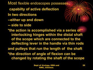
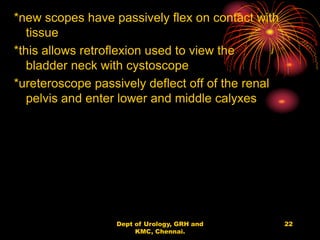
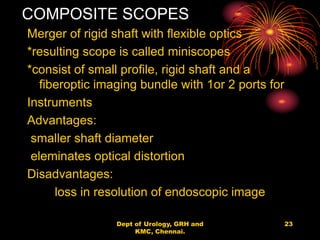
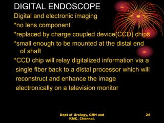
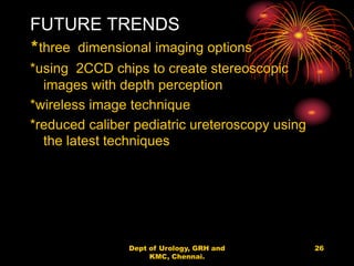
Low Rate Call Girls Pune Esha 9907093804 Short 1500 Night 6000 Best call girl...
Optics in urology
1. OPTICS IN UROLOGY
Dept of Urology
Govt Royapettah Hospital and Kilpauk Medical College
Chennai
2. Moderators:
Professors:
• Prof. Dr. G. Sivasankar, M.S., M.Ch.,
• Prof. Dr. A. Senthilvel, M.S., M.Ch.,
Asst Professors:
• Dr. J. Sivabalan, M.S., M.Ch.,
• Dr. R. Bhargavi, M.S., M.Ch.,
• Dr. S. Raju, M.S., M.Ch.,
• Dr. K. Muthurathinam, M.S., M.Ch.,
• Dr. D. Tamilselvan, M.S., M.Ch.,
• Dr. K. Senthilkumar, M.S., M.Ch.
Dept of Urology, GRH and KMC,
Chennai.
2
4. RIGID ENDOSCOPES
HISTORY:
1806 – Bozzini’s Lichtleiter–direct vision
urethral tube
--candle light is used
1879 –Nitze&leiter – simple lens system into
theviewing tube
-- version of modern
cystoscope
1900 – heated platinum wires & light bulbs
used
1960 – modern fiberoptic lighting introduced
- rigid endoscopy
4
Dept of Urology, GRH and
KMC, Chennai.
5. 20th century: Hopkins rod-lens
system
-- replacing the thin relay lenses
within
the shaft of the telescope
with long,
contoured glasses
-- 9 fold increase in light
transmission
--improved refractive index
--increased viewing angle
--decreased profile of telescope
shaft 5
Dept of Urology, GRH and
KMC, Chennai.
7. OPTICS
Rigid endoscopes
-- use a telescope that consist of a series
of lenses along the shaft
--to relay a single optical image from the
imaging lens at the distal tip of the
endoscope back to the eye of the urologist
In early days
-- image was transmitted through the
hollow tube
via thin relay and field lenses placed at
precise distance within the air filled
assembly tube
7
Dept of Urology, GRH and
KMC, Chennai.
8. ROD LENS SYSTEM
In late1950’s Hopkin introduced this system
--air spaces are reduced with long rods of
glasses
ground,contoured and polished at both ends
--images can be magnified at the ocular lens
--degree of magnification depend on diameter
of viewing lens
-- size of magnified image
through a small caliber (ureteroscope) is
smaller than large bore caliber (cystoscope)
--addition of video cameras & monitor based
endoscopes has eliminated the discrepancy
in image size
--binocular view 8
Dept of Urology, GRH and
KMC, Chennai.
9. --objective lenses with angles ranging from
0-120 degree
--cystoscopes –interchangeable lenses
0-5 degree– direct vision internal
urethertomy
for urethral stricture
12-30degree– transurethral prostate surgery
30-70degree—bladder
For rigid ureteroscopes & nephroscopes:
-direct vision lens is used
-rarely necessary to exchange lens
-available only on larger ureteroscope &
nephroscope
- seldom used
9
Dept of Urology, GRH and
KMC, Chennai.
10. CONSTRUCTION
Cystoscopes mostly uses straight
viewing telescopes
--access to instrumentation channel is via
angled ports inferior to telescope
--accessories such as stone graspers or
forceps introduction is difficult
Rigid ureteroscope & Nephroscope
--right angled or offset oblique ocular
configuration
--direct access to accessories will be easy
--with the use of video cameras this is
becoming less an issue
10
Dept of Urology, GRH and
KMC, Chennai.
14. FLEXIBLE ENDOSCOPES
--costlier
--complex of construction
--shaft consist of 3 fiberoptic bundles :
Light bundle:
2 noncoherent bundles of fiber that transmit
light
Imaging bundle:
single coherent bundle of glass fibers
constitute the imaging bundle
Images :
-- images obtained is not a single image
--composite matrix of each fiber within the
bundle
-- image is analogous to a newspaper
photograph 14
Dept of Urology, GRH and
KMC, Chennai.
16. HONEYCOMB effect
*composed of multiple dots
merging into a single
reconstructed image .
--Each optical fiber diameter is 8
micrometer
--Each fiber is composed of glass
possessing higher refractive
index than the cladding that
surrounds it
--this composition allows excellent
light & image preservation over
long distance 16
Dept of Urology, GRH and
KMC, Chennai.
18. The coherent bundle consist of
thousand of
individual fibers with identical
arrangement
*Each image of each fiber is transmitted
along the entire bundle to the ocular lens
at eyepiece
*Even though optical fibers are flexible
they may be damaged by
- excessive flexion
- force
- Pressure on the shaft of the
scope
18
Dept of Urology, GRH and
KMC, Chennai.
19. * If individual fiber is damaged the
image will be a black dot
*many broken fibers leads to
indeterminate image
19
Dept of Urology, GRH and
KMC, Chennai.
20. Most flexible endoscopes possesses
capabilty of active deflection
In two directions
--either up and down
-- side to side
*the action is accomplished via a series of
interlocking hinges within the distal shaft
of the scope which are connected to the
deflecting lever in the handle via thin rods
and pulleys that run the length of the shaft
*the direction of angle of flexion can be
changed by rotating the shaft of the scope
20
Dept of Urology, GRH and
KMC, Chennai.
22. *new scopes have passively flex on contact with
tissue
*this allows retroflexion used to view the
bladder neck with cystoscope
*ureteroscope passively deflect off of the renal
pelvis and enter lower and middle calyxes
22
Dept of Urology, GRH and
KMC, Chennai.
23. COMPOSITE SCOPES
Merger of rigid shaft with flexible optics
*resulting scope is called miniscopes
*consist of small profile, rigid shaft and a
fiberoptic imaging bundle with 1or 2 ports for
Instruments
Advantages:
smaller shaft diameter
eleminates optical distortion
Disadvantages:
loss in resolution of endoscopic image
23
Dept of Urology, GRH and
KMC, Chennai.
24. DIGITAL ENDOSCOPE
Digital and electronic imaging
*no lens component
*replaced by charge coupled device(CCD) chips
*small enough to be mounted at the distal end
of shaft
*CCD chip will relay digitalized information via a
single fiber back to a distal processor which will
reconstruct and enhance the image
electronically on a television monitor
24
Dept of Urology, GRH and
KMC, Chennai.
25. • Advantages:
1.superior quality image
2.To know generational distortion
3.Space saved
4.Light weight
5.Larger instrumental channels
6.Larger working elements used
7.No need for additional light source and
camera setup
8.Smaller size
9.Increased flexibility
10.Improved durability
25
Dept of Urology, GRH and
KMC, Chennai.
26. FUTURE TRENDS
*three dimensional imaging options
*using 2CCD chips to create stereoscopic
images with depth perception
*wireless image technique
*reduced caliber pediatric ureteroscopy using
the latest techniques
26
Dept of Urology, GRH and
KMC, Chennai.