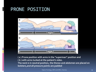
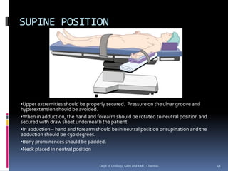
POSITIONING IN UROLOGICAL
PROCEDURES
Dept of Urology
Govt Royapettah Hospital and Kilpauk Medical College
Chennai
Moderators:
Professors:
▪ Prof. Dr. G. Sivasankar, M.S., M.Ch.,
▪ Prof. Dr. A. Senthilvel, M.S., M.Ch.,
Asst Professors:
▪ Dr. J. Sivabalan, M.S., M.Ch.,
▪ Dr. R. Bhargavi, M.S., M.Ch.,
▪ Dr. S. Raju, M.S., M.Ch.,
▪ Dr. K. Muthurathinam, M.S., M.Ch.,
▪ Dr. D.Tamilselvan, M.S., M.Ch.,
▪ Dr. K. Senthilkumar, M.S., M.Ch.
Dept of Urology, GRH and KMC, Chennai. 2
Introduction
▪ Strategic positioning to allow access to pelvis,
retroperitoneum and the perineum
▪ Anesthesiologists, urologist, and nurses –
shared responsibility
 To ensure patient safety
 To provide optimal surgical exposure
 To avoid iatrogenic injuries
3
Dept of Urology, GRH and KMC, Chennai.
Various Positions
▪ Lithotomy
▪ Lateral decubitus & Jackknife position
▪ Prone position
▪ Supine Position
▪ Trendelenburg Position
4
Dept of Urology, GRH and KMC, Chennai.
Lithotomy position
▪ Most frequently used position
 Transurethral cystoscopic procedures
 Open urological procedures where access to
perineum and anus is necessary
5
Dept of Urology, GRH and KMC, Chennai.
Lithotomy Position
▪ Patient’s anterior superior iliac spine should be placed over
the break in the bed.
▪ Stirrups should be anchored level with the patient’s knees
and angled toward the contralateral shoulder.
▪ Upper extremities can either be tucked at the patient’s side
in neutral position or abducted <90 degrees with the arms
either supinated or neutral
▪ When raising and lowering the legs in and out of the
stirrups, it should be done in unison by at least two OR
staffs – to avoid torsion on the lumbar spine and possible
dislocation of either hip.
6
Dept of Urology, GRH and KMC, Chennai.
▪ The goal of leg positioning in the stirrups is
for
 Hips to be flexed 80 – 100 degrees from the trunk
 Legs abducted <30 – 45 degrees from the midline
 Knees being bend until parallel with the torso
▪ Stirrups should be padded circumferentially
around the lower extremities to avoid
compression injuries
7
Dept of Urology, GRH and KMC, Chennai.
Lithotomy positioning I
8
Dept of Urology, GRH and KMC, Chennai.
Lithotomy positioning II
9
Dept of Urology, GRH and KMC, Chennai.
LITHOTOMY POSITION
Hips are flexed <100°; knees are flexed with legs parallel to patient’s torso.
Legs abducted < 30 to 45 degrees from midline
10
Dept of Urology, GRH and KMC, Chennai.
LITHOTOMY POSITION
Lower extremities are suspended in candy cane stirrups and externally
rotated avoiding compression by stirrups on lateral aspect of legs
11
Dept of Urology, GRH and KMC, Chennai.
With arms tucked, risk
for finger trauma or
crush injury exists when
adjusting lower portion
of bed in lithotomy
position
12
Dept of Urology, GRH and KMC, Chennai.
▪ When lowering the legs out of lithotomy, the
knees should be brought together at midline,
followed by unflexing the legs back to supine
position.
▪ Preoperative examination focused on
potential limitations to hip, knee and ankle
movement should be noted.
▪ Multiple versions of stirrups – Candy canes,
calf rests, boots, shepherd’s crook foot straps,
or Bierhoff knee crutch stirrups
13
Dept of Urology, GRH and KMC, Chennai.
Various types of Lithotomy
Stirrups
14
Dept of Urology, GRH and KMC, Chennai.
Exaggerated Lithotomy
position
▪ Involves flexing the hips beyond 100 degrees.
▪ Should be avoided due to excessive traction it
places on the sciatic and peroneal nerves.
▪ When necessary, it should be for a limited
duration of time and only during those points in
surgical procedure when it is absolutely crucial.
▪ A buttress should be placed under the lower
back in these instances to relieve some of the
nerve and spinal traction that the patient may
experience.
15
Dept of Urology, GRH and KMC, Chennai.
Exaggerated Lithotomy
Position
16
Dept of Urology, GRH and KMC, Chennai.
Complications
▪ Peripheral neuropathies
▪ Lumbar spine torsion
▪ Dislocation of hip
▪ Finger trauma
17
Dept of Urology, GRH and KMC, Chennai.
Neuropathies in Lithotomy
Position
▪ Common peroneal nerve
 Most common neuropathy in lithotomy position
 Compression between lateral head of fibula and stirrup
 Common in low BMI, prolonged duration >3h
 Lack of dorsiflexion of foot / Paresthesia or numbness
 Full recovery is normally observed
▪ Sciatic nerve
 Most susceptible to stretch injury – exaggerated lithotomy
position
 Hyperflexion of hip + extension of knee → greatest risk for
stretch neuropathy
18
Dept of Urology, GRH and KMC, Chennai.
▪ Obturator nerve
 Stretched when hips are flexed beyond 100 degrees
 Stretched and compressed against the pubic ramus of the
pelvis as it exits the obturator foramen
 Also at risk when legs are first abducted and then flexed at
the hip and knee when placed into the stirrups
▪ PosteriorTibial nerve
 Sensory and motor supply to plantar surface of the foot
 Compression in the tarsal tunnel
 Due to improper padding of lithotomy boots
19
Dept of Urology, GRH and KMC, Chennai.
▪ Femoral Nerve
 Due to hyperflexion of thigh
 Inguinal ligament is streched & compresses nerve
▪ Lateral femoral cutaneous nerve
 Sensory innervation to lateral thigh
 “Meralgia paresthetica”
 Compression of lateral thigh against candy cane stirrup rod
▪ Saphenous nerve
 Supplies sensory innervation to medial aspect of foot
 Compression – against medial tibial condyle
20
Dept of Urology, GRH and KMC, Chennai.
Lithotomy Position – Nerve
Injuries
21
Dept of Urology, GRH and KMC, Chennai.
Lateral Decubitus &
Jackknife position
▪ Optimal surgical exposure for access to the
adrenal glands, kidney, and collecting system
▪ Beneficial position for removal of stones
located in the upper ureter and renal pelvis
requiring an open procedure, as well as
nephrectomies of non-malignant disease.
22
Dept of Urology, GRH and KMC, Chennai.
▪ First anesthetized in the supine position, and
then with the help of other members of the OR
staff, the patient is turned to the lateral
decubitus position.
▪ For extraperitoneal surgical procedures, the
patient is turned a full 90°; however, if surgical
exposure requires access to the intraperitoneal
space, then turning 45° lateral may be adequate.
▪ Maintaining the patient firmly on their side
without displacement by anchoring tape over
towels placed at the patient’s shoulder and waist
to the OR bed.
23
Dept of Urology, GRH and KMC, Chennai.
Jackknife position
▪ Jackknife is a modification of the lateral decubitus
position, in which the OR table is flexed at its
midpoint underneath the patient’s iliac crest.
▪ Provides stretch between the nondependent iliac
crest and the costal margin on the operative side,
creating a maximal surgical exposure.
▪ Place the table into reverseTrendelenburg until the
upper torso is parallel with the ground to optimize
both hemodynamic stability and tension over the
incision site.
24
Dept of Urology, GRH and KMC, Chennai.
▪ If additional flexion is needed, a kidney rest can
be added to the OR table apparatus.
▪ The kidney rest should be anchored where the
OR table breaks and placed directly under the
dependent iliac crest.
▪ Care should be taken to ensure that the kidney
rest is not malpositioned underneath the flank or
lower costal margin.
▪ Such malpositions can result in compression of
the inferior vena cava and decreased venous
return, as well as impeding ventilation of the
dependent lung.
25
Dept of Urology, GRH and KMC, Chennai.
JACKKNIFE LATERAL DECUBITUS
POSITION
Improper placement of
kidney rest at
( a ) below the
fl ank
( b ) dependent costal
margin.
( c ) Correct positioning
below dependent iliac
crest
26
Dept of Urology, GRH and KMC, Chennai.
▪ When turning the patient to the lateral decubitus position,
maintain the head and neck in a neutral position in line with
the vertebral column.
▪ The head should be kept neutral by placing blankets and/or
a foam doughnut beneath it for support. Failure to do so
can result in lateral stretch of the neck and subsequent
stretch of the brachial plexus.
▪ Horner syndrome has also been reported as a possible
complication of excessive lateral neck flexion due to injury
of the ipsilateral stellate ganglion.
▪ Attention should also be taken with the dependent eye to
prevent external compression or possible corneal abrasion.
27
Dept of Urology, GRH and KMC, Chennai.
•The arms should be placed
perpendicular to the shoulders and
parallel to each other.
•They should be neither abducted
>90° nor flexed at the elbow >90° to
avoid stretching of the brachial
plexus and its branches.
•The nondependent arm can be
either propped up on pillows above
the dependent arm or placed on a
padded armrest positioned
perpendicular to the corresponding
shoulder
•Axillary roll is placed under
dependent thorax, caudad to the
axilla
28
Dept of Urology, GRH and KMC, Chennai.
▪ The roll should never be located in the axilla itself, as
its purpose is to distribute the weight of the thorax
away from the axilla and prevent compression of its
neurovascular bundle.
▪ Placing the pulse oximeter on the dependent arm
can be used as an indicator of axillary neurovascular
compression. If hypotension is recorded in the
dependent arm, then axillary compression must be
ruled out.
▪ Periodic checks throughout the surgical procedure
should also be performed to ensure that the axillary
roll has not become displaced during surgery.
29
Dept of Urology, GRH and KMC, Chennai.
Dependent leg is flexed
at the knee with
padding beneath the
dependent leg and
between both knees to
prevent compression at
bony prominences
30
Dept of Urology, GRH and KMC, Chennai.
Complications
Potential Complications Recommendations
Peroneal Nerve Adequately pad dependent leg against
table
Brachial Plexus Use axillary roll
Bring dependent shoulder and arm out
from under rib cage
Skin breakdown Pad pressure points
Compartment syndrome Avoid prolonged surgery
Rhabdomyolysis Higher incidence in obese or male
patients and prolonged surgery
31
Dept of Urology, GRH and KMC, Chennai.
Prone Position
▪ For access to the retroperitoneum and upper
urinary tract procedures
▪ Commonly used for
 Percutaneous nephrolithotomy
 Adrenalectomy
 Pediatric pyeloplasty via the dorsal lumbotomy
approach
32
Dept of Urology, GRH and KMC, Chennai.
▪ During positioning, attention should be paid to
avoid inadvertent extubation of the trachea and
to maintain the neck in neutral position.
▪ The arms are typically at the patient’s sides while
turning prone, and can then be tucked with
palms facing medially, or extended in the
“superman” position on arm boards with the
arms abducted less than 90° at the shoulders
and flexed at the elbows
33
Dept of Urology, GRH and KMC, Chennai.
▪ The knees and hips should be slightly flexed.
▪ All pressure points, including forehead, chin,
elbows, knees, shins, and toes, must be properly
padded.
▪ The chest and abdomen should be supported
above the operating table by bolsters or frames
to prevent compression of abdominal contents
and reduction in pulmonary compliance and
venous return.
34
Dept of Urology, GRH and KMC, Chennai.
PRONE POSITION
( a ) Prone position with arms in the “superman” position and
( b ) with arms tucked at the patient’s sides.
The neck is in neutral position, the thorax and abdomen are placed on
bolsters,and all pressure points are padded
35
Dept of Urology, GRH and KMC, Chennai.
▪ Care should also be taken to avoid compression
of breasts and male genitalia.
▪ Breasts should be placed medially to the bolsters
that support the chest.
▪ Commercially available foam headrests have
openings for the eyes, nose, and mouth to avoid
tissue injury from excessive pressure or
endotracheal tube kinking.
36
Dept of Urology, GRH and KMC, Chennai.
▪ Challenging in the morbidly obese patient, and extra
operating room staff should be available to assist with
turning the patient.
▪ Alternatively a technique of awake intubation, followed by
patient self positioning can be employed.This method has
been described for percutaneous nephrolithotomy in obese
patients.
▪ The advantage of this technique is that the patient can
achieve a comfortable position and can be questioned as to
whether additional adjustments or padding are necessary
prior to induction of anesthesia.
37
Dept of Urology, GRH and KMC, Chennai.
Complication
Potential Complications Recommendations
Occlusion of carotid or verterbral
arteries
Avoid excessive rotation of neck
Cervical spine injury Avoid excessive extension or flexion of
neck
Brachial plexus Arm elevated <90 degrees or placed at
sides.
Avoid pressure on axilla
Pressure injuries of knees, breasts, face
and feet
Pad pressure points
Upper airway edema Maintain head in neutral position
Venous air embolism Avoid lowering heads & legs
38
Dept of Urology, GRH and KMC, Chennai.
SUPINE POSITION
▪ Majority of open urological procedures, most
commonly involving penis, scrotum, inguinal lymph
nodes and urethra.
▪ Optimal exposure of pelvic and intra-abdominal
organs such as bladder and prostate.
▪ Also retroperitoneal surgeries - Adrenals, Kidney &
ureters
▪ Any urological procedure involving the manipulation
of the intestinal tract for urinary diversion or
augmentation
39
Dept of Urology, GRH and KMC, Chennai.
Supine Position
▪ Mostly the patients are asked to position
themselves especially those with prior history
of peripheral neuropathy, contractures or
other risk factors.
▪ Once anesthetized, care should be taken for
further adjustments.
40
Dept of Urology, GRH and KMC, Chennai.
SUPINE POSITION
•Upper extremities should be properly secured. Pressure on the ulnar groove and
hyperextension should be avoided.
•When in adduction, the hand and forearm should be rotated to neutral position and
secured with draw sheet underneath the patient
•In abduction – hand and forearm should be in neutral position or supination and the
abduction should be <90 degrees.
•Bony prominences should be padded.
•Neck placed in neutral position
41
Dept of Urology, GRH and KMC, Chennai.
Special Considerations
▪ Patients with chronic back pain or kyphoscoliosis
– require additional padding or even slight
flexion of the bed to avoid exacerbation of their
condition
▪ For long or extended procedures – periodically
rotate the head to redistribute the weight and
evade pressure alopecia secondary to hair follicle
ischaemia.
▪ Avoid leaning on the extremeties by OR staff →
inhibits venous return and increased risk of DVT
42
Dept of Urology, GRH and KMC, Chennai.
Complications
43
Dept of Urology, GRH and KMC, Chennai.
Peripheral Neuropathies
 Ulnar neuropathy
 Most common
 Susceptible as it courses through the medial elbow in post
condylar groove
 Median neuropathy
 Compression at cubital fossa
 Avoid hyperextension at the elbow
 When arm falls unintentionally off the edge of the table in
pronated position
 Radial nerve
 Compression - spiral groove of the humerus
 Distally placed nonivasive blood pressure cuff at elbow
 Allowing supinated arm to inadvertently hang off the side of
the table
44
Dept of Urology, GRH and KMC, Chennai.
▪ Spinal Hyperextension
 Hyperextension beyond 10 degrees → back pain
and neuropathies
▪ Brachial plexus compression injury
 Flexion of head to contralateral side
 External and dorsal rotation of arm
 Hyperabduction at shoulder joint
 To avoid → head and arms in neutral positon, with
arms abducted <90 degrees
45
Dept of Urology, GRH and KMC, Chennai.
Upper limb Nerves
46
Dept of Urology, GRH and KMC, Chennai.
Brachial plexus
47
Dept of Urology, GRH and KMC, Chennai.
Trendelenburg Position
▪ Modification of the supine position, by tilting
the table and patient head down.
▪ Often employed to displace the abdominal
viscera toward the diaphragm, providing
improved exposure to the lower abdominal
and pelvic organs.
▪ Positioning of the arms remains similar to
that in the supine position.
48
Dept of Urology, GRH and KMC, Chennai.
▪ The arms are more preferably, tucked at the patient’s side
in the neutral position, avoiding the risk of the arms sliding
off the arm boards once the patient is tilted.
▪ Shoulder braces should also be placed bilaterally over the
acromioclavicular joints, but only when the arms are tucked
at the patient’s side.
▪ Utilization of shoulder braces in combination with arm
abduction may result in brachial plexus neuropathy, due to
stretching and its potential compression against the
humeral head as it courses through the shoulder and upper
extremity.
49
Dept of Urology, GRH and KMC, Chennai.
TRENDELENBURG POSITION
Note the arms tucked at the patient’s side in the neutral position with padding
underneath the head and neck
50
Dept of Urology, GRH and KMC, Chennai.
Kidney-shaped padding
around the
acromioclavicular joints
of bilateral shoulders.
Avoid if arms are
abducted
51
Dept of Urology, GRH and KMC, Chennai.
▪ Trendelenburg position is often a tilt of the bed of <20° in
order to displace the viscera.
▪ However, there are many circumstances especially in
urologic procedures, where steepTrendelenburg
positioning of 30–45° tilt may be necessary.
▪ This steep positioning should be avoided when possible.
▪ It is important to ensure that there is a non-sliding mattress
to prevent the mattress and subsequently the patient from
sliding cephalad off the OR table.
52
Dept of Urology, GRH and KMC, Chennai.
Complications
Potential Complications Recommendations
Brachial Plexus Use kidney shaped shoulder braces /
non sliding mattress
Pad acromioclavicular joints
Ischemic optic neuropathy Limit time inTrendelenburg position
Increased intracranial pressure Monitor head and neck for excessive
edema
Head/neck venous pooling Limit time inTrendelenburg position
53
Dept of Urology, GRH and KMC, Chennai.
THANKYOU
54
Dept of Urology, GRH and KMC, Chennai.

Positioning in urological procedures

  • 1.
    POSITIONING IN UROLOGICAL PROCEDURES Deptof Urology Govt Royapettah Hospital and Kilpauk Medical College Chennai
  • 2.
    Moderators: Professors: ▪ Prof. Dr.G. Sivasankar, M.S., M.Ch., ▪ Prof. Dr. A. Senthilvel, M.S., M.Ch., Asst Professors: ▪ Dr. J. Sivabalan, M.S., M.Ch., ▪ Dr. R. Bhargavi, M.S., M.Ch., ▪ Dr. S. Raju, M.S., M.Ch., ▪ Dr. K. Muthurathinam, M.S., M.Ch., ▪ Dr. D.Tamilselvan, M.S., M.Ch., ▪ Dr. K. Senthilkumar, M.S., M.Ch. Dept of Urology, GRH and KMC, Chennai. 2
  • 3.
    Introduction ▪ Strategic positioningto allow access to pelvis, retroperitoneum and the perineum ▪ Anesthesiologists, urologist, and nurses – shared responsibility  To ensure patient safety  To provide optimal surgical exposure  To avoid iatrogenic injuries 3 Dept of Urology, GRH and KMC, Chennai.
  • 4.
    Various Positions ▪ Lithotomy ▪Lateral decubitus & Jackknife position ▪ Prone position ▪ Supine Position ▪ Trendelenburg Position 4 Dept of Urology, GRH and KMC, Chennai.
  • 5.
    Lithotomy position ▪ Mostfrequently used position  Transurethral cystoscopic procedures  Open urological procedures where access to perineum and anus is necessary 5 Dept of Urology, GRH and KMC, Chennai.
  • 6.
    Lithotomy Position ▪ Patient’santerior superior iliac spine should be placed over the break in the bed. ▪ Stirrups should be anchored level with the patient’s knees and angled toward the contralateral shoulder. ▪ Upper extremities can either be tucked at the patient’s side in neutral position or abducted <90 degrees with the arms either supinated or neutral ▪ When raising and lowering the legs in and out of the stirrups, it should be done in unison by at least two OR staffs – to avoid torsion on the lumbar spine and possible dislocation of either hip. 6 Dept of Urology, GRH and KMC, Chennai.
  • 7.
    ▪ The goalof leg positioning in the stirrups is for  Hips to be flexed 80 – 100 degrees from the trunk  Legs abducted <30 – 45 degrees from the midline  Knees being bend until parallel with the torso ▪ Stirrups should be padded circumferentially around the lower extremities to avoid compression injuries 7 Dept of Urology, GRH and KMC, Chennai.
  • 8.
    Lithotomy positioning I 8 Deptof Urology, GRH and KMC, Chennai.
  • 9.
    Lithotomy positioning II 9 Deptof Urology, GRH and KMC, Chennai.
  • 10.
    LITHOTOMY POSITION Hips areflexed <100°; knees are flexed with legs parallel to patient’s torso. Legs abducted < 30 to 45 degrees from midline 10 Dept of Urology, GRH and KMC, Chennai.
  • 11.
    LITHOTOMY POSITION Lower extremitiesare suspended in candy cane stirrups and externally rotated avoiding compression by stirrups on lateral aspect of legs 11 Dept of Urology, GRH and KMC, Chennai.
  • 12.
    With arms tucked,risk for finger trauma or crush injury exists when adjusting lower portion of bed in lithotomy position 12 Dept of Urology, GRH and KMC, Chennai.
  • 13.
    ▪ When loweringthe legs out of lithotomy, the knees should be brought together at midline, followed by unflexing the legs back to supine position. ▪ Preoperative examination focused on potential limitations to hip, knee and ankle movement should be noted. ▪ Multiple versions of stirrups – Candy canes, calf rests, boots, shepherd’s crook foot straps, or Bierhoff knee crutch stirrups 13 Dept of Urology, GRH and KMC, Chennai.
  • 14.
    Various types ofLithotomy Stirrups 14 Dept of Urology, GRH and KMC, Chennai.
  • 15.
    Exaggerated Lithotomy position ▪ Involvesflexing the hips beyond 100 degrees. ▪ Should be avoided due to excessive traction it places on the sciatic and peroneal nerves. ▪ When necessary, it should be for a limited duration of time and only during those points in surgical procedure when it is absolutely crucial. ▪ A buttress should be placed under the lower back in these instances to relieve some of the nerve and spinal traction that the patient may experience. 15 Dept of Urology, GRH and KMC, Chennai.
  • 16.
    Exaggerated Lithotomy Position 16 Dept ofUrology, GRH and KMC, Chennai.
  • 17.
    Complications ▪ Peripheral neuropathies ▪Lumbar spine torsion ▪ Dislocation of hip ▪ Finger trauma 17 Dept of Urology, GRH and KMC, Chennai.
  • 18.
    Neuropathies in Lithotomy Position ▪Common peroneal nerve  Most common neuropathy in lithotomy position  Compression between lateral head of fibula and stirrup  Common in low BMI, prolonged duration >3h  Lack of dorsiflexion of foot / Paresthesia or numbness  Full recovery is normally observed ▪ Sciatic nerve  Most susceptible to stretch injury – exaggerated lithotomy position  Hyperflexion of hip + extension of knee → greatest risk for stretch neuropathy 18 Dept of Urology, GRH and KMC, Chennai.
  • 19.
    ▪ Obturator nerve Stretched when hips are flexed beyond 100 degrees  Stretched and compressed against the pubic ramus of the pelvis as it exits the obturator foramen  Also at risk when legs are first abducted and then flexed at the hip and knee when placed into the stirrups ▪ PosteriorTibial nerve  Sensory and motor supply to plantar surface of the foot  Compression in the tarsal tunnel  Due to improper padding of lithotomy boots 19 Dept of Urology, GRH and KMC, Chennai.
  • 20.
    ▪ Femoral Nerve Due to hyperflexion of thigh  Inguinal ligament is streched & compresses nerve ▪ Lateral femoral cutaneous nerve  Sensory innervation to lateral thigh  “Meralgia paresthetica”  Compression of lateral thigh against candy cane stirrup rod ▪ Saphenous nerve  Supplies sensory innervation to medial aspect of foot  Compression – against medial tibial condyle 20 Dept of Urology, GRH and KMC, Chennai.
  • 21.
    Lithotomy Position –Nerve Injuries 21 Dept of Urology, GRH and KMC, Chennai.
  • 22.
    Lateral Decubitus & Jackknifeposition ▪ Optimal surgical exposure for access to the adrenal glands, kidney, and collecting system ▪ Beneficial position for removal of stones located in the upper ureter and renal pelvis requiring an open procedure, as well as nephrectomies of non-malignant disease. 22 Dept of Urology, GRH and KMC, Chennai.
  • 23.
    ▪ First anesthetizedin the supine position, and then with the help of other members of the OR staff, the patient is turned to the lateral decubitus position. ▪ For extraperitoneal surgical procedures, the patient is turned a full 90°; however, if surgical exposure requires access to the intraperitoneal space, then turning 45° lateral may be adequate. ▪ Maintaining the patient firmly on their side without displacement by anchoring tape over towels placed at the patient’s shoulder and waist to the OR bed. 23 Dept of Urology, GRH and KMC, Chennai.
  • 24.
    Jackknife position ▪ Jackknifeis a modification of the lateral decubitus position, in which the OR table is flexed at its midpoint underneath the patient’s iliac crest. ▪ Provides stretch between the nondependent iliac crest and the costal margin on the operative side, creating a maximal surgical exposure. ▪ Place the table into reverseTrendelenburg until the upper torso is parallel with the ground to optimize both hemodynamic stability and tension over the incision site. 24 Dept of Urology, GRH and KMC, Chennai.
  • 25.
    ▪ If additionalflexion is needed, a kidney rest can be added to the OR table apparatus. ▪ The kidney rest should be anchored where the OR table breaks and placed directly under the dependent iliac crest. ▪ Care should be taken to ensure that the kidney rest is not malpositioned underneath the flank or lower costal margin. ▪ Such malpositions can result in compression of the inferior vena cava and decreased venous return, as well as impeding ventilation of the dependent lung. 25 Dept of Urology, GRH and KMC, Chennai.
  • 26.
    JACKKNIFE LATERAL DECUBITUS POSITION Improperplacement of kidney rest at ( a ) below the fl ank ( b ) dependent costal margin. ( c ) Correct positioning below dependent iliac crest 26 Dept of Urology, GRH and KMC, Chennai.
  • 27.
    ▪ When turningthe patient to the lateral decubitus position, maintain the head and neck in a neutral position in line with the vertebral column. ▪ The head should be kept neutral by placing blankets and/or a foam doughnut beneath it for support. Failure to do so can result in lateral stretch of the neck and subsequent stretch of the brachial plexus. ▪ Horner syndrome has also been reported as a possible complication of excessive lateral neck flexion due to injury of the ipsilateral stellate ganglion. ▪ Attention should also be taken with the dependent eye to prevent external compression or possible corneal abrasion. 27 Dept of Urology, GRH and KMC, Chennai.
  • 28.
    •The arms shouldbe placed perpendicular to the shoulders and parallel to each other. •They should be neither abducted >90° nor flexed at the elbow >90° to avoid stretching of the brachial plexus and its branches. •The nondependent arm can be either propped up on pillows above the dependent arm or placed on a padded armrest positioned perpendicular to the corresponding shoulder •Axillary roll is placed under dependent thorax, caudad to the axilla 28 Dept of Urology, GRH and KMC, Chennai.
  • 29.
    ▪ The rollshould never be located in the axilla itself, as its purpose is to distribute the weight of the thorax away from the axilla and prevent compression of its neurovascular bundle. ▪ Placing the pulse oximeter on the dependent arm can be used as an indicator of axillary neurovascular compression. If hypotension is recorded in the dependent arm, then axillary compression must be ruled out. ▪ Periodic checks throughout the surgical procedure should also be performed to ensure that the axillary roll has not become displaced during surgery. 29 Dept of Urology, GRH and KMC, Chennai.
  • 30.
    Dependent leg isflexed at the knee with padding beneath the dependent leg and between both knees to prevent compression at bony prominences 30 Dept of Urology, GRH and KMC, Chennai.
  • 31.
    Complications Potential Complications Recommendations PeronealNerve Adequately pad dependent leg against table Brachial Plexus Use axillary roll Bring dependent shoulder and arm out from under rib cage Skin breakdown Pad pressure points Compartment syndrome Avoid prolonged surgery Rhabdomyolysis Higher incidence in obese or male patients and prolonged surgery 31 Dept of Urology, GRH and KMC, Chennai.
  • 32.
    Prone Position ▪ Foraccess to the retroperitoneum and upper urinary tract procedures ▪ Commonly used for  Percutaneous nephrolithotomy  Adrenalectomy  Pediatric pyeloplasty via the dorsal lumbotomy approach 32 Dept of Urology, GRH and KMC, Chennai.
  • 33.
    ▪ During positioning,attention should be paid to avoid inadvertent extubation of the trachea and to maintain the neck in neutral position. ▪ The arms are typically at the patient’s sides while turning prone, and can then be tucked with palms facing medially, or extended in the “superman” position on arm boards with the arms abducted less than 90° at the shoulders and flexed at the elbows 33 Dept of Urology, GRH and KMC, Chennai.
  • 34.
    ▪ The kneesand hips should be slightly flexed. ▪ All pressure points, including forehead, chin, elbows, knees, shins, and toes, must be properly padded. ▪ The chest and abdomen should be supported above the operating table by bolsters or frames to prevent compression of abdominal contents and reduction in pulmonary compliance and venous return. 34 Dept of Urology, GRH and KMC, Chennai.
  • 35.
    PRONE POSITION ( a) Prone position with arms in the “superman” position and ( b ) with arms tucked at the patient’s sides. The neck is in neutral position, the thorax and abdomen are placed on bolsters,and all pressure points are padded 35 Dept of Urology, GRH and KMC, Chennai.
  • 36.
    ▪ Care shouldalso be taken to avoid compression of breasts and male genitalia. ▪ Breasts should be placed medially to the bolsters that support the chest. ▪ Commercially available foam headrests have openings for the eyes, nose, and mouth to avoid tissue injury from excessive pressure or endotracheal tube kinking. 36 Dept of Urology, GRH and KMC, Chennai.
  • 37.
    ▪ Challenging inthe morbidly obese patient, and extra operating room staff should be available to assist with turning the patient. ▪ Alternatively a technique of awake intubation, followed by patient self positioning can be employed.This method has been described for percutaneous nephrolithotomy in obese patients. ▪ The advantage of this technique is that the patient can achieve a comfortable position and can be questioned as to whether additional adjustments or padding are necessary prior to induction of anesthesia. 37 Dept of Urology, GRH and KMC, Chennai.
  • 38.
    Complication Potential Complications Recommendations Occlusionof carotid or verterbral arteries Avoid excessive rotation of neck Cervical spine injury Avoid excessive extension or flexion of neck Brachial plexus Arm elevated <90 degrees or placed at sides. Avoid pressure on axilla Pressure injuries of knees, breasts, face and feet Pad pressure points Upper airway edema Maintain head in neutral position Venous air embolism Avoid lowering heads & legs 38 Dept of Urology, GRH and KMC, Chennai.
  • 39.
    SUPINE POSITION ▪ Majorityof open urological procedures, most commonly involving penis, scrotum, inguinal lymph nodes and urethra. ▪ Optimal exposure of pelvic and intra-abdominal organs such as bladder and prostate. ▪ Also retroperitoneal surgeries - Adrenals, Kidney & ureters ▪ Any urological procedure involving the manipulation of the intestinal tract for urinary diversion or augmentation 39 Dept of Urology, GRH and KMC, Chennai.
  • 40.
    Supine Position ▪ Mostlythe patients are asked to position themselves especially those with prior history of peripheral neuropathy, contractures or other risk factors. ▪ Once anesthetized, care should be taken for further adjustments. 40 Dept of Urology, GRH and KMC, Chennai.
  • 41.
    SUPINE POSITION •Upper extremitiesshould be properly secured. Pressure on the ulnar groove and hyperextension should be avoided. •When in adduction, the hand and forearm should be rotated to neutral position and secured with draw sheet underneath the patient •In abduction – hand and forearm should be in neutral position or supination and the abduction should be <90 degrees. •Bony prominences should be padded. •Neck placed in neutral position 41 Dept of Urology, GRH and KMC, Chennai.
  • 42.
    Special Considerations ▪ Patientswith chronic back pain or kyphoscoliosis – require additional padding or even slight flexion of the bed to avoid exacerbation of their condition ▪ For long or extended procedures – periodically rotate the head to redistribute the weight and evade pressure alopecia secondary to hair follicle ischaemia. ▪ Avoid leaning on the extremeties by OR staff → inhibits venous return and increased risk of DVT 42 Dept of Urology, GRH and KMC, Chennai.
  • 43.
    Complications 43 Dept of Urology,GRH and KMC, Chennai.
  • 44.
    Peripheral Neuropathies  Ulnarneuropathy  Most common  Susceptible as it courses through the medial elbow in post condylar groove  Median neuropathy  Compression at cubital fossa  Avoid hyperextension at the elbow  When arm falls unintentionally off the edge of the table in pronated position  Radial nerve  Compression - spiral groove of the humerus  Distally placed nonivasive blood pressure cuff at elbow  Allowing supinated arm to inadvertently hang off the side of the table 44 Dept of Urology, GRH and KMC, Chennai.
  • 45.
    ▪ Spinal Hyperextension Hyperextension beyond 10 degrees → back pain and neuropathies ▪ Brachial plexus compression injury  Flexion of head to contralateral side  External and dorsal rotation of arm  Hyperabduction at shoulder joint  To avoid → head and arms in neutral positon, with arms abducted <90 degrees 45 Dept of Urology, GRH and KMC, Chennai.
  • 46.
    Upper limb Nerves 46 Deptof Urology, GRH and KMC, Chennai.
  • 47.
    Brachial plexus 47 Dept ofUrology, GRH and KMC, Chennai.
  • 48.
    Trendelenburg Position ▪ Modificationof the supine position, by tilting the table and patient head down. ▪ Often employed to displace the abdominal viscera toward the diaphragm, providing improved exposure to the lower abdominal and pelvic organs. ▪ Positioning of the arms remains similar to that in the supine position. 48 Dept of Urology, GRH and KMC, Chennai.
  • 49.
    ▪ The armsare more preferably, tucked at the patient’s side in the neutral position, avoiding the risk of the arms sliding off the arm boards once the patient is tilted. ▪ Shoulder braces should also be placed bilaterally over the acromioclavicular joints, but only when the arms are tucked at the patient’s side. ▪ Utilization of shoulder braces in combination with arm abduction may result in brachial plexus neuropathy, due to stretching and its potential compression against the humeral head as it courses through the shoulder and upper extremity. 49 Dept of Urology, GRH and KMC, Chennai.
  • 50.
    TRENDELENBURG POSITION Note thearms tucked at the patient’s side in the neutral position with padding underneath the head and neck 50 Dept of Urology, GRH and KMC, Chennai.
  • 51.
    Kidney-shaped padding around the acromioclavicularjoints of bilateral shoulders. Avoid if arms are abducted 51 Dept of Urology, GRH and KMC, Chennai.
  • 52.
    ▪ Trendelenburg positionis often a tilt of the bed of <20° in order to displace the viscera. ▪ However, there are many circumstances especially in urologic procedures, where steepTrendelenburg positioning of 30–45° tilt may be necessary. ▪ This steep positioning should be avoided when possible. ▪ It is important to ensure that there is a non-sliding mattress to prevent the mattress and subsequently the patient from sliding cephalad off the OR table. 52 Dept of Urology, GRH and KMC, Chennai.
  • 53.
    Complications Potential Complications Recommendations BrachialPlexus Use kidney shaped shoulder braces / non sliding mattress Pad acromioclavicular joints Ischemic optic neuropathy Limit time inTrendelenburg position Increased intracranial pressure Monitor head and neck for excessive edema Head/neck venous pooling Limit time inTrendelenburg position 53 Dept of Urology, GRH and KMC, Chennai.
  • 54.
    THANKYOU 54 Dept of Urology,GRH and KMC, Chennai.