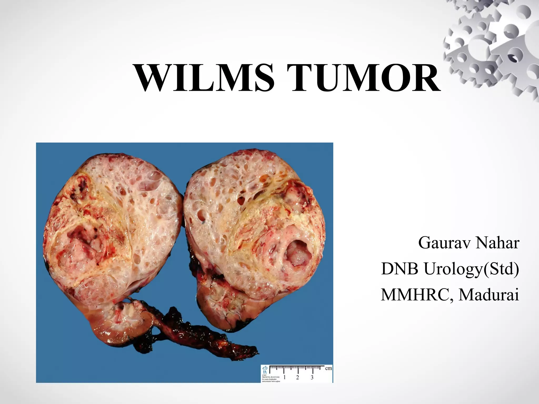
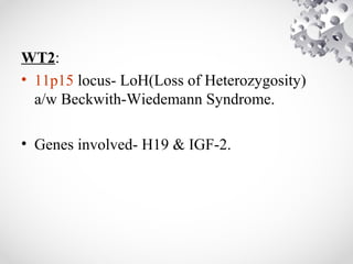
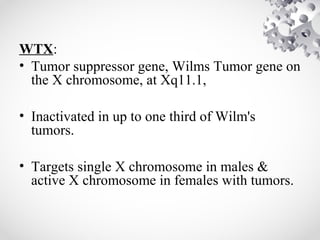
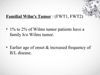
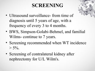
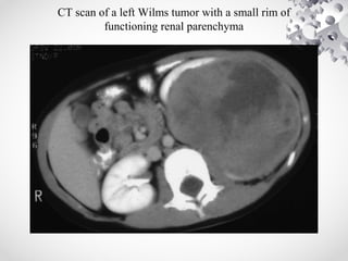
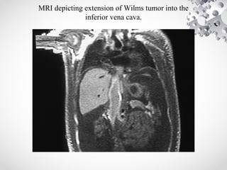
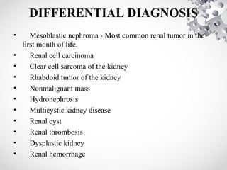
Wilms tumor, also known as nephroblastoma, is the most common malignant renal tumor of childhood. It develops from embryonic kidney tissue and accounts for 6-7% of childhood cancers. The tumor is usually diagnosed before age 5 and may be detected via abdominal mass or hematuria. Staging involves imaging like CT or MRI to determine extent. Prognosis depends on histology and stage. Treatment typically involves nephrectomy followed by chemotherapy, with radiation sometimes used. Late effects can include growth issues, infertility, and second cancers. Long term follow up is important after treatment ends.
Rhabdomyosarcoma is a rare cancer that forms in the body's soft tissues, such as muscle and connective tissue. In rhabdomyosarcoma, the cancer cells look similar to immature muscle cells.
POSTERIOR URETHRAL VALVES- Pediatric Surgery
• Dear viewers,
• Greetings from “ Surgical Educator”
• Today I have uploaded one more video in Pediatric Surgery/Pediatric Urology- “ Posterior Urethral Valves”
• Posterior Urethral Valves is the congenital cause for Bladder Outlet Obstruction, resulting in abnormal development of the kidneys as well as the bladder.
• In this video, I talked about the learning outcomes, introduction, etiopathogenesis, clinical features, investigations, differential diagnosis, treatment, follow-up and prognosis of “ Posterior Urethral Valves”
• I hope you will enjoy the video for its educational value.
• You can watch all my teaching videos in the following links
• surgicaleducator.blogspot.com youtube.com/c/surgicaleducator
• Thank you for watching the video.
WILMS’ TUMOUR-Pediatric Surgery
• Dear Viewers,
• Greetings from “Surgical Educator”
• Today I have uploaded a video on the commonest childhood solid tumour- Wilms’ tumour
• This is a malignant tumour arising from embryonal kidney cells
• It has excellent prognosis because of multi-disciplinary management
• In this video I have discussed the epidemiology, etiology, pathology, clinical features, investigations, staging, differential diagnosis and treatment of Wilms’ tumour.
• I hope you will like and enjoy watching the video
• You can watch all my surgery teaching videos in the following links:
• youtube.com/c/surgicaleducator surgicaleducator.blogspot.com
• Thank you for watching the video.
Detailed ppt on Wilma’s tumor … it includes definitions ,causes , pathophysiology, sign and symptoms, diagnostic evaluation, treatment, management with images , stages with images , nursing management
Nephroblastoma also known as Wilms tumor, is the most common renal malignancy affecting one in 10,000 children <15 years old
Children with bilateral disease are diagnosed at an earlier age (median age, girls at 31 months and boys at 24 months): Patients with associated congenital anomalies are also diagnosed at an earlier age
Accounts for 6-7% of cases of childhood cancer in the developed world and 12% in South Africa
In Tanzania the prevalence is 6.7% ( Mgaya E et al., 2000), “third from leukemia and lymphoma” (Shakilu J, 2017)
The overall survival rate of nephroblastoma approaches 90% in the developed world but in developing countries the survival rates are much less and in some sub-Saharan countries it is only 40% at 8 months after diagnosis
These may occur in 1% of infantile kidneys but typically regress during childhood
Nephrogenic rests normally occur in 1% of newborn kidneys and regress early in childhood: in contrast, they are present in 35% of kidneys with unilateral Wilms tumor and almost 100% of kidneys with bilateral disease
Has been associated with loss of function mutations of a number of tumor suppressor and transcription genes
These include mutations of the WT1, p53, FWT1, and FWT2 genes and at the 11p15.5 locus
Histologically, the classic favorable histology Wilms tumor is comprised of three cell types:
Blastemal cells – Undifferentiated cells
Stromal cells – Immature spindle cells and heterologous skeletal muscle, cartilage, osteoid or fat
Epithelial cells – Glomeruli and tubules
Grossly, Wilms tumors are usually well-circumscribed and have a pseudo-capsule
Histologically, Wilms is divided into "Favorable" and "Unfavorable" histologies
"Favorable" Histology: 90% of Wilms tumors will demonstrate "favorable" histology which generally has a better prognosis
Most children with Wilms tumor present with an abdominal mass or swelling without other signs or symptoms
The definitive diagnosis of Wilms tumor is made by histologic confirmation at the time of either surgical excision or biopsy
Stage I indicates the tumor was completely contained within the kidney without any breaks or spillage outside the renal capsule and no vascular invasion
Stage II would be a tumor that has grown outside the kidney
Stage IIIunresectable tumor
Stage IV-Metastasis
Stage V-bilateral kidney
Surgery is the main treatment
This slide explains about Germ cell tumor ovary (GCT Ovary). It explains how a various stages developmental anomaly could give rise to various types of GCT.
NVBDCP.pptx Nation vector borne disease control programSapna Thakur
NVBDCP was launched in 2003-2004 . Vector-Borne Disease: Disease that results from an infection transmitted to humans and other animals by blood-feeding arthropods, such as mosquitoes, ticks, and fleas. Examples of vector-borne diseases include Dengue fever, West Nile Virus, Lyme disease, and malaria.
Ethanol (CH3CH2OH), or beverage alcohol, is a two-carbon alcohol
that is rapidly distributed in the body and brain. Ethanol alters many
neurochemical systems and has rewarding and addictive properties. It
is the oldest recreational drug and likely contributes to more morbidity,
mortality, and public health costs than all illicit drugs combined. The
5th edition of the Diagnostic and Statistical Manual of Mental Disorders
(DSM-5) integrates alcohol abuse and alcohol dependence into a single
disorder called alcohol use disorder (AUD), with mild, moderate,
and severe subclassifications (American Psychiatric Association, 2013).
In the DSM-5, all types of substance abuse and dependence have been
combined into a single substance use disorder (SUD) on a continuum
from mild to severe. A diagnosis of AUD requires that at least two of
the 11 DSM-5 behaviors be present within a 12-month period (mild
AUD: 2–3 criteria; moderate AUD: 4–5 criteria; severe AUD: 6–11 criteria).
The four main behavioral effects of AUD are impaired control over
drinking, negative social consequences, risky use, and altered physiological
effects (tolerance, withdrawal). This chapter presents an overview
of the prevalence and harmful consequences of AUD in the U.S.,
the systemic nature of the disease, neurocircuitry and stages of AUD,
comorbidities, fetal alcohol spectrum disorders, genetic risk factors, and
pharmacotherapies for AUD.
Acute scrotum is a general term referring to an emergency condition affecting the contents or the wall of the scrotum.
There are a number of conditions that present acutely, predominantly with pain and/or swelling
A careful and detailed history and examination, and in some cases, investigations allow differentiation between these diagnoses. A prompt diagnosis is essential as the patient may require urgent surgical intervention
Testicular torsion refers to twisting of the spermatic cord, causing ischaemia of the testicle.
Testicular torsion results from inadequate fixation of the testis to the tunica vaginalis producing ischemia from reduced arterial inflow and venous outflow obstruction.
The prevalence of testicular torsion in adult patients hospitalized with acute scrotal pain is approximately 25 to 50 percent
Ozempic: Preoperative Management of Patients on GLP-1 Receptor Agonists Saeid Safari
Preoperative Management of Patients on GLP-1 Receptor Agonists like Ozempic and Semiglutide
ASA GUIDELINE
NYSORA Guideline
2 Case Reports of Gastric Ultrasound
TEST BANK for Operations Management, 14th Edition by William J. Stevenson, Ve...kevinkariuki227
TEST BANK for Operations Management, 14th Edition by William J. Stevenson, Verified Chapters 1 - 19, Complete Newest Version.pdf
Pulmonary Thromboembolism - etilogy, types, medical- Surgical and nursing man...VarunMahajani
Disruption of blood supply to lung alveoli due to blockage of one or more pulmonary blood vessels is called as Pulmonary thromboembolism. In this presentation we will discuss its causes, types and its management in depth.
263778731218 Abortion Clinic /Pills In Harare ,sisternakatoto
263778731218 Abortion Clinic /Pills In Harare ,ABORTION WOMEN’S CLINIC +27730423979 IN women clinic we believe that every woman should be able to make choices in her pregnancy. Our job is to provide compassionate care, safety,affordable and confidential services. That’s why we have won the trust from all generations of women all over the world. we use non surgical method(Abortion pills) to terminate…Dr.LISA +27730423979women Clinic is committed to providing the highest quality of obstetrical and gynecological care to women of all ages. Our dedicated staff aim to treat each patient and her health concerns with compassion and respect.Our dedicated group ABORTION WOMEN’S CLINIC +27730423979 IN women clinic we believe that every woman should be able to make choices in her pregnancy. Our job is to provide compassionate care, safety,affordable and confidential services. That’s why we have won the trust from all generations of women all over the world. we use non surgical method(Abortion pills) to terminate…Dr.LISA +27730423979women Clinic is committed to providing the highest quality of obstetrical and gynecological care to women of all ages. Our dedicated staff aim to treat each patient and her health concerns with compassion and respect.Our dedicated group of receptionists, nurses, and physicians have worked together as a teamof receptionists, nurses, and physicians have worked together as a team wwww.lisywomensclinic.co.za/
2. INTRODUCTION
• Wilms tumor - Nephroblastoma.
• Most common primary malignant renal tumor
of childhood.
• This embryonal tumor develops from remnants
of immature kidney.
3. EPIDEMIOLOGY
• Accounts for 6% to 7% of all childhood
cancers.
• Children<15 yrs: annual incidence rate 7 to 10
cases per million.
• More than 80% of cases are diagnosed before
5 years of age, with a median age of 3.5 years.
4. • B/L Wilms tumors present at earlier age.
• Incidence lower in East Asian populations &
higher in black populations compared with
North American and European whites.
• Environmental risk factors play a minor role.
5. ETIOLOGY
• Majority of Wilms tumors arise from somatic
mutations restricted to tumor tissue.
• A much smaller percentage originate from
germline mutations.
• Several genetic events result in Wilms tumor
development.
6. • 10% tumors- have coexistent congenital
anomalies and syndromes.
• 5% to 10% tumors- bilateral/multicentric.
• 1% to 2% are familial.
7.
8. WT1:
• 1st Wilms tumor gene to be identified.
• Gross deletions at chromosome 11p13.
• Associated syndromes:
1.WAGR syndrome
2.Denys-Drash syndrome
3.Frasier syndrome
9. • WT1 gene is important for normal kidney &
gonadal development.
• WT1 encodes a zinc-finger transcription factor
expressed in kidney, gonads, spleen, &
mesothelium
• WT1 is necessary for ureteric bud outgrowth
and nephrogenesis.
10. • WAGR (Wilms tumor, Aniridia, Genital
anomalies, mental Retardation) syndrome.
• Aniridia, found in 1.1% of Wilms tumor
patients, is caused by an abnormality of the
PAX6 gene located adjacent to WT1.
• Wilms tumor develops in 40% to 70% of
aniridia patients with deletions of WT1.
11. • Denys-Drash syndrome (DDS): specific
association of male pseudohermaphroditism,
renal mesangial sclerosis, and nephroblastoma.
• Caused by point mutations in zinc finger DNA
binding region of WT1.
• >90% of DDS patients harbor germline point
mutations in only one WT1 allele.
12. • WAGR and DDS patients- more likely to have
bilateral tumors & are diagnosed at a younger age.
• WAGR patients- increased risk of renal failure if they
survive into puberty.
• Genitourinary anomalies- renal fusion anomalies,
cryptorchidism, hypospadias are present in 4.5% of
patients with Wilms tumor.
14. • Beckwith-Wiedemann syndrome(BWS)
characterized by excess growth at cellular,
organ (macroglossia, nephromegaly,
hepatomegaly), or body segment
(hemihypertrophy) levels.
• Adrenocortical neoplasms and hepatoblastoma
also occur in BWS.
• Most cases sporadic; 15% heritable- AD.
15. WTX:
• Tumor suppressor gene, Wilms Tumor gene on
the X chromosome, at Xq11.1,
• Inactivated in up to one third of Wilm's
tumors.
• Targets single X chromosome in males &
active X chromosome in females with tumors.
16. Familial Wilm's Tumor : (FWT1, FWT2)
• 1% to 2% of Wilms tumor patients have a
family h/o Wilms tumor.
• Earlier age of onset & increased frequency of
B/L disease.
17. • TP53 mutations- increased frequency in
anaplastic histology.
• LoH at 1p and 16q are associated with an
increased risk of tumor relapse and death.
18. SCREENING
• Ultrasound surveillance- from time of
diagnosis until 5 years of age, with a
frequency of every 3 to 4 months.
• BWS, Simpson-Golabi-Behmel, and familial
Wilms- continue to 7 years.
• Screening recommended when WT incidence
> 5%.
• Screening of contralateral kidney after
nephrectomy for U/L Wilm's.
19. • CT or MRI if USG shows any suspicious lesion.
• 7-fold increased risk of Wilm's tumor in HK.
• an increased risk of müllerian duct anomalies in girls
with Wilms tumor- Approx.10% girls will have
abnormalities, such as duplication of cervix or uterus,
or bicornuate uterus.
20. PATHOLOGY
Favorable-Histology Wilms Tumor(FH):
• Wilms tumor compresses adjacent normal
renal parenchyma, forming an "intrarenal
pseudocapsule."
• Tremendous histologic diversity.
• 90% of all renal tumors have favorable
histology.
21. • “Classical” Wilms tumor is characterized by
islands of compact undifferentiated blastema,
presence of variable epithelial differentiation
in the form of embryonic tubules, rosettes, and
glomeruloid structures,
separated by a significant stromal component.
22.
23. • Predominantly epithelial differentiation- low
degree of aggressiveness, majority are stage I
tumors,
• But may be more resistant to therapy, if they
present as advanced-stage disease.
25. Anaplastic Wilms Tumor:
• Anaplasia is characterized by the presence of
three abnormalities:
1.nuclear enlargement to three or more times the
diameter of adjacent cells,
2.hyperchromasia of enlarged nuclei, and
3.abnormal mitotic figures.
• Rarely seen in children< 3 years.
26. • Resistant to chemotherapy.
• Poor prognosis.
• Further divided into focal & diffuse patterns.
27. Nephrogenis Rests:
• Precursor lesions; still most don't form Wilm's
tumor.
• A rest can undergo maturation, sclerosis,
involution, or complete disappearance.
• Two types based on location: Perilobar &
Intralobar(PLNRs & ILNRs).
28. • Perilobar NRs- found only in the lobar
periphery, elaborated late in embryogenesis.
• Subcortical, sharply demarcated, and contain
predominantly blastema & tubules.
• Usually found in BWS, linked to 11p15 locus.
29. • Intralobar NRs found anywhere within the
lobe, renal sinus and wall of PCS.
• Result of earlier gestational aberrations.
• ILNRs are commonly stroma rich.
• Typically seen in aniridia, WAGR, DDS or
other features a/w WT1.
30.
31. CLINICAL PRESENTATION
• A palpable smooth abdominal mass- 90%.
Incidentally discovered.
• Abdominal pain, gross hematuria & fever- less
frequent.
• Tumor rupture with hemorrhage into peritoneal
cavity- mimics acute abdomen.
• Extension into renal vein & IVC- varicocoele,
hepatomegaly due to hepatic vein obstruction, ascites,
and congestive heart failure- <10%.
32. • Hypertension- common at diagnosis, d/t
elevated plasma renin levels; resolves shortly
after removal.
• Acquired von Willebrand disease found in 8%
of newly diagnosed Wilms tumor.
33. IMAGING
• FOUR FIELD CHEST RADIOGRAPHY:
may show lung metastasis.
RENAL ULTRASOUND:
1st study to evaluate child with abd.mass.
demonstrate solid nature of the lesion.
Doppler USG helps to exclude intracaval
tumor extension, & its proximal extent.
34. Solid renal tumor: CT demonstrates that lesion is amenable to renal-sparing
surgery
35. • CT SCAN:
helps to determine origin of the tumor, lymph
node involvement, B/L kidney involvement,
invasion into major vessels (IVC), and liver
metastases.
CT chest to rule out lung metastasis.
36. CT scan of a left Wilms tumor with a small rim of
functioning renal parenchyma
37. • MRI ABDOMEN:
Most sensitive imaging modality for caval
patency, to determine tumor extension into
IVC.
low signal intensity on T1-weighted images
and high signal intensity on T2-weighted
images.
40. DIFFERENTIAL DIAGNOSIS
• Mesoblastic nephroma - Most common renal tumor in the
first month of life.
• Renal cell carcinoma
• Clear cell sarcoma of the kidney
• Rhabdoid tumor of the kidney
• Nonmalignant mass
• Hydronephrosis
• Multicystic kidney disease
• Renal cyst
• Renal thrombosis
• Dysplastic kidney
• Renal hemorrhage
42. PROGNOSTIC FACTORS
• Most Important determinants of outcome:
histopathology & tumor stage.
• Chromosomal Abnormalities: LOH for
chromosome 16q and/or 1p (20% of Wilms
tumors) a/w increased risk for relapse & death.
• High telomerase activity- an unfavourable
prognostic feature.
• DNA Content: Aneuploidy & DNA index .
1.5- strongly a/w anaplastic histology.
• Cytokines: VEGF angiogenic cytokine.
43. TREAMENT
• Usual approach- nephrectomy followed by
chemotherapy, with or without postoperative
radiotherapy.
• Multiple RCTs to determine therapeutic protocols by:
1. National Wilm's Tumor Study Group/Children's
Oncology Group(NWTSG/COG),
2. International Society of Pediatric Oncology(SIOP),
and
3. United Kingdom Children’s Cancer Study Group
(UKCCSG) .
44.
45. COG AREN0321 protocol for high risk Wilms
tumor
• Focal anaplastic stage I-III Wilms tumors and diffuse
anaplastic stage I Wilms tumors - Nephrectomy followed by
vincristine, actinomycin-D, and doxorubicin in addition to
local radiotherapy
• Focal anaplastic stage IV Wilms tumors and diffuse
anaplastic stage II-III tumors –Patients undergo the same
treatment, with the addition of cyclophosphamide, etoposide,
and carboplatin
• Stage IV diffuse anaplastic Wilms tumors - More
aggressive treatment is delivered; nephrectomy is followed by
initial irinotecan and vincristine administration, which in turn
is followed by actinomycin-D, doxorubicin,
cyclophosphamide, carboplatin, etoposide, and radiotherapy.
46. SURGICAL CONSIDERATIONS:
• Radical nephrectomy by transperitoneal approach.
• Thorough exploration of the abdominal cavity to
exclude local tumor extension, liver and nodal
metastases, or peritoneal seeding.
• Accurate staging to determine appropriate
chemotherapy & need for radiation therapy.
• Selective sampling of suspicious nodes is essential.
• Formal RPLND is not recommended.
47. • Risk factors for local tumor recurrence
(Shamberger):
1.tumor spillage,
2.unfavorable histology,
3.incomplete tumor removal, and
4.absence of any lymph node sampling.
48. PREOPERATIVE CHEMOTHERAPY:
• Situations where preoperative chemotherapy is
recommended:-
1. Children for whom renal-sparing surgery is planned,
2. Tumors are inoperable at surgical exploration, and
3. There is tumor extension into IVC above hepatic
veins.
• An inoperable tumor should be considered stage III
and treated accordingly.
49. • Inoperability should not be based on
preoperative imaging studies, which can
overestimate local tumor extension.
• Pretreatment with chemotherapy almost
always reduces the bulk of tumor and renders
it resectable.
• Majority of reduction in tumor volume occurs
in first 4 weeks of chemotherapy.
50. A, MRI of a Wilms tumor that was pretreated with chemotherapy.
B, After 6 weeks of chemotherapy, the tumor is much smaller in size
51. MANAGEMENT OF LUNG METASTASIS:
• CXR negative but CT positive: tissue
diagnosis of lung nodules because several
conditions (eg, histoplasmosis, atelectasis,
pseudotumor, intrapulmonary lymph node,
pneumonia) can mimic pulmonary metastases.
52. • WT FH with lung mets, no other mets/1p or
16q deletion: 6 weeks of actinomycin-D,
doxorubicin, and vincristine.
Complete resolution- No radiation required.
No resolution- cyclophosphamide and
etoposide in addition + radiation therapy.
53. MANAGEMENT OF B/L WILMS TUMORS:
• No initial radical nephrectomy.
• Preoperative chemotherapy for 6 weeks.
• tumors amenable to renal-sparing procedures
can proceed with surgery.
• Tumors not responding- B/L open biopsy &
additional chemo based on biopsy findings.
54. • Proceed to Sx at 12 weeks of therapy (no
benefit beyond 12 weeks).
• Partial nephrectomy, tumor enucleation or
wedge excision of tumor.
• In FH tumors, even with positive margins or
large B/L residual masses, adjuvant therapy
provides a good outcome.
55. LATE EFFECTS OF Rx
RADIATION:
• Musculoskeletal problems like scoliosis.
• Reduction in stature.
• Hypogonadism & temporary azoospermia.
• Delayed sexual maturation.
• Ovarian failure.
• Adverse pregnancy outcomes with increased
risk for miscarriage, LBW, prematurity &
congenital malformations.
• Increased risk of 2nd malignant neoplasms.