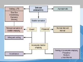
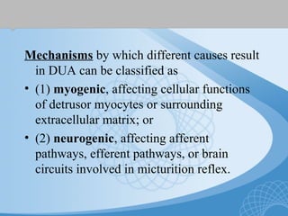
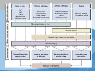
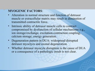
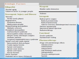
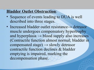
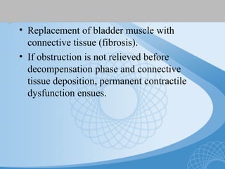
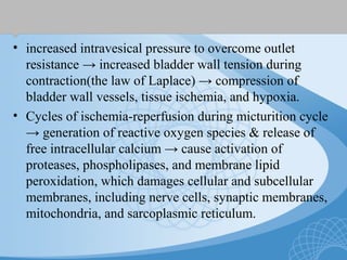
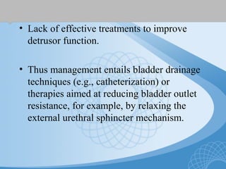
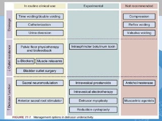
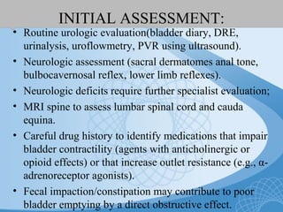
UNDERACTIVE
DETRUSOR
Gaurav Nahar
DNB Urology Resident
MMHRC, Madurai
INTRODUCTION
• Impaired ability to empty the bladder may
be d/t:
1.increased resistance of bladder outlet and/or
2.a reduction in the ability to generate an
efficient bladder contraction, referred to as
detrusor underactivity (DUA) by ICS.
TERMINOLOGY & DEFINITION
• Impaired detrusor
contractility
• Detrusor areflexia
• Detrusor failure
• ICS Standardization
document 2002: DUA
defined on the basis of a
urodynamic study (UDS)
as “a contraction of
reduced strength and/or
duration, resulting in
prolonged bladder
emptying and/or failure
to achieve complete
bladder emptying within
a normal time span."
SYMPTOMS
• Difficult to define because of absence of pathognomonic
symptoms.
• Symptoms diverse & overlap with those of OAB & BOO.
• Voiding phase: hesitancy, intermittency, straining & weak
stream.
• Storage phase:Urinary frequency, nocturia, loss of normal
urge to void & infrequent voiding
• Post-voiding: feeling of incomplete bladder emptying.
• Large PVR- incontinence(esp.during sleep).
EPIDEMIOLOGY
• Population prevalence of DUA is not
known, because of the lack of a noninvasive
marker.
• Storage LUTS are present in 45.7% of men
and 66.8% of women.
• Voiding LUTS documented in 57.1% of
men and 48% of women.
• Postmicturition symptoms: similar
prevalence in both men and women.
• DUA affects 9% to 28% of men under 50
years of age and 48% in those over 70 years
undergoing UDS and is more prevalent
among the institutionalized elderly.
• In women DUA is found in 12% to 45%
undergoing UDS and is more prevalent
among the institutionalized elderly.
• Detrusor hyperactivity impaired
contractility (DHIC): DUA with
concomitant DOA.
• DUA often coexists with other LUT
dysfunctions in the elderly; BOO in males
& Urodynamic SUI in females.
ETIOPATHOGENESIS
• In clinical practice, no obvious cause found in
majority.
• May occur secondary to common age-related
changes & decline in detrusor function.
• More pronounced decline in detrusor
contractility in men because of bladder wall
changes (increased connective tissue and reduced
smooth muscle) occurring as a result of BOO.
Mechanisms by which different causes result
in DUA can be classified as
• (1) myogenic, affecting cellular functions
of detrusor myocytes or surrounding
extracellular matrix; or
• (2) neurogenic, affecting afferent
pathways, efferent pathways, or brain
circuits involved in micturition reflex.
MYOGENIC FACTORS:
• Alteration in normal structure and function of detrusor
muscle or extracellular matrix may result in diminution of
transmitted contractile force.
• Intrinsic ability of detrusor muscle cells to contract
compromised by dysfunction of cellular mechanisms e.g.,
ion storage/exchange, excitation-contraction coupling,
calcium storage, energy generation.
• Degeneration pattern in DUA: widespread disrupted
detrusor myoctyes and axonal degeneration.
• Whether detrusor myocyte disruption is the cause of DUA
or a consequence of a pathologic insult is not clear.
NEUROGENIC FACTORS:
Brain Circuits:
• CNS control mechanism governing
micturition reflex: perception and
integration of storage and voiding, if
disturbed, may result in DUA.
• Areas of brainstem and cortex implicated:
insula, hypothalamus, limbic system,
periaqueductal gray(PAG), and PMC.
Bladder Efferent Pathways:
• Interruption or impairment of efferent
signaling in sacral cord, sacral roots, and
pelvic nerves → absent or reduced detrusor
contraction.
• Evidence suggest that a reduction in
autonomic innervation occurs in human
bladders as a consequence of normal aging.
Bladder and Urethral Afferent Pathways:
• Intact bladder sensation is critical for
functioning of efferent limb of micturition
reflex.
• Bladder afferents monitor both volumes
during bladder filling in the storage phase
and magnitude of detrusor contractions
during voiding phase.
• Urethral afferents perceive both flow
through urethra and detrusor contraction.
• An impairment in afferent function(from
bladder or urethra) reduce or prematurely
end the micturition reflex, leading to
impairment or loss of voiding efficiency
• Normal aging is associated with a decline in
sensory function in the LUT.
Bladder Outlet Obstruction:
• Sequence of events leading to DUA is well
described into three stages.
• Increased bladder outlet resistance→ detrusor
muscle undergoes compensatory hypertrophy
and hyperplasia → blood supply also increases
(Contractile function almost normal; bladder in
compensated stage) → slowly detrusor
contractile function declines & bladder
emptying is impaired, marking the
decompensation phase.
• Replacement of bladder muscle with
connective tissue (fibrosis).
• If obstruction is not relieved before
decompensation phase and connective
tissue deposition, permanent contractile
dysfunction ensues.
• increased intravesical pressure to overcome outlet
resistance → increased bladder wall tension during
contraction(the law of Laplace) → compression of
bladder wall vessels, tissue ischemia, and hypoxia.
• Cycles of ischemia-reperfusion during micturition cycle
→ generation of reactive oxygen species & release of
free intracellular calcium → cause activation of
proteases, phospholipases, and membrane lipid
peroxidation, which damages cellular and subcellular
membranes, including nerve cells, synaptic membranes,
mitochondria, and sarcoplasmic reticulum.
Diabetes Mellitus:
• May impair detrusor function through a
combination of myogenic and neurogenic
mechanisms:- Diabetes-induced bladder
dysfunction (DBD) or diabetic
cystopathy.
• Autonomic neuropathy occurring as a
result of axonal degeneration and segmental
demyelination resulting in diminished
bladder sensation.
Underlying mechanisms (d/t hyperglycemia):
• activation of polyol pathway,
• increases in generation of free radicals,
• activation of protein kinase C,
• formation of advanced glycated end
products,
• reduction in nerve growth factor essential
for maintaining normal sympathetic and
sensory nerve function.
• Detrusor myocyte dysfunction in DBD-
explained by DBD temporal theory →
initially osmotic diuresis induced by
hyperglycemia causes bladder wall
stretching & increased intravesical pressure
→ compensatory bladder hypertrophy. as
the disease progresses, accumulation of
toxic products of oxidative stress results in
bladder decompensation → poor bladder
sensation and impaired voiding function.
NEUROLOGIC DISEASE OR INJURY:
• CVA- neurogenic DOA most common, 75%
acontractile detrusors & AUR d/t "cerebral shock"
• Parkinson's disease- DUA(20%) far less common
than DOA.
• Multisystem atrophy/Shy-Drager syndrome- 52-
92% DUA on UDS d/t atrophy of efferent
parasympathetic nerves.
• Multiple Sclerosis- DUA in 20%.
• Radical pelvic surgery can lead to injury to the
pelvic plexus(located near anterolateral wall of
lower rectum) or postganglionic fibers that
traverse lateral wall of upper vagina.
DIAGNOSIS
• Only accepted diagnostic modality-
invasive UDS.
• No universal diagnostic criteria.
Detrusor Contraction Strength:
• Current methods of estimating detrusor voiding
function almost exclusively focus on detrusor
contraction strength.
Detrusor Contraction Speed:
• A bladder that contracts more slowly could in
theory result in clinical symptoms, although this
is not part of ICS definition.
Detrusor Contraction Duration:
• A detrusor contraction of reduced duration is
suggested by ICS definition.
Bladder Sensation:
• Assessment of bladder sensation relevant to evaluation of
DUA because afferent nerves play a central role in the
initiation and maintenance of a detrusor contraction.
• Most commonly undertaken using patient's perceptions
of bladder filling (first sensation, first desire, strong
desire, and capacity).
Ambulatory Urodynamics:
• failure to void during UDS- anxiety or a so-called
bashful bladder; arises d/t poor pelvic floor relaxation
and reflex detrusor inhibition, or true DUA or
acontractile detrusor.
• Careful history & Ambulatory UDS are useful.
MANAGEMENT
GOALS:
• To reduce symptoms & improve quality of life.
• Reduce the risk of complications of impaired
bladder emptying viz.UTIs, bladder stones,
ureteric reflux leading to back-pressure on the
upper urinary tract, and skin damage from urinary
overflow incontinence associated with chronic
retention.
• Lack of effective treatments to improve
detrusor function.
• Thus management entails bladder drainage
techniques (e.g., catheterization) or
therapies aimed at reducing bladder outlet
resistance, for example, by relaxing the
external urethral sphincter mechanism.
INITIAL ASSESSMENT:
• Routine urologic evaluation(bladder diary, DRE,
urinalysis, uroflowmetry, PVR using ultrasound).
• Neurologic assessment (sacral dermatomes anal tone,
bulbocavernosal reflex, lower limb reflexes).
• Neurologic deficits require further specialist evaluation;
• MRI spine to assess lumbar spinal cord and cauda
equina.
• Careful drug history to identify medications that impair
bladder contractility (agents with anticholinergic or
opioid effects) or that increase outlet resistance (e.g., α-
adrenoreceptor agonists).
• Fecal impaction/constipation may contribute to poor
bladder emptying by a direct obstructive effect.
CONSERVATIVE MANAGEMENT
• Scheduled voiding: to increase the frequency of voids in
patients with sensory impairment.
• Double voiding to improve bladder emptying may help
reduce bothersome frequency,
• Bladder expression techniques such as Valsalva voiding
or Credé maneuver used in only very specific neurogenic
situations (i.e., DUA with incompetent sphincter);
otherwise not recommended because of risk for generating
high vesical pressure, causing vesicoureteral reflux or
reflux into prostate & seminal vesicles.
• Pelvic floor physiotherapy and biofeedback to
successfully treat children and adults with dysfunctional
voiding
• CISC is the preferred method of
establishing bladder drainage.
• Safe and effective, with lower infection
rates than with indwelling catheters.
• An indwelling urethral catheter is best
avoided in the long term, &
• A suprapubic catheter is the best long-term
option in patients unwilling or unable to
perform CISC.
PHARMACOTHERAPY
Parasympathomimetics:
• Acetylcholine- principal neurotransmitter mediating
bladder contraction, acting on muscarinic (M3) receptors.
Parasympathomimetic agents, including direct muscarinic
receptor agonists or anticholinesterases, have been used to
increase bladder contractility.
• Bethanechol and carbachol, most common compounds, are
quaternary amines selective for muscarinic receptor but not
receptor subtype selective.
• More likely to be effective if the problem is reduced or
absent contractile stimulus (e.g., reduced efferent input,
impaired acetylcholine release from parasympathetic
nerves, increased acetylcholine breakdown.
• If underlying cause is reduced tissue responsiveness to
stimulation (e.g., detrusor muscle cell dysfunction, bladder
wall fibrosis), loss of detrusor muscle,
parasympathomimetics are less likely to benefit.
• Significant dose-dependent systemic side
effects- nausea, bronchospasm, abdominal
cramping, diarrhea, increased salivation,
flushing, and visual disturbance.
• A rare but potentially lethal side effect is
severe cardiac depression resulting in
cardiac arrest.
α-Adrenoreceptor Antagonists:
• Combination therapy with an α-adrenoreceptor
antagonist and a parasympathomimetic has long
been considered a therapeutic possibility.
Prostanoids:
• Subclass of signaling molecules implicated in the
micturition reflex.
• Efficacy not clear.
• Five principal endogenous prostanoids (PGE2,
PGF2α, PGI2, PGD2, and thromboxane A.
ELECTRICAL STIMULATION
• Sacral root stimulator for patients with complete
spinal cord injury to activate anterior sacral roots and
achieve volitional bladder emptying. For the procedure
to be successful there is a need for intact peripheral
efferents and the absence of myogenic dysfunction.
• Intravesical electrotherapy (IVE). Bladder is filled
with saline, and current is passed through an electrode
(cathode) at the tip of the catheter; circuit is completed
by a neutral electrode applied to the skin in an area of
normal sensation.
• Daily sessions of stimulation are undertaken, of 1 hour
or more, with 10 to 15 sessions.
• Controversial therapy with significant time & resource
requirements.
• Sacral Neuromodulation: good efficacy in
non-obstructive urinary retention & DUA.
• SNM inhibits urethral afferent signals and
allows restoration of normal afferent flow to
the brain and resumption of normal bladder
sensation and detrusor contractions.
BOTULINUM TOXIN
• Botulinum neurotoxin A (BoNT-A) injected
into urethral sphincter reduces outlet
resistance & improves bladder emptying in
detrusor-sphincter dyssynergia
• Rationale for its use in DUA is to relax the
urethral sphincter mechanism, thereby
overcoming reflex inhibition of detrusor
function, or to facilitate Valsalva-induced
voiding.
• Short-lived action, hence not licensed for use.
SURGERY
Bladder Outlet Surgery:
• Compared to conservative treatment, outlet surgery
confers no significant improvement in symptoms or
urodynamic parameters in DUA.
• Predictors of poor outcome- low voiding pressures (<45
cm H2O), older age (>80 years), and high residual
volume (>1500 mL).
• In the absence of any other effective treatments some
advocate surgery in younger, medically fit patient who
wishes to become catheter free.
• Resection or incision of bladder neck in women with
DUA is not recommended, because this may lead to
incontinence or bladder neck stenosis.
Urinary Diversion:
• When CSIC is not possible and patient
wishes to avoid a suprapubic catheter, as in
young women with idiopathic urinary
retention refractory to SNM, a continent
catheterizable stoma is an option.
• Incontinent diversion (e.g., ileal conduit) is
occasionally performed when there are
signs of renal deterioration resulting from
obstruction to the intramural ureters
secondary to a thick-walled bladder.
RECONSTRUCTIVE SURGERY
Latissimus dorsi detrusor myoplasty:
• muscle is harvested, and its pedicle is
anastomosed to inferior epigastric vessels,
with nerve coapted to intercostal branch.
Muscle is wrapped in a spiral configuration
around the bladder, covering over three
quarters of its surface, then anchored to
pelvic floor fascia and ligaments.
• Complications: in one-third-
thromboembolism, wound infection, pelvic
abscess.
THANK YOU !!!

UNDERACTIVE DETRUSOR

  • 1.
  • 2.
    INTRODUCTION • Impaired abilityto empty the bladder may be d/t: 1.increased resistance of bladder outlet and/or 2.a reduction in the ability to generate an efficient bladder contraction, referred to as detrusor underactivity (DUA) by ICS.
  • 3.
    TERMINOLOGY & DEFINITION •Impaired detrusor contractility • Detrusor areflexia • Detrusor failure • ICS Standardization document 2002: DUA defined on the basis of a urodynamic study (UDS) as “a contraction of reduced strength and/or duration, resulting in prolonged bladder emptying and/or failure to achieve complete bladder emptying within a normal time span."
  • 5.
    SYMPTOMS • Difficult todefine because of absence of pathognomonic symptoms. • Symptoms diverse & overlap with those of OAB & BOO. • Voiding phase: hesitancy, intermittency, straining & weak stream. • Storage phase:Urinary frequency, nocturia, loss of normal urge to void & infrequent voiding • Post-voiding: feeling of incomplete bladder emptying. • Large PVR- incontinence(esp.during sleep).
  • 7.
    EPIDEMIOLOGY • Population prevalenceof DUA is not known, because of the lack of a noninvasive marker. • Storage LUTS are present in 45.7% of men and 66.8% of women. • Voiding LUTS documented in 57.1% of men and 48% of women. • Postmicturition symptoms: similar prevalence in both men and women.
  • 8.
    • DUA affects9% to 28% of men under 50 years of age and 48% in those over 70 years undergoing UDS and is more prevalent among the institutionalized elderly. • In women DUA is found in 12% to 45% undergoing UDS and is more prevalent among the institutionalized elderly. • Detrusor hyperactivity impaired contractility (DHIC): DUA with concomitant DOA.
  • 9.
    • DUA oftencoexists with other LUT dysfunctions in the elderly; BOO in males & Urodynamic SUI in females.
  • 10.
    ETIOPATHOGENESIS • In clinicalpractice, no obvious cause found in majority. • May occur secondary to common age-related changes & decline in detrusor function. • More pronounced decline in detrusor contractility in men because of bladder wall changes (increased connective tissue and reduced smooth muscle) occurring as a result of BOO.
  • 11.
    Mechanisms by whichdifferent causes result in DUA can be classified as • (1) myogenic, affecting cellular functions of detrusor myocytes or surrounding extracellular matrix; or • (2) neurogenic, affecting afferent pathways, efferent pathways, or brain circuits involved in micturition reflex.
  • 13.
    MYOGENIC FACTORS: • Alterationin normal structure and function of detrusor muscle or extracellular matrix may result in diminution of transmitted contractile force. • Intrinsic ability of detrusor muscle cells to contract compromised by dysfunction of cellular mechanisms e.g., ion storage/exchange, excitation-contraction coupling, calcium storage, energy generation. • Degeneration pattern in DUA: widespread disrupted detrusor myoctyes and axonal degeneration. • Whether detrusor myocyte disruption is the cause of DUA or a consequence of a pathologic insult is not clear.
  • 14.
    NEUROGENIC FACTORS: Brain Circuits: •CNS control mechanism governing micturition reflex: perception and integration of storage and voiding, if disturbed, may result in DUA. • Areas of brainstem and cortex implicated: insula, hypothalamus, limbic system, periaqueductal gray(PAG), and PMC.
  • 15.
    Bladder Efferent Pathways: •Interruption or impairment of efferent signaling in sacral cord, sacral roots, and pelvic nerves → absent or reduced detrusor contraction. • Evidence suggest that a reduction in autonomic innervation occurs in human bladders as a consequence of normal aging.
  • 16.
    Bladder and UrethralAfferent Pathways: • Intact bladder sensation is critical for functioning of efferent limb of micturition reflex. • Bladder afferents monitor both volumes during bladder filling in the storage phase and magnitude of detrusor contractions during voiding phase. • Urethral afferents perceive both flow through urethra and detrusor contraction.
  • 17.
    • An impairmentin afferent function(from bladder or urethra) reduce or prematurely end the micturition reflex, leading to impairment or loss of voiding efficiency • Normal aging is associated with a decline in sensory function in the LUT.
  • 19.
    Bladder Outlet Obstruction: •Sequence of events leading to DUA is well described into three stages. • Increased bladder outlet resistance→ detrusor muscle undergoes compensatory hypertrophy and hyperplasia → blood supply also increases (Contractile function almost normal; bladder in compensated stage) → slowly detrusor contractile function declines & bladder emptying is impaired, marking the decompensation phase.
  • 20.
    • Replacement ofbladder muscle with connective tissue (fibrosis). • If obstruction is not relieved before decompensation phase and connective tissue deposition, permanent contractile dysfunction ensues.
  • 21.
    • increased intravesicalpressure to overcome outlet resistance → increased bladder wall tension during contraction(the law of Laplace) → compression of bladder wall vessels, tissue ischemia, and hypoxia. • Cycles of ischemia-reperfusion during micturition cycle → generation of reactive oxygen species & release of free intracellular calcium → cause activation of proteases, phospholipases, and membrane lipid peroxidation, which damages cellular and subcellular membranes, including nerve cells, synaptic membranes, mitochondria, and sarcoplasmic reticulum.
  • 22.
    Diabetes Mellitus: • Mayimpair detrusor function through a combination of myogenic and neurogenic mechanisms:- Diabetes-induced bladder dysfunction (DBD) or diabetic cystopathy. • Autonomic neuropathy occurring as a result of axonal degeneration and segmental demyelination resulting in diminished bladder sensation.
  • 23.
    Underlying mechanisms (d/thyperglycemia): • activation of polyol pathway, • increases in generation of free radicals, • activation of protein kinase C, • formation of advanced glycated end products, • reduction in nerve growth factor essential for maintaining normal sympathetic and sensory nerve function.
  • 24.
    • Detrusor myocytedysfunction in DBD- explained by DBD temporal theory → initially osmotic diuresis induced by hyperglycemia causes bladder wall stretching & increased intravesical pressure → compensatory bladder hypertrophy. as the disease progresses, accumulation of toxic products of oxidative stress results in bladder decompensation → poor bladder sensation and impaired voiding function.
  • 25.
    NEUROLOGIC DISEASE ORINJURY: • CVA- neurogenic DOA most common, 75% acontractile detrusors & AUR d/t "cerebral shock" • Parkinson's disease- DUA(20%) far less common than DOA. • Multisystem atrophy/Shy-Drager syndrome- 52- 92% DUA on UDS d/t atrophy of efferent parasympathetic nerves. • Multiple Sclerosis- DUA in 20%. • Radical pelvic surgery can lead to injury to the pelvic plexus(located near anterolateral wall of lower rectum) or postganglionic fibers that traverse lateral wall of upper vagina.
  • 26.
    DIAGNOSIS • Only accepteddiagnostic modality- invasive UDS. • No universal diagnostic criteria.
  • 28.
    Detrusor Contraction Strength: •Current methods of estimating detrusor voiding function almost exclusively focus on detrusor contraction strength. Detrusor Contraction Speed: • A bladder that contracts more slowly could in theory result in clinical symptoms, although this is not part of ICS definition. Detrusor Contraction Duration: • A detrusor contraction of reduced duration is suggested by ICS definition.
  • 29.
    Bladder Sensation: • Assessmentof bladder sensation relevant to evaluation of DUA because afferent nerves play a central role in the initiation and maintenance of a detrusor contraction. • Most commonly undertaken using patient's perceptions of bladder filling (first sensation, first desire, strong desire, and capacity). Ambulatory Urodynamics: • failure to void during UDS- anxiety or a so-called bashful bladder; arises d/t poor pelvic floor relaxation and reflex detrusor inhibition, or true DUA or acontractile detrusor. • Careful history & Ambulatory UDS are useful.
  • 30.
    MANAGEMENT GOALS: • To reducesymptoms & improve quality of life. • Reduce the risk of complications of impaired bladder emptying viz.UTIs, bladder stones, ureteric reflux leading to back-pressure on the upper urinary tract, and skin damage from urinary overflow incontinence associated with chronic retention.
  • 31.
    • Lack ofeffective treatments to improve detrusor function. • Thus management entails bladder drainage techniques (e.g., catheterization) or therapies aimed at reducing bladder outlet resistance, for example, by relaxing the external urethral sphincter mechanism.
  • 33.
    INITIAL ASSESSMENT: • Routineurologic evaluation(bladder diary, DRE, urinalysis, uroflowmetry, PVR using ultrasound). • Neurologic assessment (sacral dermatomes anal tone, bulbocavernosal reflex, lower limb reflexes). • Neurologic deficits require further specialist evaluation; • MRI spine to assess lumbar spinal cord and cauda equina. • Careful drug history to identify medications that impair bladder contractility (agents with anticholinergic or opioid effects) or that increase outlet resistance (e.g., α- adrenoreceptor agonists). • Fecal impaction/constipation may contribute to poor bladder emptying by a direct obstructive effect.
  • 34.
    CONSERVATIVE MANAGEMENT • Scheduledvoiding: to increase the frequency of voids in patients with sensory impairment. • Double voiding to improve bladder emptying may help reduce bothersome frequency, • Bladder expression techniques such as Valsalva voiding or Credé maneuver used in only very specific neurogenic situations (i.e., DUA with incompetent sphincter); otherwise not recommended because of risk for generating high vesical pressure, causing vesicoureteral reflux or reflux into prostate & seminal vesicles. • Pelvic floor physiotherapy and biofeedback to successfully treat children and adults with dysfunctional voiding
  • 35.
    • CISC isthe preferred method of establishing bladder drainage. • Safe and effective, with lower infection rates than with indwelling catheters. • An indwelling urethral catheter is best avoided in the long term, & • A suprapubic catheter is the best long-term option in patients unwilling or unable to perform CISC.
  • 36.
    PHARMACOTHERAPY Parasympathomimetics: • Acetylcholine- principalneurotransmitter mediating bladder contraction, acting on muscarinic (M3) receptors. Parasympathomimetic agents, including direct muscarinic receptor agonists or anticholinesterases, have been used to increase bladder contractility. • Bethanechol and carbachol, most common compounds, are quaternary amines selective for muscarinic receptor but not receptor subtype selective. • More likely to be effective if the problem is reduced or absent contractile stimulus (e.g., reduced efferent input, impaired acetylcholine release from parasympathetic nerves, increased acetylcholine breakdown. • If underlying cause is reduced tissue responsiveness to stimulation (e.g., detrusor muscle cell dysfunction, bladder wall fibrosis), loss of detrusor muscle, parasympathomimetics are less likely to benefit.
  • 37.
    • Significant dose-dependentsystemic side effects- nausea, bronchospasm, abdominal cramping, diarrhea, increased salivation, flushing, and visual disturbance. • A rare but potentially lethal side effect is severe cardiac depression resulting in cardiac arrest.
  • 38.
    α-Adrenoreceptor Antagonists: • Combinationtherapy with an α-adrenoreceptor antagonist and a parasympathomimetic has long been considered a therapeutic possibility. Prostanoids: • Subclass of signaling molecules implicated in the micturition reflex. • Efficacy not clear. • Five principal endogenous prostanoids (PGE2, PGF2α, PGI2, PGD2, and thromboxane A.
  • 39.
    ELECTRICAL STIMULATION • Sacralroot stimulator for patients with complete spinal cord injury to activate anterior sacral roots and achieve volitional bladder emptying. For the procedure to be successful there is a need for intact peripheral efferents and the absence of myogenic dysfunction. • Intravesical electrotherapy (IVE). Bladder is filled with saline, and current is passed through an electrode (cathode) at the tip of the catheter; circuit is completed by a neutral electrode applied to the skin in an area of normal sensation. • Daily sessions of stimulation are undertaken, of 1 hour or more, with 10 to 15 sessions. • Controversial therapy with significant time & resource requirements.
  • 40.
    • Sacral Neuromodulation:good efficacy in non-obstructive urinary retention & DUA. • SNM inhibits urethral afferent signals and allows restoration of normal afferent flow to the brain and resumption of normal bladder sensation and detrusor contractions.
  • 41.
    BOTULINUM TOXIN • Botulinumneurotoxin A (BoNT-A) injected into urethral sphincter reduces outlet resistance & improves bladder emptying in detrusor-sphincter dyssynergia • Rationale for its use in DUA is to relax the urethral sphincter mechanism, thereby overcoming reflex inhibition of detrusor function, or to facilitate Valsalva-induced voiding. • Short-lived action, hence not licensed for use.
  • 42.
    SURGERY Bladder Outlet Surgery: •Compared to conservative treatment, outlet surgery confers no significant improvement in symptoms or urodynamic parameters in DUA. • Predictors of poor outcome- low voiding pressures (<45 cm H2O), older age (>80 years), and high residual volume (>1500 mL). • In the absence of any other effective treatments some advocate surgery in younger, medically fit patient who wishes to become catheter free. • Resection or incision of bladder neck in women with DUA is not recommended, because this may lead to incontinence or bladder neck stenosis.
  • 43.
    Urinary Diversion: • WhenCSIC is not possible and patient wishes to avoid a suprapubic catheter, as in young women with idiopathic urinary retention refractory to SNM, a continent catheterizable stoma is an option. • Incontinent diversion (e.g., ileal conduit) is occasionally performed when there are signs of renal deterioration resulting from obstruction to the intramural ureters secondary to a thick-walled bladder.
  • 44.
    RECONSTRUCTIVE SURGERY Latissimus dorsidetrusor myoplasty: • muscle is harvested, and its pedicle is anastomosed to inferior epigastric vessels, with nerve coapted to intercostal branch. Muscle is wrapped in a spiral configuration around the bladder, covering over three quarters of its surface, then anchored to pelvic floor fascia and ligaments. • Complications: in one-third- thromboembolism, wound infection, pelvic abscess.
  • 45.