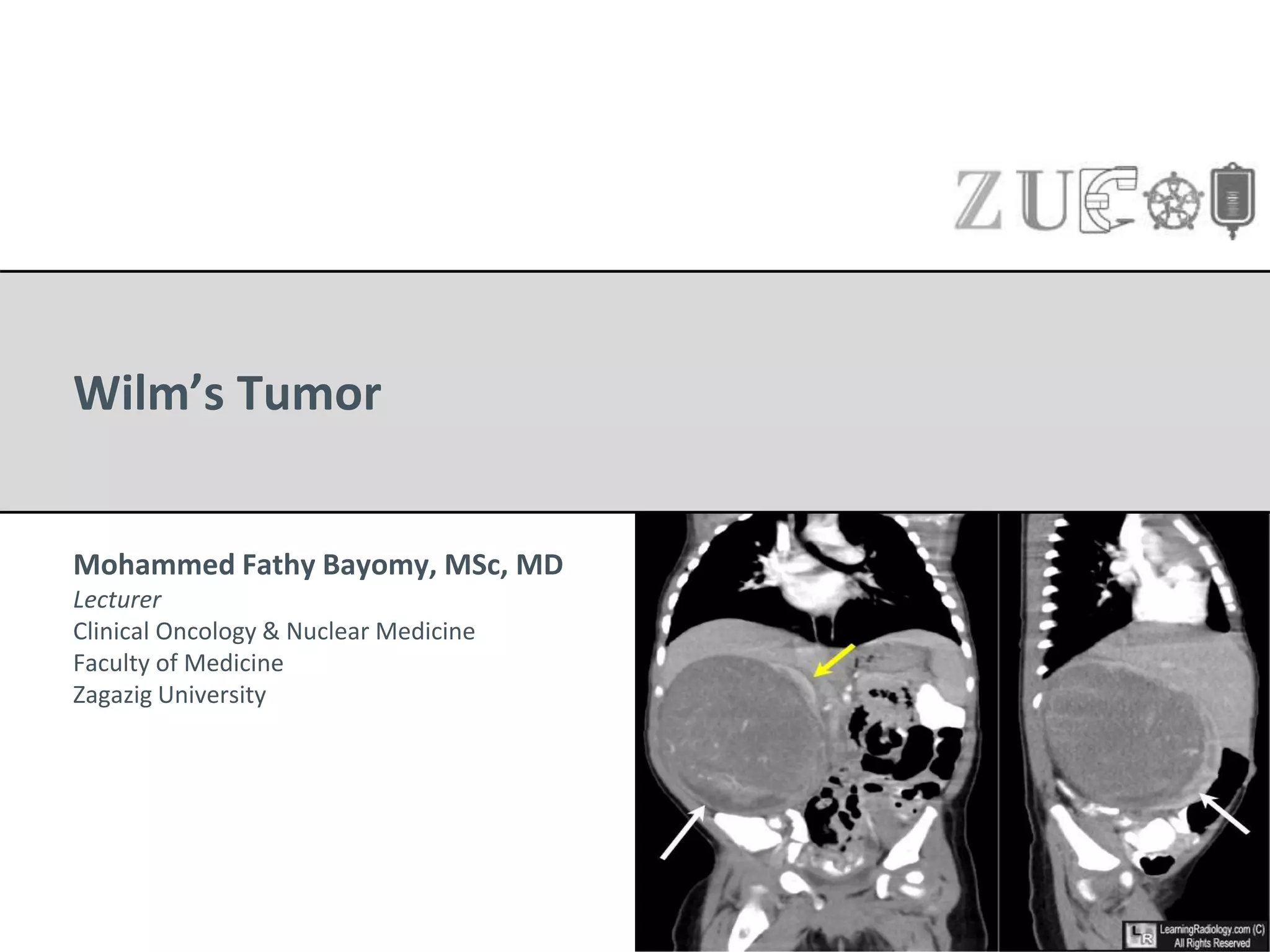
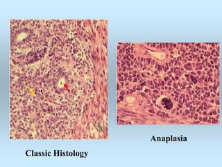
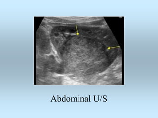
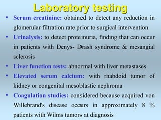
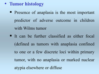
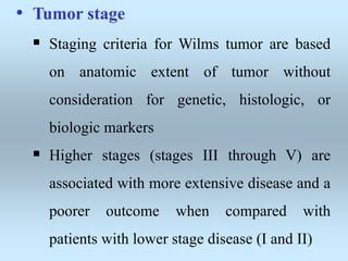
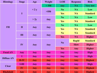
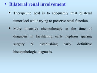
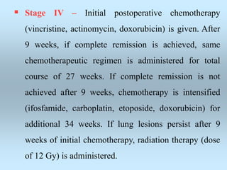
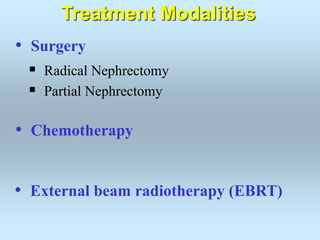
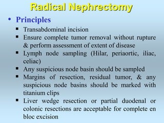
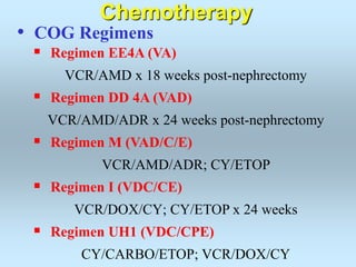
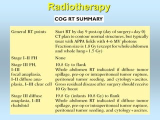
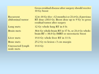
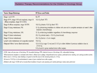
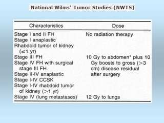
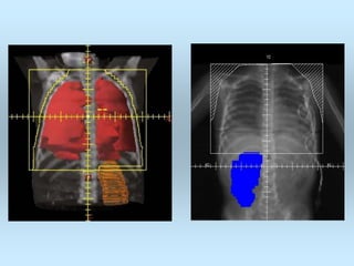
Wilm’s Tumor
Mohammed Fathy Bayomy, MSc, MD
Lecturer
Clinical Oncology & Nuclear Medicine
Faculty of Medicine
Zagazig University
Introduction
• Wilms tumor is the most common renal
malignancy in children & fourth most common
childhood cancer
• 5% of all childhood cancers, 5% bilateral
• Average age of incidence is 3 years
• 1/3 hereditary (autosomal dominant) & 2/3
sporadic
• In few children occurs as part of a congenital
malformation syndrome (WAGR, Denys-
Drash, Beckwith-Wiedemann)
Epidemiology
Incidence (US)
• Annual incidence of renal tumors is about 8 cases
per million children younger than 15 years
• Accounting for 7 % of all childhood malignancies
& for 500 new cases per year in North America
• Wilms tumor is the most common renal
malignancy in children under the age of 15 years,
accounting for about 95 percent of all cases.
Age
• 2/3 of Wilms tumor are diagnosed before five years
of age, and 95 percent before 10 years of age
• Unilateral involvement, median age at diagnosis is 40
months in girls & 35 months in boys
• bilateral disease are diagnosed at earlier age (median
age, girls at 30 months and boys at 25 months)
• Patients with associated congenital anomalies, such
as aniridia or genitourinary abnormalities, are also
diagnosed at earlier age
Race
• Risk of developing Wilms tumor varies among
ethnic groups, with a greater risk in African-
Americans and lower risk in the Asian population
• Epigenetic differences may contribute to lower
rate of disease in Asian children, as demonstrated
by study that reported infrequent loss of IGF2
imprinting in tumors from Asian patients
Congenital anomalies
• Wilms tumor is primarily a sporadic disease, &
only 1 to 2 percent of individuals with Wilms
tumor have relative with the disease
• In some children, Wilms tumor occurs as part of
multiple malformation syndrome
• These syndromes include WAGR, Denys-Drash,
and Beckwith-Wiedemann syndromes.
• WAGR syndrome
 Syndrome of Wilms tumor, Aniridia, Genitourinary
(GU) anomalies, and intellectual disability (mental
Retardation)
 Children with this syndrome have constitutional
chromosomal deletion of WT1 gene located at
11p13
 WT1 gene product is transcription factor involved
in both gonadal & renal development
 Children with WT1 deletions, including WAGR
syndrome, have greater than 20 percent risk of
developing Wilms tumor
• Denys-Drash syndrome
 Triad of progressive renal disease, male
pseudohermaphroditism, and Wilms tumor
 Affected individuals have germline point mutation
in eighth or ninth exon of WT1 gene, which results
in an amino acid substitution, and almost all
patients (90 percent) will develop Wilms tumor
 Underlying renal pathology is diffuse mesangial
sclerosis, which presents in infancy with
proteinuria & progresses to nephrotic syndrome &
renal failure.
• Beckwith-Wiedemann syndrome
 5 to 10 percent chance of developing Wilms tumors
 Caused by microduplication mutations in 11p15.5
region (WT2), site of cluster of imprinting genes
 Major clinical features: macrosomia, macroglossia,
omphalocele, prominent eyes, ear creases, large
kidneys, pancreatic hyperplasia, and
hemihypertrophy.
• Other congenital anomalies
 Perlman syndrome: autosomal recessive
overgrowth syndrome due to germline mutation of
DIS3L2, characterized by fetal gigantism,
visceromegaly, unusual face, bilateral renal
hamartomas with nephroblastomatosis, and Wilms
tumor
 Familial Wilms tumor: rare and is associated with
mutations in BRCA2 or TP53 (Li-Fraumeni
syndrome)
 Sotos syndrome: overgrowth syndrome associated
with facial, extremity, and cognitive abnormalities, 2
to 3 percent risk of Wilms tumor in children
 Simpson-Golabi-Behmel syndrome: X-linked
genetic disorder caused by mutations in gene
encoding glypican-3, which maps to chromosome
Xq26, characterized with pre- & postnatal overgrowth
with organomegaly, distinctive course facial
appearance described as bulldog appearance,
congenital heart disease, polydactyly, 7.5 percent
chance of developing Wilms tumor
 Isolated genitourinary abnormalities: Boys with
Wilms tumor may have cryptorchidism or
hypospadias, while 10 percent of girls with Wilms
tumor have congenital uterine anomalies, Other
kidney abnormalities, such as renal ectopia or
duplicated collecting systems, can also be seen.
 Isolated hemihypertrophy
Pathogenesis
• Wilms tumor appears to be caused by abnormal
renal development, resulting in proliferation of
metanephric blastema without normal tubular
& glomerular differentiation
• Wilms tumor is thought to arise from foci of
persistent metanephric cells referred to as
nephrogenic rests or nephroblastomatosis
• Nephrogenic rests normally occur in 1 percent
of newborn kidneys & regress early in
childhood. In contrast, they are present in 35
percent of kidneys with unilateral Wilms tumor
& almost 100 percent of kidneys with bilateral
disease
• Intralobar
 Earlier presentation
 Seen with WAGR,
DDS
• Perilobar
 Later presentation
 Seen with BW
Nephrogenic Rests
Genetics
• Wilms tumor has been associated with loss of
function mutations of a number of tumor
suppressor genes.
• These include mutations of WT1, p53, FWT1,
FWT2 genes, & WT2 (11p15.5 locus)
• The role of these gene mutations in the
pathogenesis of Wilms tumor remains
unknown.
• WT1 gene
 Located on chromosome 11p13
 WT1 gene product is expressed in developing
kidney, testis, and ovary
 It appears to play role in development &
differentiation of genitourinary tissues
 11p13 deletion in WAGR syndrome encompasses
several contiguous genes, including WT1 & PAX 6
(associated with aniridia) genes
 Patients with Denys-Drash syndrome have a point
mutation in the eighth or ninth exon of WT1 gene
REMEMBER: WT1 = differentiation
WT1 = PROLIFERATION!!
• WT2 gene
 Located on chromosome 11p15.5 locus
 Contains cluster of imprinted genes
 Imprinted genes are those that demonstrate
selective gene expression based upon parental
origin, such that either paternal or maternal-
inherited gene copy is expressed, but not both.
 As example, in patients with BWS, the maternal
copy of the Beckwith-Wiedemann (BW) gene is
silenced during gametogenesis & only paternal
copy is expressed.
 As a result, offspring with BWS receive mutation
passed from their father and those who inherit BW
gene mutation from their mother are asymptomatic
carriers, who can pass mutation to their offspring.
 Somatic 11p15 defects have been found in Wilms
tumor cells, perilobar nephrogenic rests associated
with Wilms tumors, and some normal renal cells
surrounding Wilms tumors suggesting 11p15
mutations may play an early role in nonsyndromic
Wilms tumorigenesis
• p53 gene
 Tumor suppressor gene is located on chromosome
17p13.1
 It encodes nuclear protein, which acts as
transcription factor & blocks progression of cell
cycle late in G1 phase
 p53 gene mutation is seen infrequently in patients
with Wilms tumor and is associated with both
favorable histologic features
• Familial WT genes (FWT)
 Familial Wilms tumor accounts for 1 to 2 percent of
cases
 Mode of inheritance appears to be autosomal
dominant with variable penetrance
 In these families, there is no association with WT1
gene mutations
 Linkages have been demonstrated to FWT1 gene
locus at 17q12-21, FWT2 gene locus at 19q13.3-
q13.4, & at 11p15.5 locus
Pathology
• Most Wilms tumors are solitary lesions. 10 % have
multifocal loci within single kidney
• 5-7 % of patients have bilateral renal involvement
• Sharply (well) defined masses with pseudocapsule
• Uniform, pale gray to tan
• Divided by prominent fibrous septa in to lobules
• Variable size & weight (60-6350 g with mean of 550 g)
• May be cystic, hemorrhagic or necrotic
Gross picture
• Classic favorable histology Wilms tumor is comprised
of three cell types: Blastemal cells – Undifferentiated
cells, Stromal cells – Immature spindle cells &
heterologous skeletal muscle, cartilage, osteoid, or fat,
Epithelial cells – Glomeruli & tubules
• Wilms tumors contain only one (monophasic) or two
cell types (biphasic).
• Anaplasia: presence of multipolar polypoid mitotic
figures & marked nuclear enlargement with
hyperchromasia
Microscopic picture
Anaplasia
Classic Histology
• Clear Cell Sarcoma (CCSK)
 CCSK is not Wilms’ tumor variant, but it is
important primary renal tumor associated with
significantly higher rate of relapse & death than
favorable histology Wilms’ tumor. In addition to
pulmonary metastases, clear cell sarcoma also
spreads to bone, brain, soft tissue
 Classic pattern of CCSK is defined by nests or
cords of cells separated by regularly spaced
arborizing fibrovascular septa
• Rhabdoid Tumor of Kidney (RTK)
 Initially thought to be rhabdomyosarcomatoid
variant of Wilms’ tumor, it is distinctive & highly
malignant tumor type
 Most distinctive features of rhabdoid tumors of
kidney are rather large cells with large vesicular
nuclei, prominent single nucleolus, & in some cells,
presence of globular eosinophilic cytoplasmic
inclusions
 Cell of origin is unknown
 Distinct clinical presentation with fever, hematuria,
young age (mean 11 months), & high tumor stage
at presentation suggests a diagnosis of rhabdoid
tumor of kidney (RTK)
 RTK tends to metastasize not only to lungs, but also
to brain.
 As many as 10% to 15% of patients with RTK also
have central nervous system lesions
Clinical
Presentation
• Most children with Wilms tumor present
with abdominal mass or swelling, without other
signs or symptoms
• Other symptoms can include abdominal pain
(30-40%), hematuria (12-25%), fever,
hypertension (25%)
• Patients with subcapsular hemorrhage can
present with rapid abdominal enlargement,
anemia, hypertension, & sometimes fever.
• Although the lung is the most common
metastatic site, children rarely present with
respiratory symptoms.
Painless abdominal mass
• Physical examination: firm, non-tender,
smooth mass that is eccentrically located, rarely
crosses midline
• Once Wilms tumor is suspected, subsequent
abdominal examinations should be performed
carefully. Vigorous palpation may rupture renal
capsule, resulting in tumor spillage, which
increases tumor stage & need for more
intensive therapy
• Assessment for associated anomalies, such as
aniridia, hemihypertrophy, genitourinary
anomalies
Diagnostic
Evaluation
• Definitive diagnosis of Wilms tumor is made by
histologic confirmation at the time of either surgical
excision or biopsy.
• Children who are suspected of having Wilms tumor
should be referred to a pediatric cancer center for
evaluation and diagnosis.
• Imaging is useful to differentiate Wilms tumor from
other causes of abdominal masses
• Goal of imaging: to establish presence of renal tumor ,
guide management decisions prior to confirmation of
histologic diagnosis, such as surgical approach & need
for preoperative chemotherapy
• Important information includes confirming presence
and function of contralateral kidney, determining
whether there is also tumor in contralateral kidney, size
& extent of tumor, and presence of lung metastases.
Imaging
• Abdominal ultrasonography
 The initial imaging study
 It will detect hydronephrosis and multicystic kidney
disease, which may present as abdominal masses or
swelling
 In patients with a suspected renal tumor, Doppler
ultrasonography should be performed to detect
tumor infiltration of renal vein and inferior vena
cava, and to assess patency of blood flow.
Abdominal U/S
• Computed tomography (CT)
 Recommended to further evaluate nature & extent
of mass
 CT also may detect small lesions of tumor or
nephrogenic rests in opposite kidney, which were
not detected by ultrasonography
 CT had relatively low sensitivity at detecting tumor
rupture and that ascites beyond cul-de-sac was most
predictive imaging finding of rupture
CT Chest/Abdomen/Pelvis define
extent of lesion, pulmonary metastasis
CT Abdomen define extent of lesion
MRI with tumor thrombus
extending into IVC
• Imaging of chest
 Needed to determine whether there are lung
metastases
 CT & chest radiography are used to detect
metastases
 CT appears to be more sensitive than chest
radiography
CT chest show pulmonary met.
• Preoperative imaging
 Plays critical role to surgeon’s determination if
tumor is operable at diagnosis.
 Guidelines define criteria for when it is more
appropriate to do upfront biopsy & prenephrectomy
chemotherapy include: tumor thrombus above level
of hepatic veins; pulmonary compromise from
massive tumor or extensive pulmonary metastases;
resection requiring removal of contiguous
structures (other than adrenal gland); or surgeon’s
judgment that attempting nephrectomy would result
in significant morbidity, tumor spill, or residual
tumor
• Serum creatinine: obtained to detect any reduction in
glomerular filtration rate prior to surgical intervention
• Urinalysis: to detect proteinuria, finding that can occur
in patients with Denys- Drash syndrome & mesangial
sclerosis
• Liver function tests: abnormal with liver metastases
• Elevated serum calcium: with rhabdoid tumor of
kidney or congenital mesoblastic nephroma
• Coagulation studies: considered because acquired von
Willebrand's disease occurs in approximately 8 %
patients with Wilms tumors at diagnosis
Laboratory testing
Differential
Diagnosis
• Differential diagnosis of Wilms tumor includes
neuroblastoma & other renal tumors
1- Neuroblastoma
 Differentiated from Wilms tumor by contrast-
enhanced computed tomography (CT) or
ultrasonography, which distinguishes renal and
nonrenal tissue
 Final diagnostic confirmation is based upon
tissue histology at the time of either biopsy or
surgical excision
2- Congenital mesoblastic nephroma
 Usually detected within the first year of life or
by prenatal ultrasonography
 Divided into classic & cellular subtypes
 Associated with hypertension, elevated
concentrations of calcium & renin
3- Renal Medullary Carcinoma
 Highly lethal tumor that is virtually restricted to
patients with sickle cell hemoglobinopathy,
most commonly sickle cell trait
 Highly invasive tumor with early metastases
4- Renal Cell Carcinoma (RCC)
 Rare in childhood, affect adolescent males
 Chromosomal translocation at Xp11.2
 Xp11.2 translocation carcinoma appears to have
poorer prognosis than non-Xp11.2 translocation
carcinoma in children and young adults
 Highest risk was observed among survivors of
neuroblastoma who received renal-directed
radiation therapy & platinum-based
chemotherapy
5- Clear cell sarcoma of kidney (CCSK)
6- Rhabdoid tumor of kidney (RTK)
Staging
Based upon surgical evaluation prior
to administration of chemotherapy
Based upon surgical evaluation after administration
of chemotherapy to reduce size of tumor
Prognostic
Factors
• Tumor histology
 Presence of anaplasia is the most important
predictor of adverse outcome in children
with Wilms tumor
 It can be further classified as either focal
(defined as tumors with anaplasia confined
to one or a few discrete loci within primary
tumor, with no anaplasia or marked nuclear
atypia elsewhere or diffuse
• Tumor stage
 Staging criteria for Wilms tumor are based
on anatomic extent of tumor without
consideration for genetic, histologic, or
biologic markers
 Higher stages (stages III through V) are
associated with more extensive disease and a
poorer outcome when compared with
patients with lower stage disease (I and II)
• Age
 In past, children less than 24 months of age
generally had lower relapse rates than older
patients, which resulted in a better clinical
outcome
 With improved therapeutic interventions, effect
of age as prognostic factor has been reduced
 Adults with Wilms tumor have same survival
rate as children with similar tumor histology
and stage when treated with same therapeutic
regimens
• Biologic markers
 Several biological markers appear to be
predictive of outcome.
 The most promising of these is loss of
heterozygosity (LOH) at chromosome 16q,
1p, 11p15 in tumor cells
 Patients with these molecular markers have
increased rates of relapse and mortality
COG Risk Group
Classification
Histology Stage Age Weight LOH Response Risk
FH
I
< 2 y
< 550 Any NA Very low
>550
No NA Low
Yes NA Standard
> 2y Any
No NA Low
Yes NA Standard
II Any Any
No NA Low
Yes NA Standard
III Any Any
No NA Standard
Yes NA Higher
IV Any Any
No
Rapid Standard
Slow Higher
Yes Any Higher
Focal AN Any Any Any Any Any High
Diffuse AN
I Any Any Any Any High
II-IV Any Any Any Any Highest
Clear
I-III Any Any Any Any High
IV Any Any Any Any Highest
Rhabdoid Any Any Any Any Any Highest
Treatment
• Most children with newly diagnosed Wilms tumor are
treated on research protocols, & risk-based therapy is
assigned based on results of initial staging, histological,
& molecular studies.
• These regimens conducted by cooperative groups
incorporate multimodal therapy (surgery, chemotherapy,
and radiation), & have resulted in dramatic
improvements in outcome in these children with overall
five-year survival rates approaching 90 percent
Principles
• The two major research groups that care for the largest
number of patients with Wilms tumor are:
 Children's Oncology Group (COG) & National
Wilms Tumor Study (NWTS) group includes
almost all of pediatric oncology centers in United
States & Canada. NWTS has been incorporated into
 International Society of Pediatric Oncology
(SIOP) includes European pediatric oncology
centers
• Unilateral renal involvement
 NWTS & SIOP differ in their management of
unilateral Wilms tumor (stages I through IV Wilms
tumor), specifically regarding timing of
chemotherapy relative to surgical excision
 NWTS encourages use of primary surgical
resection prior to chemotherapy administration.
 SIOP primarily use initial course of chemotherapy
followed by surgical resection & staging four
weeks after administration of chemotherapy
 Both treatment approaches have excellent &
comparable clinical outcomes
• Bilateral renal involvement
 Therapeutic goal is to adequately treat bilateral
tumor loci while trying to preserve renal function
 More intensive chemotherapy at the time of
diagnosis in facilitating early nephron sparing
surgery & establishing early definitive
histopathologic diagnosis
Treatment Recommendations
• NWTS & COG protocol
 Stage I favorable histology & tumor weights < 550
grams – Primary surgical resection, not require
adjuvant chemotherapy, not require RT
 Stages I/II with favorable histology – Primary
surgical resection is followed by 19 weeks of
(vincristine, dactinomycin), not require RT
 Stage I/II with LOH 1p, 16q – Primary surgical
resection is followed by 19 weeks of (vincristine,
dactinomycin, doxorubicin)
 Stage III – Primary surgical resection is followed by
24 weeks of triple drug chemotherapy (vincristine,
dactinomycin, doxorubicin). Radiation therapy (dose
of 10.8 Gy) based on lymph node involvement or
extent of peritoneal contamination is administered to
affected flank, hemi-abdomen, or complete abdomen
 Stage III disease with LOH 1p, 16q – receive
chemotherapy (vincristine, dactinomycin, doxorubicin
/cyclophosphamide, etoposide)
 Stage IV – Primary surgical resection is followed by 24
weeks of triple drug chemotherapy (vincristine,
dactinomycin, doxorubicin). Radiation therapy (dose of 12
Gy) is administered to whole lung field only if lung
metastases do not completely respond after 6 weeks of
chemotherapy. If lung metastases are completely resected
prior to enrollment, then whole lung radiation will start at
week one. In patients with evidence of spread of disease to
regional lymph nodes in hilum &/or para-aortic nodes,
radiation therapy (dose of 10.8 Gy) is administered to loco-
regional area including appropriate half of abdomen as
indicated.
Preoperative Therapy
Postoperative Therapy
• Preoperative SIOP protocol
 Preoperative chemotherapy (vincristine, dactinomycin)
are administered to patients with renal tumors for 4
weeks prior to surgery
 If there is evidence of metastatic disease beyond
abdominal peritoneal region (e.g. lung metastases),
triple drug chemotherapy (vincristine, dactinomycin,
doxorubicin) is given for 6 weeks preoperatively.
 Following preoperative chemotherapy, surgical
excision & staging are performed.
• Postoperative SIOP protocol
 Stage I – Postoperative chemotherapy (vincristine,
dactinomycin) is administered for 4 weeks. No
radiation therapy is given.
 Stage II – Postoperative chemotherapy (vincristine,
actinomycin, doxorubicin) is administered for 27
weeks. If there is tumor involvement of regional lymph
nodes, radiation therapy (dose of 15 Gy) is given.
 Stage III – Postoperative chemotherapy (vincristine,
actinomycin, doxorubicin is administered for 27
weeks. Radiation therapy (dose of 15 Gy) is given.
 Stage IV – Initial postoperative chemotherapy
(vincristine, actinomycin, doxorubicin) is given. After
9 weeks, if complete remission is achieved, same
chemotherapeutic regimen is administered for total
course of 27 weeks. If complete remission is not
achieved after 9 weeks, chemotherapy is intensified
(ifosfamide, carboplatin, etoposide, doxorubicin) for
additional 34 weeks. If lung lesions persist after 9
weeks of initial chemotherapy, radiation therapy (dose
of 12 Gy) is administered.
• Surgery
 Radical Nephrectomy
 Partial Nephrectomy
Treatment Modalities
• External beam radiotherapy (EBRT)
• Chemotherapy
• Principles
 Transabdominal incision
 Ensure complete tumor removal without rupture
& perform assessment of extent of disease
 Lymph node sampling (Hilar, periaortic, iliac,
celiac)
 Any suspicious node basin should be sampled
 Margins of resection, residual tumor, & any
suspicious node basins should be marked with
titanium clips
 Liver wedge resection or partial duodenal or
colonic resections are acceptable for complete en
bloc excision
Radical Nephrectomy
Chemotherapy
• COG Regimens
 Regimen EE4A (VA)
VCR/AMD x 18 weeks post-nephrectomy
 Regimen DD 4A (VAD)
VCR/AMD/ADR x 24 weeks post-nephrectomy
 Regimen M (VAD/C/E)
VCR/AMD/ADR; CY/ETOP
 Regimen I (VDC/CE)
VCR/DOX/CY; CY/ETOP x 24 weeks
 Regimen UH1 (VDC/CPE)
CY/CARBO/ETOP; VCR/DOX/CY
Radiotherapy
• Primary site
 Clinical target volume (CTV) is tumor bed (tumor
& kidney) + adjacent paraaortic nodes with a 1-cm
margin
 Medial border of RT field is extended medially
encompassing entire vertebral width to minimize
development of scoliosis
 Initial dose: 1000 cGy in 150- to 180-cGy
fractions
 Boost dose: 1000 cGy in 150- to 180-cGy
fractions to areas of gross disease.
• Whole abdomen
 Diffuse peritoneal implants or diffuse spillage
of tumor: 1000 cGy in 150- to 180-cGy fractions
 Gross peritoneal deposits: 2000 cGy in 150- to
180-cGy fractions, with remaining kidney
receiving not more than 1400 cGy
• Whole lung
 CTV is entire lungs, 1-cm margin is added to
create planning target volume (PTV)
 Dose: 12 Gy in 180cGy fractions (eight fractions)
Follow-up
Wilm's tumor

Wilm's tumor

  • 1.
    Wilm’s Tumor Mohammed FathyBayomy, MSc, MD Lecturer Clinical Oncology & Nuclear Medicine Faculty of Medicine Zagazig University
  • 2.
  • 3.
    • Wilms tumoris the most common renal malignancy in children & fourth most common childhood cancer • 5% of all childhood cancers, 5% bilateral • Average age of incidence is 3 years • 1/3 hereditary (autosomal dominant) & 2/3 sporadic • In few children occurs as part of a congenital malformation syndrome (WAGR, Denys- Drash, Beckwith-Wiedemann)
  • 4.
  • 5.
    Incidence (US) • Annualincidence of renal tumors is about 8 cases per million children younger than 15 years • Accounting for 7 % of all childhood malignancies & for 500 new cases per year in North America • Wilms tumor is the most common renal malignancy in children under the age of 15 years, accounting for about 95 percent of all cases.
  • 6.
    Age • 2/3 ofWilms tumor are diagnosed before five years of age, and 95 percent before 10 years of age • Unilateral involvement, median age at diagnosis is 40 months in girls & 35 months in boys • bilateral disease are diagnosed at earlier age (median age, girls at 30 months and boys at 25 months) • Patients with associated congenital anomalies, such as aniridia or genitourinary abnormalities, are also diagnosed at earlier age
  • 7.
    Race • Risk ofdeveloping Wilms tumor varies among ethnic groups, with a greater risk in African- Americans and lower risk in the Asian population • Epigenetic differences may contribute to lower rate of disease in Asian children, as demonstrated by study that reported infrequent loss of IGF2 imprinting in tumors from Asian patients
  • 8.
    Congenital anomalies • Wilmstumor is primarily a sporadic disease, & only 1 to 2 percent of individuals with Wilms tumor have relative with the disease • In some children, Wilms tumor occurs as part of multiple malformation syndrome • These syndromes include WAGR, Denys-Drash, and Beckwith-Wiedemann syndromes.
  • 9.
    • WAGR syndrome Syndrome of Wilms tumor, Aniridia, Genitourinary (GU) anomalies, and intellectual disability (mental Retardation)  Children with this syndrome have constitutional chromosomal deletion of WT1 gene located at 11p13  WT1 gene product is transcription factor involved in both gonadal & renal development  Children with WT1 deletions, including WAGR syndrome, have greater than 20 percent risk of developing Wilms tumor
  • 10.
    • Denys-Drash syndrome Triad of progressive renal disease, male pseudohermaphroditism, and Wilms tumor  Affected individuals have germline point mutation in eighth or ninth exon of WT1 gene, which results in an amino acid substitution, and almost all patients (90 percent) will develop Wilms tumor  Underlying renal pathology is diffuse mesangial sclerosis, which presents in infancy with proteinuria & progresses to nephrotic syndrome & renal failure.
  • 11.
    • Beckwith-Wiedemann syndrome 5 to 10 percent chance of developing Wilms tumors  Caused by microduplication mutations in 11p15.5 region (WT2), site of cluster of imprinting genes  Major clinical features: macrosomia, macroglossia, omphalocele, prominent eyes, ear creases, large kidneys, pancreatic hyperplasia, and hemihypertrophy.
  • 12.
    • Other congenitalanomalies  Perlman syndrome: autosomal recessive overgrowth syndrome due to germline mutation of DIS3L2, characterized by fetal gigantism, visceromegaly, unusual face, bilateral renal hamartomas with nephroblastomatosis, and Wilms tumor  Familial Wilms tumor: rare and is associated with mutations in BRCA2 or TP53 (Li-Fraumeni syndrome)
  • 13.
     Sotos syndrome:overgrowth syndrome associated with facial, extremity, and cognitive abnormalities, 2 to 3 percent risk of Wilms tumor in children  Simpson-Golabi-Behmel syndrome: X-linked genetic disorder caused by mutations in gene encoding glypican-3, which maps to chromosome Xq26, characterized with pre- & postnatal overgrowth with organomegaly, distinctive course facial appearance described as bulldog appearance, congenital heart disease, polydactyly, 7.5 percent chance of developing Wilms tumor
  • 14.
     Isolated genitourinaryabnormalities: Boys with Wilms tumor may have cryptorchidism or hypospadias, while 10 percent of girls with Wilms tumor have congenital uterine anomalies, Other kidney abnormalities, such as renal ectopia or duplicated collecting systems, can also be seen.  Isolated hemihypertrophy
  • 15.
  • 16.
    • Wilms tumorappears to be caused by abnormal renal development, resulting in proliferation of metanephric blastema without normal tubular & glomerular differentiation • Wilms tumor is thought to arise from foci of persistent metanephric cells referred to as nephrogenic rests or nephroblastomatosis
  • 18.
    • Nephrogenic restsnormally occur in 1 percent of newborn kidneys & regress early in childhood. In contrast, they are present in 35 percent of kidneys with unilateral Wilms tumor & almost 100 percent of kidneys with bilateral disease
  • 19.
    • Intralobar  Earlierpresentation  Seen with WAGR, DDS • Perilobar  Later presentation  Seen with BW Nephrogenic Rests
  • 20.
  • 21.
    • Wilms tumorhas been associated with loss of function mutations of a number of tumor suppressor genes. • These include mutations of WT1, p53, FWT1, FWT2 genes, & WT2 (11p15.5 locus) • The role of these gene mutations in the pathogenesis of Wilms tumor remains unknown.
  • 22.
    • WT1 gene Located on chromosome 11p13  WT1 gene product is expressed in developing kidney, testis, and ovary  It appears to play role in development & differentiation of genitourinary tissues  11p13 deletion in WAGR syndrome encompasses several contiguous genes, including WT1 & PAX 6 (associated with aniridia) genes  Patients with Denys-Drash syndrome have a point mutation in the eighth or ninth exon of WT1 gene
  • 23.
    REMEMBER: WT1 =differentiation
  • 24.
  • 25.
    • WT2 gene Located on chromosome 11p15.5 locus  Contains cluster of imprinted genes  Imprinted genes are those that demonstrate selective gene expression based upon parental origin, such that either paternal or maternal- inherited gene copy is expressed, but not both.  As example, in patients with BWS, the maternal copy of the Beckwith-Wiedemann (BW) gene is silenced during gametogenesis & only paternal copy is expressed.
  • 26.
     As aresult, offspring with BWS receive mutation passed from their father and those who inherit BW gene mutation from their mother are asymptomatic carriers, who can pass mutation to their offspring.  Somatic 11p15 defects have been found in Wilms tumor cells, perilobar nephrogenic rests associated with Wilms tumors, and some normal renal cells surrounding Wilms tumors suggesting 11p15 mutations may play an early role in nonsyndromic Wilms tumorigenesis
  • 27.
    • p53 gene Tumor suppressor gene is located on chromosome 17p13.1  It encodes nuclear protein, which acts as transcription factor & blocks progression of cell cycle late in G1 phase  p53 gene mutation is seen infrequently in patients with Wilms tumor and is associated with both favorable histologic features
  • 28.
    • Familial WTgenes (FWT)  Familial Wilms tumor accounts for 1 to 2 percent of cases  Mode of inheritance appears to be autosomal dominant with variable penetrance  In these families, there is no association with WT1 gene mutations  Linkages have been demonstrated to FWT1 gene locus at 17q12-21, FWT2 gene locus at 19q13.3- q13.4, & at 11p15.5 locus
  • 29.
  • 30.
    • Most Wilmstumors are solitary lesions. 10 % have multifocal loci within single kidney • 5-7 % of patients have bilateral renal involvement • Sharply (well) defined masses with pseudocapsule • Uniform, pale gray to tan • Divided by prominent fibrous septa in to lobules • Variable size & weight (60-6350 g with mean of 550 g) • May be cystic, hemorrhagic or necrotic Gross picture
  • 33.
    • Classic favorablehistology Wilms tumor is comprised of three cell types: Blastemal cells – Undifferentiated cells, Stromal cells – Immature spindle cells & heterologous skeletal muscle, cartilage, osteoid, or fat, Epithelial cells – Glomeruli & tubules • Wilms tumors contain only one (monophasic) or two cell types (biphasic). • Anaplasia: presence of multipolar polypoid mitotic figures & marked nuclear enlargement with hyperchromasia Microscopic picture
  • 34.
  • 35.
    • Clear CellSarcoma (CCSK)  CCSK is not Wilms’ tumor variant, but it is important primary renal tumor associated with significantly higher rate of relapse & death than favorable histology Wilms’ tumor. In addition to pulmonary metastases, clear cell sarcoma also spreads to bone, brain, soft tissue  Classic pattern of CCSK is defined by nests or cords of cells separated by regularly spaced arborizing fibrovascular septa
  • 36.
    • Rhabdoid Tumorof Kidney (RTK)  Initially thought to be rhabdomyosarcomatoid variant of Wilms’ tumor, it is distinctive & highly malignant tumor type  Most distinctive features of rhabdoid tumors of kidney are rather large cells with large vesicular nuclei, prominent single nucleolus, & in some cells, presence of globular eosinophilic cytoplasmic inclusions
  • 37.
     Cell oforigin is unknown  Distinct clinical presentation with fever, hematuria, young age (mean 11 months), & high tumor stage at presentation suggests a diagnosis of rhabdoid tumor of kidney (RTK)  RTK tends to metastasize not only to lungs, but also to brain.  As many as 10% to 15% of patients with RTK also have central nervous system lesions
  • 38.
  • 39.
    • Most childrenwith Wilms tumor present with abdominal mass or swelling, without other signs or symptoms • Other symptoms can include abdominal pain (30-40%), hematuria (12-25%), fever, hypertension (25%) • Patients with subcapsular hemorrhage can present with rapid abdominal enlargement, anemia, hypertension, & sometimes fever. • Although the lung is the most common metastatic site, children rarely present with respiratory symptoms.
  • 40.
  • 41.
    • Physical examination:firm, non-tender, smooth mass that is eccentrically located, rarely crosses midline • Once Wilms tumor is suspected, subsequent abdominal examinations should be performed carefully. Vigorous palpation may rupture renal capsule, resulting in tumor spillage, which increases tumor stage & need for more intensive therapy • Assessment for associated anomalies, such as aniridia, hemihypertrophy, genitourinary anomalies
  • 42.
  • 43.
    • Definitive diagnosisof Wilms tumor is made by histologic confirmation at the time of either surgical excision or biopsy. • Children who are suspected of having Wilms tumor should be referred to a pediatric cancer center for evaluation and diagnosis.
  • 44.
    • Imaging isuseful to differentiate Wilms tumor from other causes of abdominal masses • Goal of imaging: to establish presence of renal tumor , guide management decisions prior to confirmation of histologic diagnosis, such as surgical approach & need for preoperative chemotherapy • Important information includes confirming presence and function of contralateral kidney, determining whether there is also tumor in contralateral kidney, size & extent of tumor, and presence of lung metastases. Imaging
  • 45.
    • Abdominal ultrasonography The initial imaging study  It will detect hydronephrosis and multicystic kidney disease, which may present as abdominal masses or swelling  In patients with a suspected renal tumor, Doppler ultrasonography should be performed to detect tumor infiltration of renal vein and inferior vena cava, and to assess patency of blood flow.
  • 46.
  • 47.
    • Computed tomography(CT)  Recommended to further evaluate nature & extent of mass  CT also may detect small lesions of tumor or nephrogenic rests in opposite kidney, which were not detected by ultrasonography  CT had relatively low sensitivity at detecting tumor rupture and that ascites beyond cul-de-sac was most predictive imaging finding of rupture
  • 48.
    CT Chest/Abdomen/Pelvis define extentof lesion, pulmonary metastasis
  • 49.
    CT Abdomen defineextent of lesion
  • 50.
    MRI with tumorthrombus extending into IVC
  • 51.
    • Imaging ofchest  Needed to determine whether there are lung metastases  CT & chest radiography are used to detect metastases  CT appears to be more sensitive than chest radiography
  • 52.
    CT chest showpulmonary met.
  • 53.
    • Preoperative imaging Plays critical role to surgeon’s determination if tumor is operable at diagnosis.  Guidelines define criteria for when it is more appropriate to do upfront biopsy & prenephrectomy chemotherapy include: tumor thrombus above level of hepatic veins; pulmonary compromise from massive tumor or extensive pulmonary metastases; resection requiring removal of contiguous structures (other than adrenal gland); or surgeon’s judgment that attempting nephrectomy would result in significant morbidity, tumor spill, or residual tumor
  • 54.
    • Serum creatinine:obtained to detect any reduction in glomerular filtration rate prior to surgical intervention • Urinalysis: to detect proteinuria, finding that can occur in patients with Denys- Drash syndrome & mesangial sclerosis • Liver function tests: abnormal with liver metastases • Elevated serum calcium: with rhabdoid tumor of kidney or congenital mesoblastic nephroma • Coagulation studies: considered because acquired von Willebrand's disease occurs in approximately 8 % patients with Wilms tumors at diagnosis Laboratory testing
  • 55.
  • 56.
    • Differential diagnosisof Wilms tumor includes neuroblastoma & other renal tumors 1- Neuroblastoma  Differentiated from Wilms tumor by contrast- enhanced computed tomography (CT) or ultrasonography, which distinguishes renal and nonrenal tissue  Final diagnostic confirmation is based upon tissue histology at the time of either biopsy or surgical excision
  • 57.
    2- Congenital mesoblasticnephroma  Usually detected within the first year of life or by prenatal ultrasonography  Divided into classic & cellular subtypes  Associated with hypertension, elevated concentrations of calcium & renin 3- Renal Medullary Carcinoma  Highly lethal tumor that is virtually restricted to patients with sickle cell hemoglobinopathy, most commonly sickle cell trait  Highly invasive tumor with early metastases
  • 58.
    4- Renal CellCarcinoma (RCC)  Rare in childhood, affect adolescent males  Chromosomal translocation at Xp11.2  Xp11.2 translocation carcinoma appears to have poorer prognosis than non-Xp11.2 translocation carcinoma in children and young adults  Highest risk was observed among survivors of neuroblastoma who received renal-directed radiation therapy & platinum-based chemotherapy
  • 59.
    5- Clear cellsarcoma of kidney (CCSK) 6- Rhabdoid tumor of kidney (RTK)
  • 60.
  • 61.
    Based upon surgicalevaluation prior to administration of chemotherapy
  • 63.
    Based upon surgicalevaluation after administration of chemotherapy to reduce size of tumor
  • 64.
  • 65.
    • Tumor histology Presence of anaplasia is the most important predictor of adverse outcome in children with Wilms tumor  It can be further classified as either focal (defined as tumors with anaplasia confined to one or a few discrete loci within primary tumor, with no anaplasia or marked nuclear atypia elsewhere or diffuse
  • 66.
    • Tumor stage Staging criteria for Wilms tumor are based on anatomic extent of tumor without consideration for genetic, histologic, or biologic markers  Higher stages (stages III through V) are associated with more extensive disease and a poorer outcome when compared with patients with lower stage disease (I and II)
  • 67.
    • Age  Inpast, children less than 24 months of age generally had lower relapse rates than older patients, which resulted in a better clinical outcome  With improved therapeutic interventions, effect of age as prognostic factor has been reduced  Adults with Wilms tumor have same survival rate as children with similar tumor histology and stage when treated with same therapeutic regimens
  • 68.
    • Biologic markers Several biological markers appear to be predictive of outcome.  The most promising of these is loss of heterozygosity (LOH) at chromosome 16q, 1p, 11p15 in tumor cells  Patients with these molecular markers have increased rates of relapse and mortality
  • 69.
  • 70.
    Histology Stage AgeWeight LOH Response Risk FH I < 2 y < 550 Any NA Very low >550 No NA Low Yes NA Standard > 2y Any No NA Low Yes NA Standard II Any Any No NA Low Yes NA Standard III Any Any No NA Standard Yes NA Higher IV Any Any No Rapid Standard Slow Higher Yes Any Higher Focal AN Any Any Any Any Any High Diffuse AN I Any Any Any Any High II-IV Any Any Any Any Highest Clear I-III Any Any Any Any High IV Any Any Any Any Highest Rhabdoid Any Any Any Any Any Highest
  • 71.
  • 72.
    • Most childrenwith newly diagnosed Wilms tumor are treated on research protocols, & risk-based therapy is assigned based on results of initial staging, histological, & molecular studies. • These regimens conducted by cooperative groups incorporate multimodal therapy (surgery, chemotherapy, and radiation), & have resulted in dramatic improvements in outcome in these children with overall five-year survival rates approaching 90 percent Principles
  • 73.
    • The twomajor research groups that care for the largest number of patients with Wilms tumor are:  Children's Oncology Group (COG) & National Wilms Tumor Study (NWTS) group includes almost all of pediatric oncology centers in United States & Canada. NWTS has been incorporated into  International Society of Pediatric Oncology (SIOP) includes European pediatric oncology centers
  • 74.
    • Unilateral renalinvolvement  NWTS & SIOP differ in their management of unilateral Wilms tumor (stages I through IV Wilms tumor), specifically regarding timing of chemotherapy relative to surgical excision  NWTS encourages use of primary surgical resection prior to chemotherapy administration.  SIOP primarily use initial course of chemotherapy followed by surgical resection & staging four weeks after administration of chemotherapy  Both treatment approaches have excellent & comparable clinical outcomes
  • 75.
    • Bilateral renalinvolvement  Therapeutic goal is to adequately treat bilateral tumor loci while trying to preserve renal function  More intensive chemotherapy at the time of diagnosis in facilitating early nephron sparing surgery & establishing early definitive histopathologic diagnosis
  • 76.
  • 80.
    • NWTS &COG protocol  Stage I favorable histology & tumor weights < 550 grams – Primary surgical resection, not require adjuvant chemotherapy, not require RT  Stages I/II with favorable histology – Primary surgical resection is followed by 19 weeks of (vincristine, dactinomycin), not require RT  Stage I/II with LOH 1p, 16q – Primary surgical resection is followed by 19 weeks of (vincristine, dactinomycin, doxorubicin)
  • 81.
     Stage III– Primary surgical resection is followed by 24 weeks of triple drug chemotherapy (vincristine, dactinomycin, doxorubicin). Radiation therapy (dose of 10.8 Gy) based on lymph node involvement or extent of peritoneal contamination is administered to affected flank, hemi-abdomen, or complete abdomen  Stage III disease with LOH 1p, 16q – receive chemotherapy (vincristine, dactinomycin, doxorubicin /cyclophosphamide, etoposide)
  • 82.
     Stage IV– Primary surgical resection is followed by 24 weeks of triple drug chemotherapy (vincristine, dactinomycin, doxorubicin). Radiation therapy (dose of 12 Gy) is administered to whole lung field only if lung metastases do not completely respond after 6 weeks of chemotherapy. If lung metastases are completely resected prior to enrollment, then whole lung radiation will start at week one. In patients with evidence of spread of disease to regional lymph nodes in hilum &/or para-aortic nodes, radiation therapy (dose of 10.8 Gy) is administered to loco- regional area including appropriate half of abdomen as indicated.
  • 83.
  • 84.
    • Preoperative SIOPprotocol  Preoperative chemotherapy (vincristine, dactinomycin) are administered to patients with renal tumors for 4 weeks prior to surgery  If there is evidence of metastatic disease beyond abdominal peritoneal region (e.g. lung metastases), triple drug chemotherapy (vincristine, dactinomycin, doxorubicin) is given for 6 weeks preoperatively.  Following preoperative chemotherapy, surgical excision & staging are performed.
  • 85.
    • Postoperative SIOPprotocol  Stage I – Postoperative chemotherapy (vincristine, dactinomycin) is administered for 4 weeks. No radiation therapy is given.  Stage II – Postoperative chemotherapy (vincristine, actinomycin, doxorubicin) is administered for 27 weeks. If there is tumor involvement of regional lymph nodes, radiation therapy (dose of 15 Gy) is given.  Stage III – Postoperative chemotherapy (vincristine, actinomycin, doxorubicin is administered for 27 weeks. Radiation therapy (dose of 15 Gy) is given.
  • 86.
     Stage IV– Initial postoperative chemotherapy (vincristine, actinomycin, doxorubicin) is given. After 9 weeks, if complete remission is achieved, same chemotherapeutic regimen is administered for total course of 27 weeks. If complete remission is not achieved after 9 weeks, chemotherapy is intensified (ifosfamide, carboplatin, etoposide, doxorubicin) for additional 34 weeks. If lung lesions persist after 9 weeks of initial chemotherapy, radiation therapy (dose of 12 Gy) is administered.
  • 87.
    • Surgery  RadicalNephrectomy  Partial Nephrectomy Treatment Modalities • External beam radiotherapy (EBRT) • Chemotherapy
  • 88.
    • Principles  Transabdominalincision  Ensure complete tumor removal without rupture & perform assessment of extent of disease  Lymph node sampling (Hilar, periaortic, iliac, celiac)  Any suspicious node basin should be sampled  Margins of resection, residual tumor, & any suspicious node basins should be marked with titanium clips  Liver wedge resection or partial duodenal or colonic resections are acceptable for complete en bloc excision Radical Nephrectomy
  • 89.
    Chemotherapy • COG Regimens Regimen EE4A (VA) VCR/AMD x 18 weeks post-nephrectomy  Regimen DD 4A (VAD) VCR/AMD/ADR x 24 weeks post-nephrectomy  Regimen M (VAD/C/E) VCR/AMD/ADR; CY/ETOP  Regimen I (VDC/CE) VCR/DOX/CY; CY/ETOP x 24 weeks  Regimen UH1 (VDC/CPE) CY/CARBO/ETOP; VCR/DOX/CY
  • 90.
  • 94.
    • Primary site Clinical target volume (CTV) is tumor bed (tumor & kidney) + adjacent paraaortic nodes with a 1-cm margin  Medial border of RT field is extended medially encompassing entire vertebral width to minimize development of scoliosis  Initial dose: 1000 cGy in 150- to 180-cGy fractions  Boost dose: 1000 cGy in 150- to 180-cGy fractions to areas of gross disease.
  • 96.
    • Whole abdomen Diffuse peritoneal implants or diffuse spillage of tumor: 1000 cGy in 150- to 180-cGy fractions  Gross peritoneal deposits: 2000 cGy in 150- to 180-cGy fractions, with remaining kidney receiving not more than 1400 cGy
  • 98.
    • Whole lung CTV is entire lungs, 1-cm margin is added to create planning target volume (PTV)  Dose: 12 Gy in 180cGy fractions (eight fractions)
  • 100.