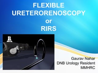
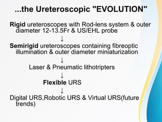
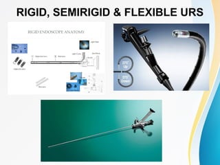
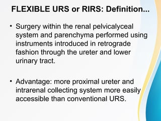
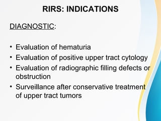
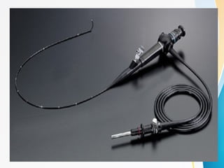
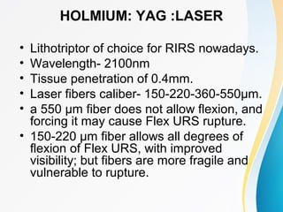
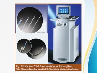
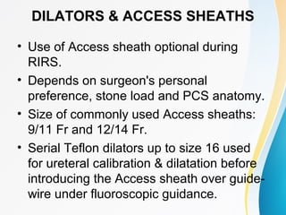
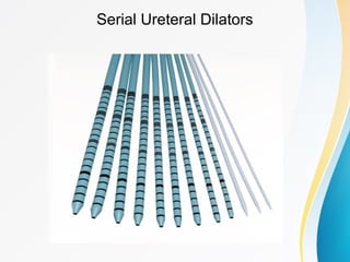
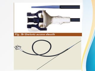
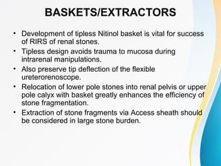
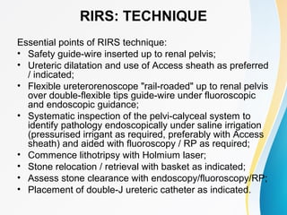
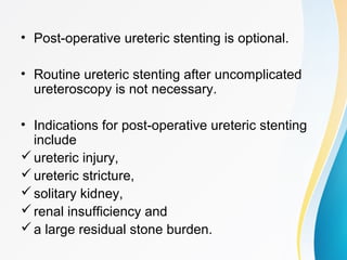
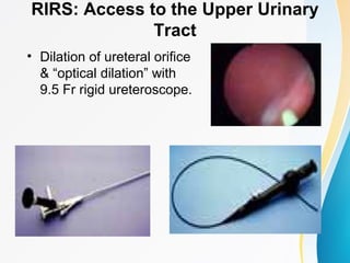
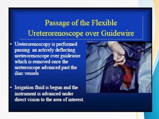
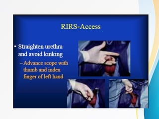
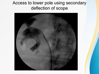
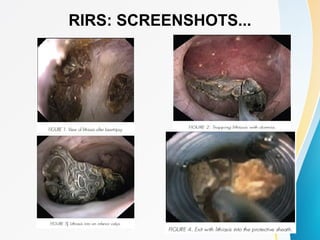
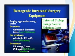
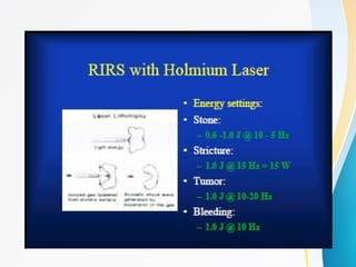
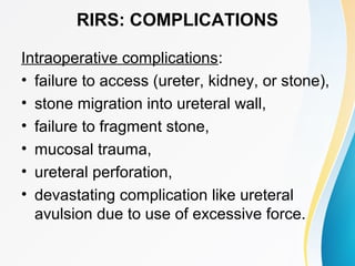
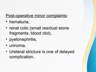
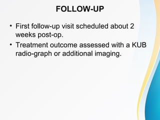
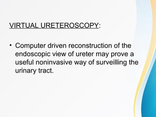
FLEXIBLE
URETERORENOSCOPY
or
RIRS
Gaurav Nahar
DNB Urology Resident
MMHRC
INTRODUCTION
• Trend in surgical practice today is towards
developing minimally invasive techniques.
• Endoscopic surgery using minimally
invasive techniques has been the frontier
of medical innovation in last two decades.
• Development of small calibre flexible
instruments permits endoscopic surgery of
urinary tract to treat a wide variety of
lesions/conditions within the kidney.
Rising trend for URS...Why???
• Shorter hospital stay
• Lower costs
• Improved cosmesis
• Earlier return to work
• Suboptimal results with ESWL(for renal
stone disease)
FLASHBACK...
• 1912: 1st Ureteroscopy by Hugh Hampton
Young ("Father of Modern Urology"); a
rigid 12Fr cystoscope was advanced into
massively dilated ureter of a pt. with PUV.
• Late 1950's: development of first fibreoptic
endoscope.
• 1964: first flexible ureterorenoscopy by
Marshall via a ureterotomy using a 9Fr
flexible endoscope for diagnostic
purposes.
• 1977: Goodman & Lyon reported first rigid
ureteroscopy for therapeutic purposes.
• 1990: Fuchs & Fuchs reported the first
large series (208 patients) of renal calculi
treated by flexible ureteroscopy.
• 1998: contemporary RIRS using flexible
ureterorenoscope and Holmium laser for
treatment of renal stones by Grasso &
Chalik.
...the Ureteroscopic "EVOLUTION"
Rigid ureteroscopes with Rod-lens system & outer
diameter 12-13.5Fr & US/EHL probe
↓
Semirigid ureteroscopes containing fibreoptic
illumination & outer diameter miniaturization
↓
Laser & Pneumatic lithotripters
↓
Flexible URS
↓
Digital URS,Robotic URS & Virtual URS(future
trends)
RIGID, SEMIRIGID & FLEXIBLE URS
FLEXIBLE URS or RIRS: Definition...
• Surgery within the renal pelvicalyceal
system and parenchyma performed using
instruments introduced in retrograde
fashion through the ureter and lower
urinary tract.
• Advantage: more proximal ureter and
intrarenal collecting system more easily
accessible than conventional URS.
RIRS: INDICATIONS
DIAGNOSTIC:
• Evaluation of hematuria
• Evaluation of positive upper tract cytology
• Evaluation of radiographic filling defects or
obstruction
• Surveillance after conservative treatment
of upper tract tumors
THERAPEUTIC:
Stone Disease:
• Failed ESWL (stones <1.5 cm)
• RIRS assisted ESWL (stones upto 2.5 cm)
• Radiolucent stones (stones <1.5 cm)
• Concomitant renal and ureteral stones (renal stone <1
cm)
• Calyceal diverticular stones
• Stones with nephrocalcinosis
• Stones with associated anatomic obstruction
• Stones with supravesical diversion
• Rarely, partial staghorn stones
Others:
• Treatment of PUJ obstruction
• Treatment of anastomotic strictures
• Treatment of urothelial tumors
• Fulguration of bleeding vessels
• Retrograde percutaneous renal puncture
RIRS: CONTRAINDICATIONS
• Patients presenting with ‘high-spiking’
fever & rigors, mental status changes, or
other signs of serious infection.
• Better benefit from a PCN.
• Irrigating pressures within ureter during
endoscopy can lead to pyelovenous and/
or pyelolymphatic back-flow, placing the
patient at risk of septicemia.
RIRS: SPECIALLY USEFUL
• Urolithiasis m/m in prepubertal children.
• Pregnant females.
• Bleeding diathesis or
Anticoagulant/Antiplatelet therapy.
RIRS: INSTRUMENTATION
Instrumentation for RIRS:
1.Newly designed flexible instrument with dual
deflection
2.Energy sources (EHL, Holmium:YAG laser with
small caliber probes)
3.Flexible accessories including guidewires,
dilators, access sheaths, baskets, graspers,
ureteric catheters and forceps
4.Suction pump
5.Video camera unit
6.Fluoroscopy unit
FLEXIBLE URETERORENOSCOPE
• Standard fibreoptic flexible
ureterorenoscopes have a tip size in the
range of 6.75 - 9Fr.
• actively deflectable (primary deflection)
with 120 to 170 degrees of deflection in
one direction and 170 to 270 degrees in
other. Secondary deflection will be passive
or active.
• active secondary deflection allows better
manoeuvrability in LC.
• Working channels of Fr 3.6 - 4 and
standard instruments (e.g. baskets) are Fr
2.2 - 3 in size.
HOLMIUM: YAG :LASER
• Lithotriptor of choice for RIRS nowadays.
• Wavelength- 2100nm
• Tissue penetration of 0.4mm.
• Laser fibers caliber- 150-220-360-550μm.
• a 550 μm fiber does not allow flexion, and
forcing it may cause Flex URS rupture.
• 150-220 μm fiber allows all degrees of
flexion of Flex URS, with improved
visibility; but fibers are more fragile and
vulnerable to rupture.
• Laser energy is delivered via quartz fibres
to stone surface, where it is absorbed and
turned into heat energy that pulverises the
stone into dust by a "photothermal" effect.
• Stone fragment retrieval with basket /
grasping forceps not necessary.
• Can be safely done in anticoagulated pts.
DILATORS & ACCESS SHEATHS
• Use of Access sheath optional during
RIRS.
• Depends on surgeon's personal
preference, stone load and PCS anatomy.
• Size of commonly used Access sheaths:
9/11 Fr and 12/14 Fr.
• Serial Teflon dilators up to size 16 used
for ureteral calibration & dilatation before
introducing the Access sheath over guide-
wire under fluoroscopic guidance.
Serial Ureteral Dilators
Advantages of Access Sheath:
• Facilitate repeated introduction and withdrawal
of endoscope required for large stone burden;
reduces operative time.
• minimizes distal ureteric trauma, wear on the
ureteroscope.
• Avoid build-up of pressure within PCS especially
when pressurised irrigating fluid is used to
improve vision.
Disadvantages of Access Sheath:
• Requirement of pre-stenting 2-4 weeks due to
less spcious ureters.
• Chances of ureteral injury.
BASKETS/EXTRACTORS
• Development of tipless Nitinol basket is vital for success
of RIRS of renal stones.
• Tipless design avoids trauma to mucosa during
intrarenal manipulations.
• Also preserve tip deflection of the flexible
ureterorenoscope.
• Relocation of lower pole stones into renal pelvis or upper
pole calyx with basket greatly enhances the efficiency of
stone fragmentation.
• Extraction of stone fragments via Access sheath should
be considered in large stone burden.
RIRS: PREOPERATIVE ASSESSMENT
• CT Urogram/IVU to assess stone load, stone
location and pelvi-calyceal anatomy.
• Informed consent.
• Counselling on treatment options, procedure and
potential complications, with possibilities of
requiring postop stenting, second-look
procedure, auxiliary procedure and failed
procedure are all thoroughly explained.
• Preoperative urine cultures.
• General anesthesia & prophylactic induction
antibiotic.
RIRS: PATIENT POSITION
• Dorsal lithotomy position.
• Padding of all pressure points.
RIRS: TECHNIQUE
Essential points of RIRS technique:
• Safety guide-wire inserted up to renal pelvis;
• Ureteric dilatation and use of Access sheath as preferred
/ indicated;
• Flexible ureterorenoscope "rail-roaded" up to renal pelvis
over double-flexible tips guide-wire under fluoroscopic
and endoscopic guidance;
• Systematic inspection of the pelvi-calyceal system to
identify pathology endoscopically under saline irrigation
(pressurised irrigant as required, preferably with Access
sheath) and aided with fluoroscopy / RP as required;
• Commence lithotripsy with Holmium laser;
• Stone relocation / retrieval with basket as indicated;
• Assess stone clearance with endoscopy/fluoroscopy/RP;
• Placement of double-J ureteric catheter as indicated.
• Post-operative ureteric stenting is optional.
• Routine ureteric stenting after uncomplicated
ureteroscopy is not necessary.
• Indications for post-operative ureteric stenting
include
ureteric injury,
ureteric stricture,
solitary kidney,
renal insufficiency and
a large residual stone burden.
RIRS: Access to the Upper Urinary
Tract
• Dilation of ureteral orifice
& “optical dilation” with
9.5 Fr rigid ureteroscope.
Access to lower pole using secondary
deflection of scope
RIRS: SCREENSHOTS...
ALGORITHM FOR RENAL STONE
DISEASE
RIRS: COMPLICATIONS
Intraoperative complications:
• failure to access (ureter, kidney, or stone),
• stone migration into ureteral wall,
• failure to fragment stone,
• mucosal trauma,
• ureteral perforation,
• devastating complication like ureteral
avulsion due to use of excessive force.
Post-operative minor complaints:
• hematuria,
• renal colic (small residual stone
fragments, blood clot),
• pyelonephritis,
• urinoma.
• Ureteral stricture is one of delayed
complication.
FOLLOW-UP
• First follow-up visit scheduled about 2
weeks post-op.
• Treatment outcome assessed with a KUB
radio-graph or additional imaging.
LIMITATIONS OF RIRS
Technical difficulties due to deficient quality
of instruments used, such as
ureteroscopes offering
• scanty visibility,
• poor illumination,
• a small working channel,
• deficient quality of forceps and baskets,
etc. and
• limited life of instruments with high cost to
Service, especially if used by several
urologists.
FUTURE TRENDS...
DIGITAL URS:
• Humphreys MR et al demonstrated the beauty of
digital ureterorenoscopy.
• The distal tip objective is a CMOS imaging
sensor coupled to a prism, utilising light emitting
diodes(LED) as the light source, which gives
superb vision within the PCS.
• Further studies required to show improved
outcome.
FLEXIBLE ROBOTIC URS:
• Potential advantages- an increased range
of motion, instrument stability, and
improved ergonomics.
• Avoids excessive strain on the surgeon
who keeps the flexible uretroscope in
deflection for prolonged periods to
approach the target.
VIRTUAL URETEROSCOPY:
• Computer driven reconstruction of the
endoscopic view of ureter may prove a
useful noninvasive way of surveilling the
urinary tract.
CONCLUSION
• RIRS is an integral part of armamentarium of
urologists.
• A valuable tool both in the treatment of renal
stones refractory to ESWL and also for
diagnosis of upper tract pathology.
• With a refinement of technique, complications
like stricture, perforation and extravasation can
be minimized.
• Future developments of RIRS are digital imaging
to improve the quality of vision and robotics to
improve manoeuvrability.
THANK
YOU !!!

Flexible Uretero-renoscopy or RIRS

  • 1.
  • 2.
    INTRODUCTION • Trend insurgical practice today is towards developing minimally invasive techniques. • Endoscopic surgery using minimally invasive techniques has been the frontier of medical innovation in last two decades. • Development of small calibre flexible instruments permits endoscopic surgery of urinary tract to treat a wide variety of lesions/conditions within the kidney.
  • 3.
    Rising trend forURS...Why??? • Shorter hospital stay • Lower costs • Improved cosmesis • Earlier return to work • Suboptimal results with ESWL(for renal stone disease)
  • 4.
    FLASHBACK... • 1912: 1stUreteroscopy by Hugh Hampton Young ("Father of Modern Urology"); a rigid 12Fr cystoscope was advanced into massively dilated ureter of a pt. with PUV. • Late 1950's: development of first fibreoptic endoscope. • 1964: first flexible ureterorenoscopy by Marshall via a ureterotomy using a 9Fr flexible endoscope for diagnostic purposes.
  • 5.
    • 1977: Goodman& Lyon reported first rigid ureteroscopy for therapeutic purposes. • 1990: Fuchs & Fuchs reported the first large series (208 patients) of renal calculi treated by flexible ureteroscopy. • 1998: contemporary RIRS using flexible ureterorenoscope and Holmium laser for treatment of renal stones by Grasso & Chalik.
  • 6.
    ...the Ureteroscopic "EVOLUTION" Rigidureteroscopes with Rod-lens system & outer diameter 12-13.5Fr & US/EHL probe ↓ Semirigid ureteroscopes containing fibreoptic illumination & outer diameter miniaturization ↓ Laser & Pneumatic lithotripters ↓ Flexible URS ↓ Digital URS,Robotic URS & Virtual URS(future trends)
  • 7.
    RIGID, SEMIRIGID &FLEXIBLE URS
  • 8.
    FLEXIBLE URS orRIRS: Definition... • Surgery within the renal pelvicalyceal system and parenchyma performed using instruments introduced in retrograde fashion through the ureter and lower urinary tract. • Advantage: more proximal ureter and intrarenal collecting system more easily accessible than conventional URS.
  • 9.
    RIRS: INDICATIONS DIAGNOSTIC: • Evaluationof hematuria • Evaluation of positive upper tract cytology • Evaluation of radiographic filling defects or obstruction • Surveillance after conservative treatment of upper tract tumors
  • 10.
    THERAPEUTIC: Stone Disease: • FailedESWL (stones <1.5 cm) • RIRS assisted ESWL (stones upto 2.5 cm) • Radiolucent stones (stones <1.5 cm) • Concomitant renal and ureteral stones (renal stone <1 cm) • Calyceal diverticular stones • Stones with nephrocalcinosis • Stones with associated anatomic obstruction • Stones with supravesical diversion • Rarely, partial staghorn stones
  • 11.
    Others: • Treatment ofPUJ obstruction • Treatment of anastomotic strictures • Treatment of urothelial tumors • Fulguration of bleeding vessels • Retrograde percutaneous renal puncture
  • 12.
    RIRS: CONTRAINDICATIONS • Patientspresenting with ‘high-spiking’ fever & rigors, mental status changes, or other signs of serious infection. • Better benefit from a PCN. • Irrigating pressures within ureter during endoscopy can lead to pyelovenous and/ or pyelolymphatic back-flow, placing the patient at risk of septicemia.
  • 13.
    RIRS: SPECIALLY USEFUL •Urolithiasis m/m in prepubertal children. • Pregnant females. • Bleeding diathesis or Anticoagulant/Antiplatelet therapy.
  • 14.
    RIRS: INSTRUMENTATION Instrumentation forRIRS: 1.Newly designed flexible instrument with dual deflection 2.Energy sources (EHL, Holmium:YAG laser with small caliber probes) 3.Flexible accessories including guidewires, dilators, access sheaths, baskets, graspers, ureteric catheters and forceps 4.Suction pump 5.Video camera unit 6.Fluoroscopy unit
  • 15.
    FLEXIBLE URETERORENOSCOPE • Standardfibreoptic flexible ureterorenoscopes have a tip size in the range of 6.75 - 9Fr. • actively deflectable (primary deflection) with 120 to 170 degrees of deflection in one direction and 170 to 270 degrees in other. Secondary deflection will be passive or active. • active secondary deflection allows better manoeuvrability in LC. • Working channels of Fr 3.6 - 4 and standard instruments (e.g. baskets) are Fr 2.2 - 3 in size.
  • 17.
    HOLMIUM: YAG :LASER •Lithotriptor of choice for RIRS nowadays. • Wavelength- 2100nm • Tissue penetration of 0.4mm. • Laser fibers caliber- 150-220-360-550μm. • a 550 μm fiber does not allow flexion, and forcing it may cause Flex URS rupture. • 150-220 μm fiber allows all degrees of flexion of Flex URS, with improved visibility; but fibers are more fragile and vulnerable to rupture.
  • 18.
    • Laser energyis delivered via quartz fibres to stone surface, where it is absorbed and turned into heat energy that pulverises the stone into dust by a "photothermal" effect. • Stone fragment retrieval with basket / grasping forceps not necessary. • Can be safely done in anticoagulated pts.
  • 20.
    DILATORS & ACCESSSHEATHS • Use of Access sheath optional during RIRS. • Depends on surgeon's personal preference, stone load and PCS anatomy. • Size of commonly used Access sheaths: 9/11 Fr and 12/14 Fr. • Serial Teflon dilators up to size 16 used for ureteral calibration & dilatation before introducing the Access sheath over guide- wire under fluoroscopic guidance.
  • 21.
  • 23.
    Advantages of AccessSheath: • Facilitate repeated introduction and withdrawal of endoscope required for large stone burden; reduces operative time. • minimizes distal ureteric trauma, wear on the ureteroscope. • Avoid build-up of pressure within PCS especially when pressurised irrigating fluid is used to improve vision. Disadvantages of Access Sheath: • Requirement of pre-stenting 2-4 weeks due to less spcious ureters. • Chances of ureteral injury.
  • 24.
    BASKETS/EXTRACTORS • Development oftipless Nitinol basket is vital for success of RIRS of renal stones. • Tipless design avoids trauma to mucosa during intrarenal manipulations. • Also preserve tip deflection of the flexible ureterorenoscope. • Relocation of lower pole stones into renal pelvis or upper pole calyx with basket greatly enhances the efficiency of stone fragmentation. • Extraction of stone fragments via Access sheath should be considered in large stone burden.
  • 26.
    RIRS: PREOPERATIVE ASSESSMENT •CT Urogram/IVU to assess stone load, stone location and pelvi-calyceal anatomy. • Informed consent. • Counselling on treatment options, procedure and potential complications, with possibilities of requiring postop stenting, second-look procedure, auxiliary procedure and failed procedure are all thoroughly explained. • Preoperative urine cultures. • General anesthesia & prophylactic induction antibiotic.
  • 27.
    RIRS: PATIENT POSITION •Dorsal lithotomy position. • Padding of all pressure points.
  • 28.
    RIRS: TECHNIQUE Essential pointsof RIRS technique: • Safety guide-wire inserted up to renal pelvis; • Ureteric dilatation and use of Access sheath as preferred / indicated; • Flexible ureterorenoscope "rail-roaded" up to renal pelvis over double-flexible tips guide-wire under fluoroscopic and endoscopic guidance; • Systematic inspection of the pelvi-calyceal system to identify pathology endoscopically under saline irrigation (pressurised irrigant as required, preferably with Access sheath) and aided with fluoroscopy / RP as required; • Commence lithotripsy with Holmium laser; • Stone relocation / retrieval with basket as indicated; • Assess stone clearance with endoscopy/fluoroscopy/RP; • Placement of double-J ureteric catheter as indicated.
  • 29.
    • Post-operative uretericstenting is optional. • Routine ureteric stenting after uncomplicated ureteroscopy is not necessary. • Indications for post-operative ureteric stenting include ureteric injury, ureteric stricture, solitary kidney, renal insufficiency and a large residual stone burden.
  • 30.
    RIRS: Access tothe Upper Urinary Tract • Dilation of ureteral orifice & “optical dilation” with 9.5 Fr rigid ureteroscope.
  • 33.
    Access to lowerpole using secondary deflection of scope
  • 34.
  • 37.
    ALGORITHM FOR RENALSTONE DISEASE
  • 38.
    RIRS: COMPLICATIONS Intraoperative complications: •failure to access (ureter, kidney, or stone), • stone migration into ureteral wall, • failure to fragment stone, • mucosal trauma, • ureteral perforation, • devastating complication like ureteral avulsion due to use of excessive force.
  • 39.
    Post-operative minor complaints: •hematuria, • renal colic (small residual stone fragments, blood clot), • pyelonephritis, • urinoma. • Ureteral stricture is one of delayed complication.
  • 40.
    FOLLOW-UP • First follow-upvisit scheduled about 2 weeks post-op. • Treatment outcome assessed with a KUB radio-graph or additional imaging.
  • 41.
    LIMITATIONS OF RIRS Technicaldifficulties due to deficient quality of instruments used, such as ureteroscopes offering • scanty visibility, • poor illumination, • a small working channel, • deficient quality of forceps and baskets, etc. and • limited life of instruments with high cost to Service, especially if used by several urologists.
  • 42.
    FUTURE TRENDS... DIGITAL URS: •Humphreys MR et al demonstrated the beauty of digital ureterorenoscopy. • The distal tip objective is a CMOS imaging sensor coupled to a prism, utilising light emitting diodes(LED) as the light source, which gives superb vision within the PCS. • Further studies required to show improved outcome.
  • 44.
    FLEXIBLE ROBOTIC URS: •Potential advantages- an increased range of motion, instrument stability, and improved ergonomics. • Avoids excessive strain on the surgeon who keeps the flexible uretroscope in deflection for prolonged periods to approach the target.
  • 46.
    VIRTUAL URETEROSCOPY: • Computerdriven reconstruction of the endoscopic view of ureter may prove a useful noninvasive way of surveilling the urinary tract.
  • 48.
    CONCLUSION • RIRS isan integral part of armamentarium of urologists. • A valuable tool both in the treatment of renal stones refractory to ESWL and also for diagnosis of upper tract pathology. • With a refinement of technique, complications like stricture, perforation and extravasation can be minimized. • Future developments of RIRS are digital imaging to improve the quality of vision and robotics to improve manoeuvrability.
  • 49.