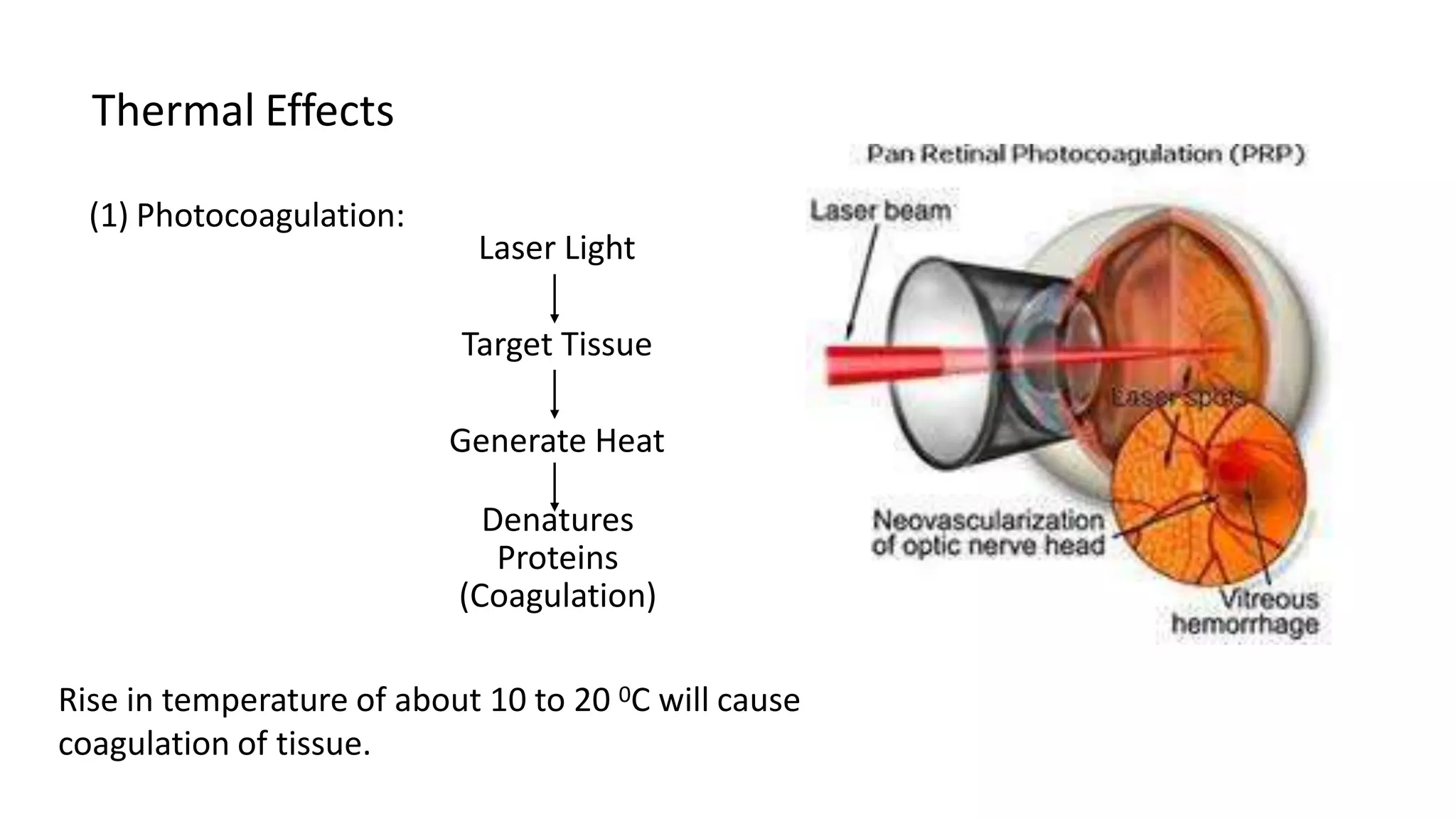
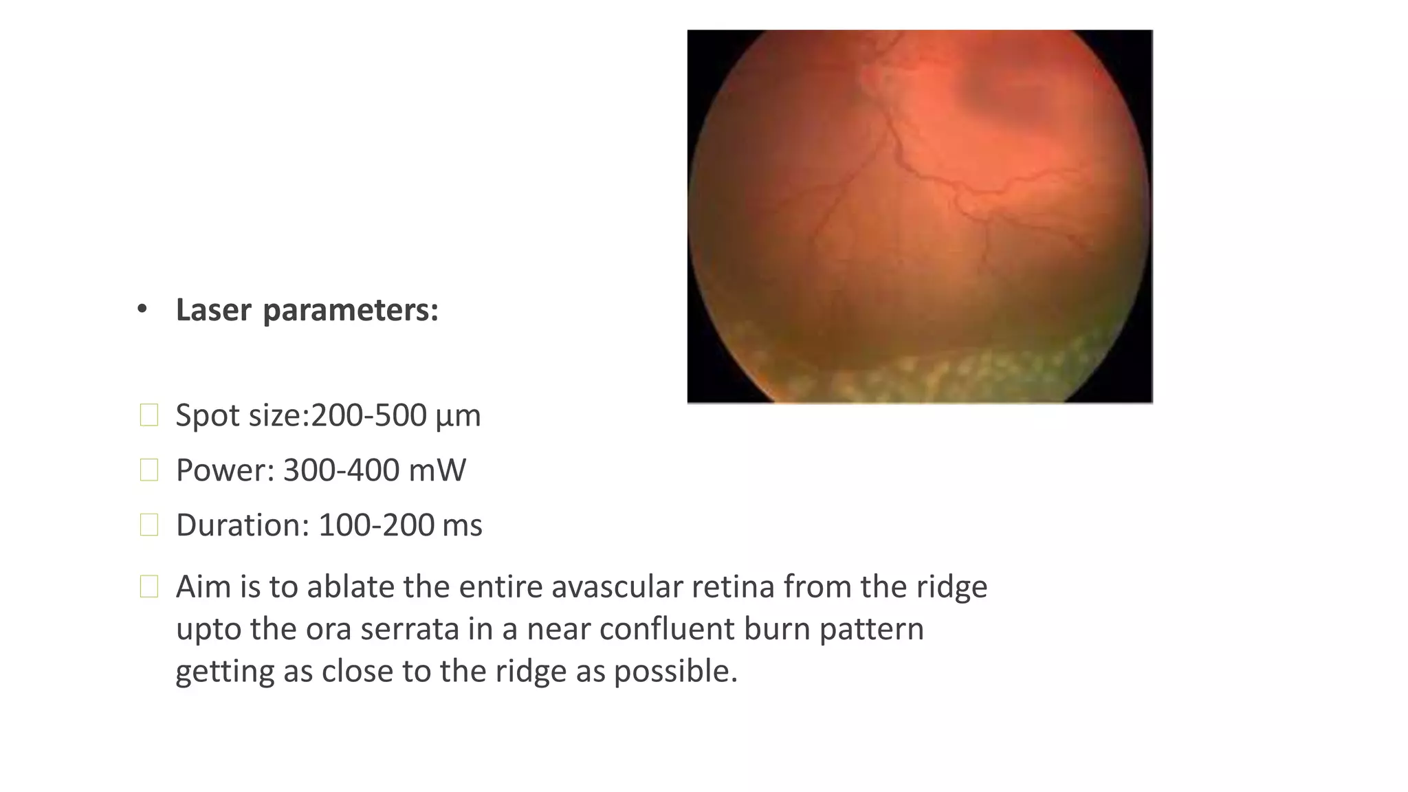
Laser retinal therapy uses lasers to treat various retinal conditions:
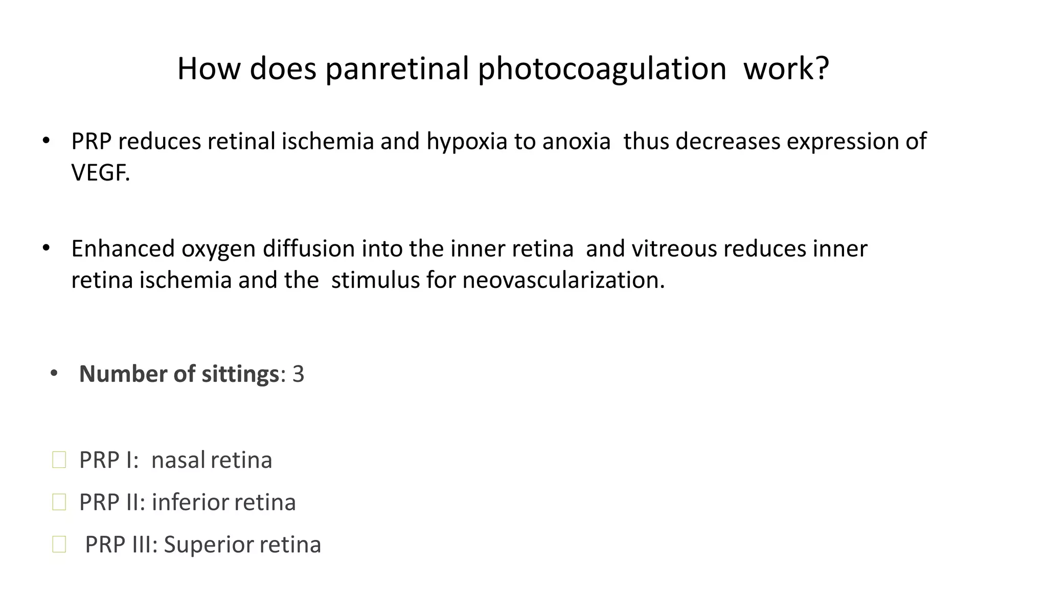
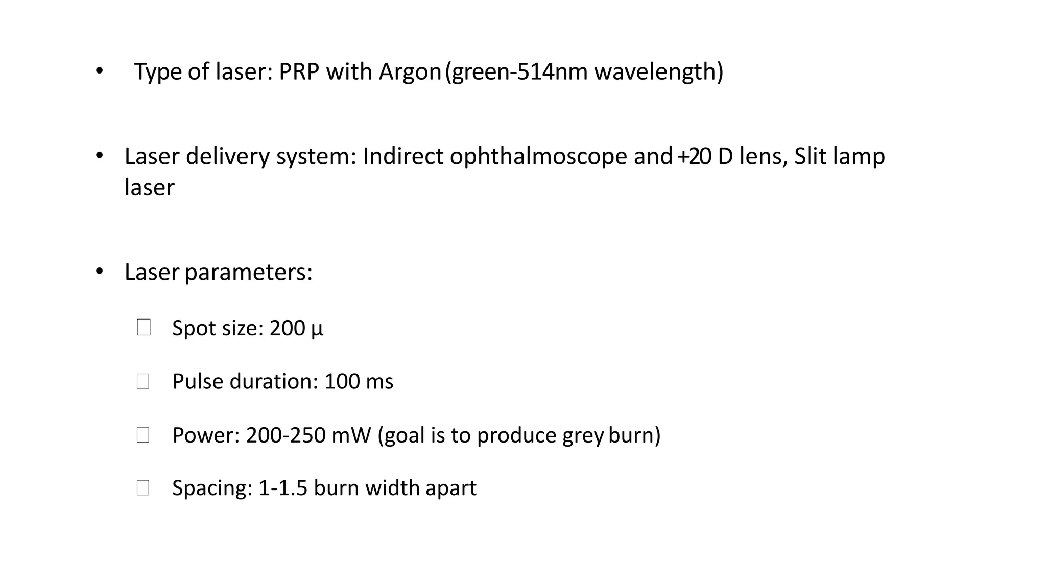
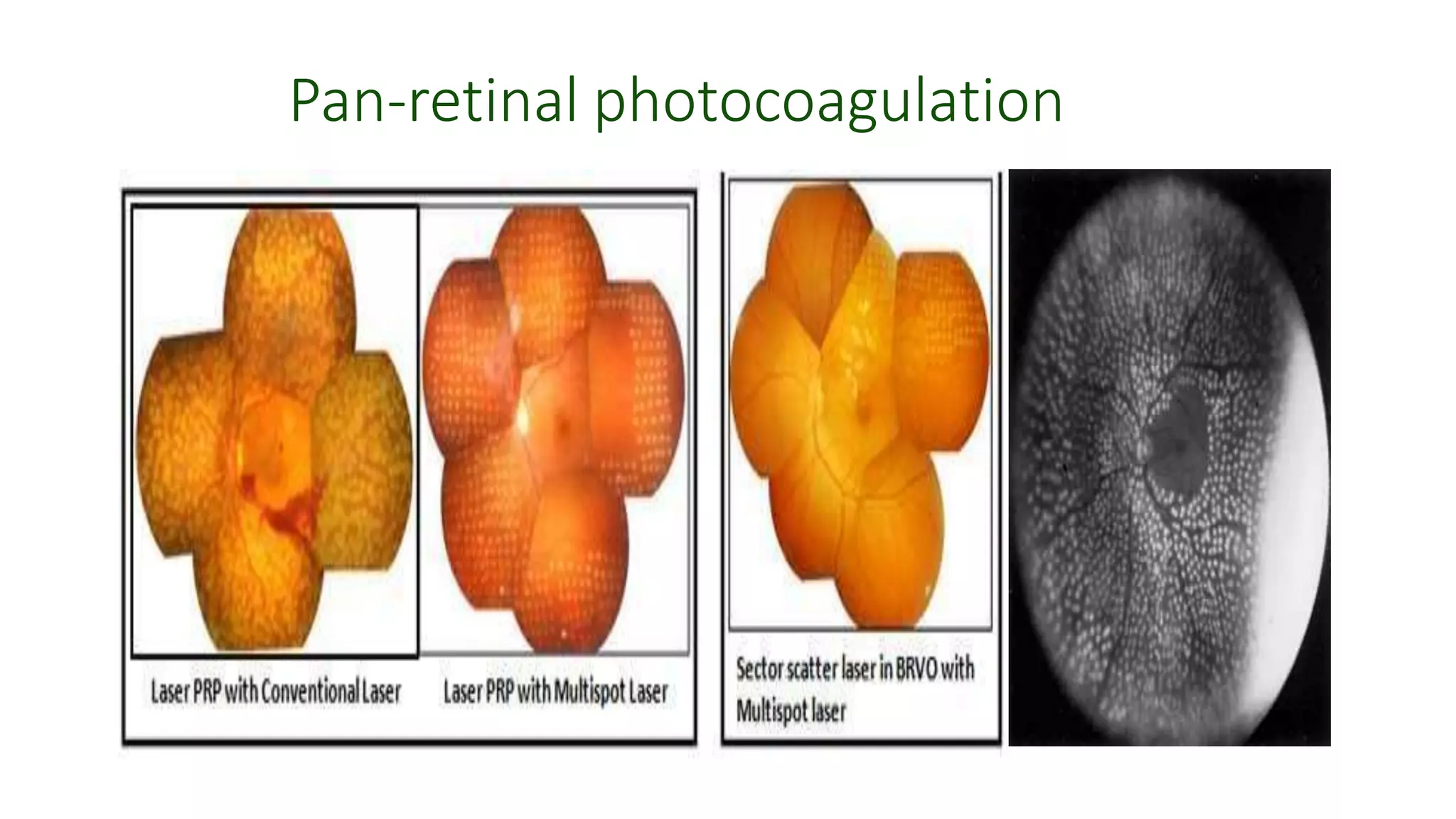
1. Pan-retinal photocoagulation uses lasers like argon to treat diabetic retinopathy by reducing retinal ischemia.
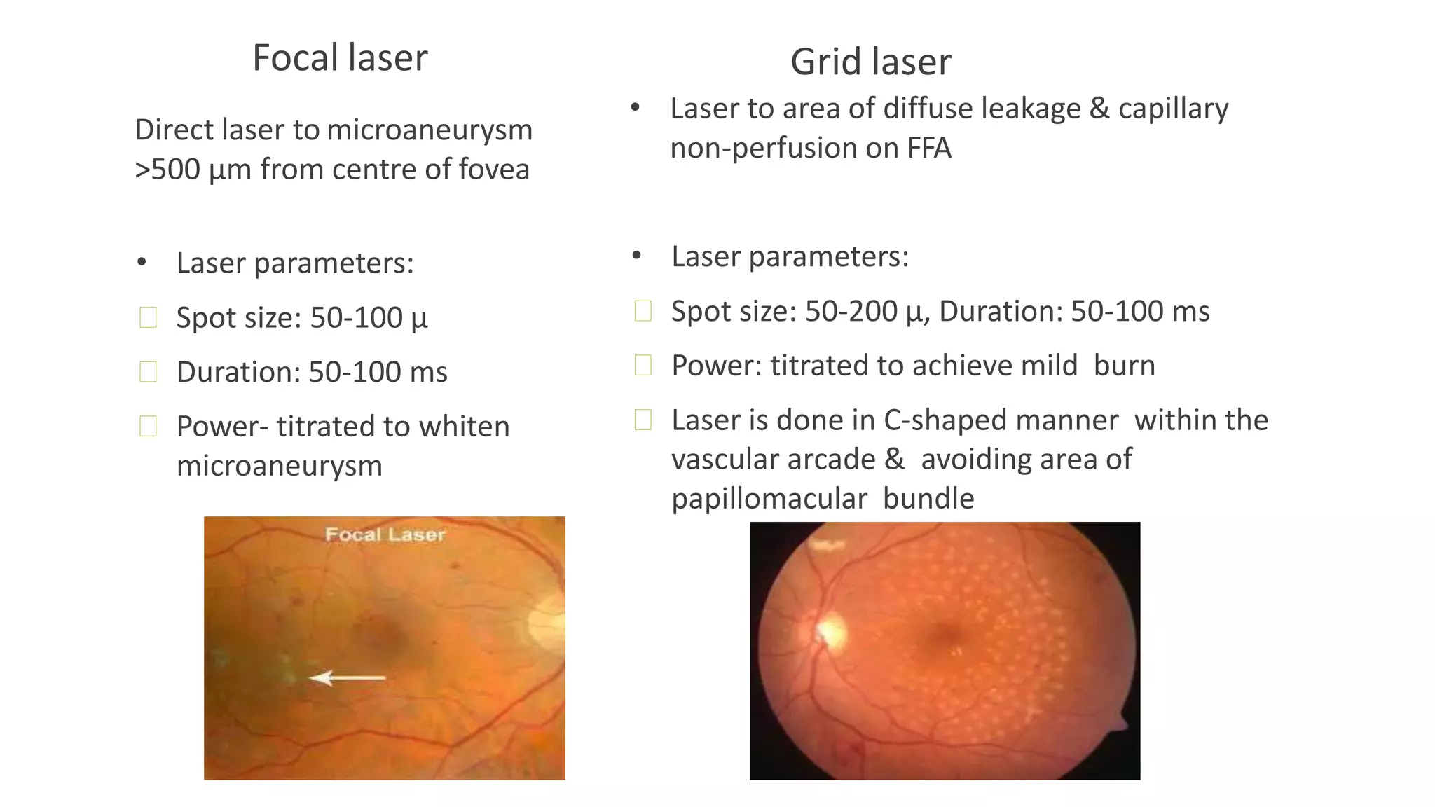
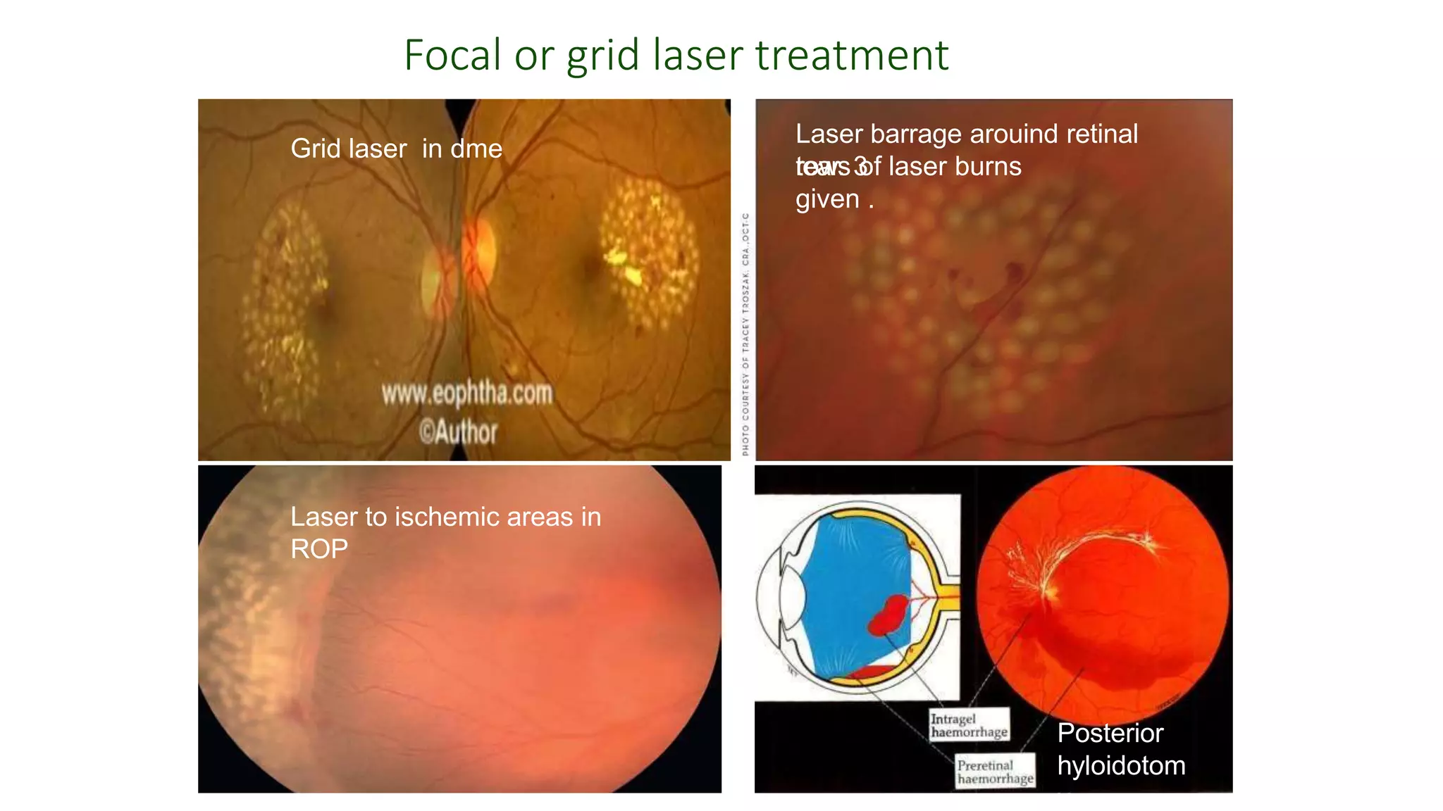
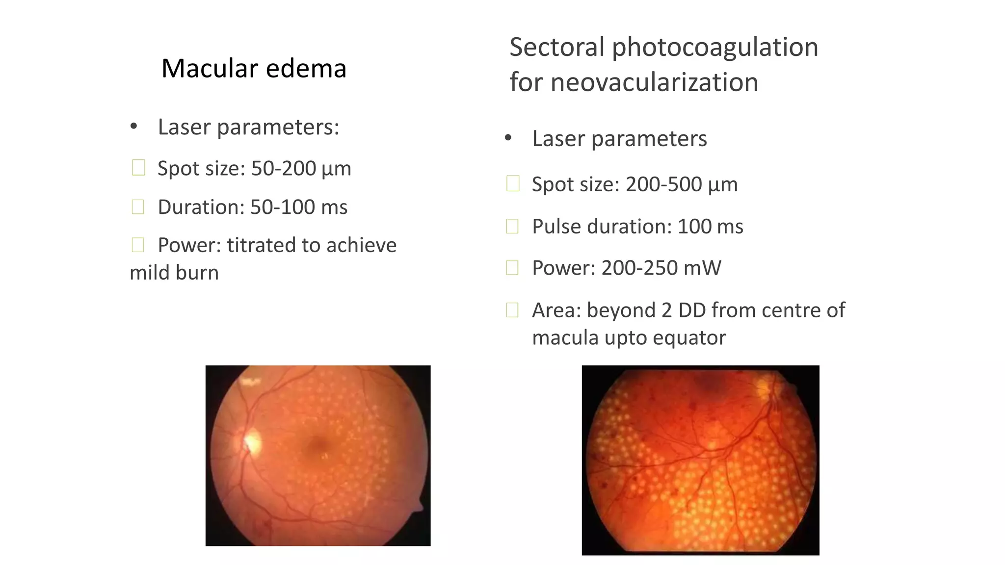
2. Macular edema is treated with focal or grid laser photocoagulation targeted at leaking microaneurysms or diffuse areas of leakage.
3. Retinal tears and detachments are treated preventatively with laser barrage surrounding tears to stimulate proliferation and adhesion.
Congenital pit is an atypical coloboma usually located on the temporal edge of the disc, associated with irregular defects in the juxtapapillary choroid and pigment epithelium. Macular fibers passing through this area often are affected and corresponding changes in the retinal ganglion cell layer and in the visual field occur.
Lasers in ophthalmology - Dr. Parag Apteparag apte
A full presentation of one hour of all types of lasers in ophthalmology for under graduates and post graduates after going through all the uploaded slides till today. This includes laser photocoagulation, laser iridotomy, and laser capsulotomy in detail
Light is an integral part of our life. Advances in technology are increasing and changing the ways that the patient experience dental treatment. One of the milestones in technological advancements in dentistry is the use of lasers The early 20th century saw one of the greatest inventions in science & technology, in that LASERS which later went on to became a gift to health sciences. Albert Einstein is usually credited for the development of the laser theory. He was the first one to coin the term “Stimulated Emission” in his publication “Zur Quantentheorie der Strahlung”, published in 1917 in the “Physikalische Zeitschrift”
Lasers are devices that produce beams of coherent and very high intensity light. The word LASER is an acronym for “Light Amplification by Stimulated\Emission of Radiation”. A crystal or gas is excited to emit light photons of a characteristic wavelength that are amplified and filtered to make a coherent light beam. The effect of the laser depends upon the power of the beam and the extent to which the beam absorbed. Several types of lasers are available based on the wavelengths. These range from long wavelengths (infrared), to visible wavelengths, to short wavelengths (ultraviolet), to special ultraviolet lasers called excimers. Lasers are used nowadays in many areas in the field of dentistry It is of the most captivating technologies in dental practice. Even though, introduced as an alternative to the traditional halogen curing light, laser now has become the instrument of choice, in many dental applications. Its advancements in the field of dentistry are playing a major role in patient care and well being.
MANAGEMENT OF ATRIOVENTRICULAR CONDUCTION BLOCK.pdfJim Jacob Roy
Cardiac conduction defects can occur due to various causes.
Atrioventricular conduction blocks ( AV blocks ) are classified into 3 types.
This document describes the acute management of AV block.
Ozempic: Preoperative Management of Patients on GLP-1 Receptor Agonists Saeid Safari
Preoperative Management of Patients on GLP-1 Receptor Agonists like Ozempic and Semiglutide
ASA GUIDELINE
NYSORA Guideline
2 Case Reports of Gastric Ultrasound
HOT NEW PRODUCT! BIG SALES FAST SHIPPING NOW FROM CHINA!! EU KU DB BK substit...GL Anaacs
Contact us if you are interested:
Email / Skype : kefaya1771@gmail.com
Threema: PXHY5PDH
New BATCH Ku !!! MUCH IN DEMAND FAST SALE EVERY BATCH HAPPY GOOD EFFECT BIG BATCH !
Contact me on Threema or skype to start big business!!
Hot-sale products:
NEW HOT EUTYLONE WHITE CRYSTAL!!
5cl-adba precursor (semi finished )
5cl-adba raw materials
ADBB precursor (semi finished )
ADBB raw materials
APVP powder
5fadb/4f-adb
Jwh018 / Jwh210
Eutylone crystal
Protonitazene (hydrochloride) CAS: 119276-01-6
Flubrotizolam CAS: 57801-95-3
Metonitazene CAS: 14680-51-4
Payment terms: Western Union,MoneyGram,Bitcoin or USDT.
Deliver Time: Usually 7-15days
Shipping method: FedEx, TNT, DHL,UPS etc.Our deliveries are 100% safe, fast, reliable and discreet.
Samples will be sent for your evaluation!If you are interested in, please contact me, let's talk details.
We specializes in exporting high quality Research chemical, medical intermediate, Pharmaceutical chemicals and so on. Products are exported to USA, Canada, France, Korea, Japan,Russia, Southeast Asia and other countries.
Lung Cancer: Artificial Intelligence, Synergetics, Complex System Analysis, S...Oleg Kshivets
RESULTS: Overall life span (LS) was 2252.1±1742.5 days and cumulative 5-year survival (5YS) reached 73.2%, 10 years – 64.8%, 20 years – 42.5%. 513 LCP lived more than 5 years (LS=3124.6±1525.6 days), 148 LCP – more than 10 years (LS=5054.4±1504.1 days).199 LCP died because of LC (LS=562.7±374.5 days). 5YS of LCP after bi/lobectomies was significantly superior in comparison with LCP after pneumonectomies (78.1% vs.63.7%, P=0.00001 by log-rank test). AT significantly improved 5YS (66.3% vs. 34.8%) (P=0.00000 by log-rank test) only for LCP with N1-2. Cox modeling displayed that 5YS of LCP significantly depended on: phase transition (PT) early-invasive LC in terms of synergetics, PT N0—N12, cell ratio factors (ratio between cancer cells- CC and blood cells subpopulations), G1-3, histology, glucose, AT, blood cell circuit, prothrombin index, heparin tolerance, recalcification time (P=0.000-0.038). Neural networks, genetic algorithm selection and bootstrap simulation revealed relationships between 5YS and PT early-invasive LC (rank=1), PT N0—N12 (rank=2), thrombocytes/CC (3), erythrocytes/CC (4), eosinophils/CC (5), healthy cells/CC (6), lymphocytes/CC (7), segmented neutrophils/CC (8), stick neutrophils/CC (9), monocytes/CC (10); leucocytes/CC (11). Correct prediction of 5YS was 100% by neural networks computing (area under ROC curve=1.0; error=0.0).
CONCLUSIONS: 5YS of LCP after radical procedures significantly depended on: 1) PT early-invasive cancer; 2) PT N0--N12; 3) cell ratio factors; 4) blood cell circuit; 5) biochemical factors; 6) hemostasis system; 7) AT; 8) LC characteristics; 9) LC cell dynamics; 10) surgery type: lobectomy/pneumonectomy; 11) anthropometric data. Optimal diagnosis and treatment strategies for LC are: 1) screening and early detection of LC; 2) availability of experienced thoracic surgeons because of complexity of radical procedures; 3) aggressive en block surgery and adequate lymph node dissection for completeness; 4) precise prediction; 5) adjuvant chemoimmunoradiotherapy for LCP with unfavorable prognosis.
- Video recording of this lecture in English language: https://youtu.be/lK81BzxMqdo
- Video recording of this lecture in Arabic language: https://youtu.be/Ve4P0COk9OI
- Link to download the book free: https://nephrotube.blogspot.com/p/nephrotube-nephrology-books.html
- Link to NephroTube website: www.NephroTube.com
- Link to NephroTube social media accounts: https://nephrotube.blogspot.com/p/join-nephrotube-on-social-media.html
Couples presenting to the infertility clinic- Do they really have infertility...Sujoy Dasgupta
Dr Sujoy Dasgupta presented the study on "Couples presenting to the infertility clinic- Do they really have infertility? – The unexplored stories of non-consummation" in the 13th Congress of the Asia Pacific Initiative on Reproduction (ASPIRE 2024) at Manila on 24 May, 2024.
The prostate is an exocrine gland of the male mammalian reproductive system
It is a walnut-sized gland that forms part of the male reproductive system and is located in front of the rectum and just below the urinary bladder
Function is to store and secrete a clear, slightly alkaline fluid that constitutes 10-30% of the volume of the seminal fluid that along with the spermatozoa, constitutes semen
A healthy human prostate measures (4cm-vertical, by 3cm-horizontal, 2cm ant-post ).
It surrounds the urethra just below the urinary bladder. It has anterior, median, posterior and two lateral lobes
It’s work is regulated by androgens which are responsible for male sex characteristics
Generalised disease of the prostate due to hormonal derangement which leads to non malignant enlargement of the gland (increase in the number of epithelial cells and stromal tissue)to cause compression of the urethra leading to symptoms (LUTS
These lecture slides, by Dr Sidra Arshad, offer a quick overview of physiological basis of a normal electrocardiogram.
Learning objectives:
1. Define an electrocardiogram (ECG) and electrocardiography
2. Describe how dipoles generated by the heart produce the waveforms of the ECG
3. Describe the components of a normal electrocardiogram of a typical bipolar leads (limb II)
4. Differentiate between intervals and segments
5. Enlist some common indications for obtaining an ECG
Study Resources:
1. Chapter 11, Guyton and Hall Textbook of Medical Physiology, 14th edition
2. Chapter 9, Human Physiology - From Cells to Systems, Lauralee Sherwood, 9th edition
3. Chapter 29, Ganong’s Review of Medical Physiology, 26th edition
4. Electrocardiogram, StatPearls - https://www.ncbi.nlm.nih.gov/books/NBK549803/
5. ECG in Medical Practice by ABM Abdullah, 4th edition
6. ECG Basics, http://www.nataliescasebook.com/tag/e-c-g-basics
Tom Selleck Health: A Comprehensive Look at the Iconic Actor’s Wellness Journeygreendigital
Tom Selleck, an enduring figure in Hollywood. has captivated audiences for decades with his rugged charm, iconic moustache. and memorable roles in television and film. From his breakout role as Thomas Magnum in Magnum P.I. to his current portrayal of Frank Reagan in Blue Bloods. Selleck's career has spanned over 50 years. But beyond his professional achievements. fans have often been curious about Tom Selleck Health. especially as he has aged in the public eye.
Follow us on: Pinterest
Introduction
Many have been interested in Tom Selleck health. not only because of his enduring presence on screen but also because of the challenges. and lifestyle choices he has faced and made over the years. This article delves into the various aspects of Tom Selleck health. exploring his fitness regimen, diet, mental health. and the challenges he has encountered as he ages. We'll look at how he maintains his well-being. the health issues he has faced, and his approach to ageing .
Early Life and Career
Childhood and Athletic Beginnings
Tom Selleck was born on January 29, 1945, in Detroit, Michigan, and grew up in Sherman Oaks, California. From an early age, he was involved in sports, particularly basketball. which played a significant role in his physical development. His athletic pursuits continued into college. where he attended the University of Southern California (USC) on a basketball scholarship. This early involvement in sports laid a strong foundation for his physical health and disciplined lifestyle.
Transition to Acting
Selleck's transition from an athlete to an actor came with its physical demands. His first significant role in "Magnum P.I." required him to perform various stunts and maintain a fit appearance. This role, which he played from 1980 to 1988. necessitated a rigorous fitness routine to meet the show's demands. setting the stage for his long-term commitment to health and wellness.
Fitness Regimen
Workout Routine
Tom Selleck health and fitness regimen has evolved. adapting to his changing roles and age. During his "Magnum, P.I." days. Selleck's workouts were intense and focused on building and maintaining muscle mass. His routine included weightlifting, cardiovascular exercises. and specific training for the stunts he performed on the show.
Selleck adjusted his fitness routine as he aged to suit his body's needs. Today, his workouts focus on maintaining flexibility, strength, and cardiovascular health. He incorporates low-impact exercises such as swimming, walking, and light weightlifting. This balanced approach helps him stay fit without putting undue strain on his joints and muscles.
Importance of Flexibility and Mobility
In recent years, Selleck has emphasized the importance of flexibility and mobility in his fitness regimen. Understanding the natural decline in muscle mass and joint flexibility with age. he includes stretching and yoga in his routine. These practices help prevent injuries, improve posture, and maintain mobilit
Pulmonary Thromboembolism - etilogy, types, medical- Surgical and nursing man...VarunMahajani
Disruption of blood supply to lung alveoli due to blockage of one or more pulmonary blood vessels is called as Pulmonary thromboembolism. In this presentation we will discuss its causes, types and its management in depth.
2. What is Laser?
L :Light
A: Amplification (by)
S : Stimulated
E: Emission (of)
R: Radiation
• Term coined by Gordon Gould.
• Lase means to absorb energy in one form and to emit anew form
of light energy which is more useful.
4. • The concept of ocular therapy using light first was publicized by Meyer-
Schwickerath, who took patients to the roof of his laboratory in 1949 and
focused sunlight on their retinas to treat melanomas.
• Carbon arcs were used; by the mid-1950s, the xenon arc photocoagulator had
been developed and was made commercially available by Zeiss.
• Large size,
• poorly collimated beam,
• tendency to produce intense retinal burns.
1960 - Theodore Maiman : Built first laser by using a ruby crystal medium
.
5. PROPERTIES OF LASER LIGHT
• Monochromatic (emit only one wave length)
• Coherence (all in same phase-improve focusing)
• Polarized (in one plane-easy to pass through media)
• Collimated (in one direction & non spreading)
• High energy (Intensity measured by WattJ/s)
6. LASER PHYSICS:
• Light as electromagnetic waves, emitting radiant energy in tiny package
called ‘quanta’/photon. Each photon has a characteristic frequency and its
energy is proportional to its frequency.
• Three basic ways for photons and atoms to interact:
• Absorption
• Spontaneous Emission
• Stimulated Emission
7. Absorption
Energized electron in
higher orbit
Electron in orbit
A photon of the “right” energy gets absorbed and
“bumps” an electron into a higher energy level.
photon
9. Stimulated Emission:
photon
photon
Electron in orbit
photon
A photon strikes an excited electron. The electron falls to its lower energy
level, releasing a photon that is going in the same direction and in exact
phase with, the original photon. Note that only one photon strikes the atom
but two photons leave it—the original photon plus the emittedphoton
Energized electron in higher
orbit
10. • Now consider this ‘stimulated emission’.
• The two photons that have been produced can then generate 4 photons,
and the 4 16 etc… etc… cascade of intense monochromatic radiation.
• Thus Stimulated emission is the basis of the laser action.
11. Laser Construction
1
2
3
• A pump source or exciting medium
• A gain medium or laser medium.
• An optical resonator or laser tube.
12. Pump Source
• Provides energy to the laser system.
• Examples: electrical discharges, flash lamps and chemical
reactions.
• Ex. Excimer lasers use an electrical discharges.
13. Gain Medium
• It is a major determining factor of the wavelength of the
laser.
• It is Excited by the pump source.
• Here spontaneous and stimulated emission of
photons takes place.
• Gain medium can be solid, liquid, and gas.
14. • The energy of the emitted laser beam from gain medium is
increased(amplified) still further by causing the light beam to
traverse through the same material multiple times.
• This is accomplished by placing a mirror over each end of the
crystal or gas tube so that the distance between them is an even
multiple of the laser light’s wavelength.
• The coherent light beam is reflected back and forth
becoming more and more intense.
LASER tube or Optical Resonator
16. MODES OF LASER OUTPUT
• Continuous Wave (CW) Laser: Delivery of energy in a
continuous stream of photons.
• Pulsed Lasers: Produce energy pulses of a few tens of micro to
millisecond.
• Q Switches Lasers: Delivery of energy pulses is of extremely
short duration (nanosecond).
• A Mode-locked Lasers: Emits a train of short duration pulses
(picoseconds).
17. LASER INSTRUMENTATION
LASER Components are –
• Console: It contain laser medium and tube,
power supply and laser control system.
• Control Panel: It contain dials or push buttons or
touch screen for controlling various parameters.
• Aiming Beam
• Laser Switch
• Safety Filter
• Delivery System:
Slit Lamp Microscope
Indirect Ophthalmoscopes
Endolaser probes
18. ACCESSORY COMPONENT
Corneal Contact Lenses for Laser used-
• Single mirror goniolens for goniotomy
• Abraham lens - iriditomy
• Goldman style 3-mirror lens for photcoagulation
(PRP) lenses
• Volk-Superquad and pan 165 for PRP
• Mainster and Area centralis for focal and grid laser
• Indirect Fundus Lenses (20 D) for Indirect laser
delivery
19. • Laser light transmitted by optical fibre
Optical fiber typically consists of a core, cladding, and a jacket (buffer coating). Light
launched into the fiber with its acceptance cone is trapped within a core due to the
total internal reflection at the core/cladding interface.
20. 1- Optical fiber and electronic cable connecting laser with a slit lamp system.
2- Optical coupler projecting the beam exiting from the fiber onto the retina.
3- Contact lens.
21. CLASSIFICATION OF LASER
• Solid State
Ruby
Nd. Yag
Erbium. YAG
• Gas Ion
Argon
Krypton
He-Neon
CO2
• Metal Vapour
Cu
Gold
• Dye
Rhodamine
• Excimer
Argon Fluoride
Krypton Fluoride
Krypton Chloride
• Diode
Gallium-Aluminum-
Arsenide (GaAlAs)
22. • Light entering the eye can be reflected, absorbed , transmitted or scattered.
• Absorption depend on ocular characterstics chromophores
melanin - retinal and iris pigmented epithelia, choroid, uvea, trabecular meshwork;
haemoglobin - red blood cells;
xanthophyll - plexiform layers of the retina, especially in the macula;
rhodopsin and cone photopigments – photoreceptors
lipofuscin located primarily in the RPE layer
23. lasers Wavelength
Diode 810 nm
Krypton red 647 nm
Krypton yellow 568 nm
Frequency
doubled Nd YAG
532 nm
Argon green 514 nm
Argon blue 485 nm
TYPES OF OPHTHALMIC LASERS
25. Thermal Effects
(1) Photocoagulation:
Laser Light
Target Tissue
Generate Heat
Denatures
Proteins
(Coagulation)
Rise in temperature of about 10 to 20 0C will cause
coagulation of tissue.
27. Thermal Effects
(3)Photovaporization:
• Vaporization of tissue to CO2 and water occurswhen
its temperature rise 60—100 0C or greater.
• Commonly used CO2
Absorbed by water of cells
Visible vapor (vaporization)
Heat Cell disintegration
Cauterization Incision eg..co2 laser
28. Photochemical effcts
Photoablation:
Breaks the chemical bonds that hold tissue together
essentially vaporizing the tissue, e.g. Photorefractive
Keratectomy, Argon Fluoride (ArF), Excimer Laser.
Usually -
Visible Wavelength
Ultraviolet Yields :
Photocoagulation
Photoablation
Infrared Photodisruption,
Photocoagulation
29. PHOTOCHEMICAL EFFECT
• Photoradiation (PDT):
Also called photodynamic therapy.
e.g. Treatment of Ocular tumours andCNV
Photon + Photo sensitizer in ground state(S)
Molecular Oxygen Free Radical
S + O2 Cytotoxic Intermediate
Cell Damage, Vascular Damage , ImmunologicDamage
31. CLASSIFICATION OF
CHORIORETINAL BURN INTENSITY
• Light
• Mild
: Barely visible retinal blanching
: Faint white retinal burn
• Moderate : Dirty white retinal burn
• Heavy : Dense white retinal burn
32. SELECTION OF OPTICAL WAVELENGHT FOR COAGULATION
• Wavelengths that are highly absorbed by macular yellow (such as 488 nm) are
relatively contraindicated when treating in or near the macula.
• Absorption of these wavelengths in macular pigments may cause heating and
destruction of the nerve fiber layer, resulting in loss of vision.
• Double ND:YAG or green Argon
33. • Scattering loss in cataract or in vitreous opacities can be minimized using longer
wavelengths: yellow (577 nm) or red (640–680 nm)
• Large quantity of hemoglobin- wavelengths between 520 and 580 nm are best
suited
34. How focal laser works?
Several theories
• Laser energy removes unhealthy RPE cells which are then
replaced by more viable RPE cells.
• Photocoagulation stimulates the existing RPE cells to
absorb more fluid.
• Laser treatment may stimulate vascular endothelial
proliferation and improve the integrity of the inner blood-
retinal barrier.
35. How does panretinal photocoagulation work?
• PRP reduces retinal ischemia and hypoxia to anoxia thus decreases expression of
VEGF.
• Enhanced oxygen diffusion into the inner retina and vitreous reduces inner
retina ischemia and the stimulus for neovascularization.
• Number of sittings: 3
ꟷ PRP I: nasal retina
ꟷ PRP II: inferior retina
ꟷ PRP III: Superior retina
36. Uses
1. Diabetic Retinopathy – Pan-retinal photocoagulation.
Indications:
• High riskPDR
• Early PDR or very severe NPDRin
Patients with poor compliance
During pregnancy
Patients with systemic diseases
Pending cataract surgery
One-eyed patients
37. • Type of laser: PRP with Argon(green-514nm wavelength)
• Laser delivery system: Indirect ophthalmoscope and+20 D lens, Slit lamp
laser
• Laser parameters:
ꟷ Spot size: 200 μ
ꟷ Pulse duration: 100 ms
ꟷ Power: 200-250 mW (goal is to produce greyburn)
ꟷ Spacing: 1-1.5 burn width apart
39. 2. Diabetic maculopathy:
• Indication: Clinically significant macular edema
Basic guidelines
• All areas of macular thickening must be treated
• FFA is done to look for points of leakage
• Focal leak →focal laser photocoagulation
• Diffuse leak → grid photocoagulation
• Laser delivery system: Slit lamp
40. Focal laser
Direct laser to microaneurysm
>500 μm from centre of fovea
• Laser parameters:
ꟷ Spot size: 50-100 μ
ꟷ Duration: 50-100 ms
ꟷ Power- titrated to whiten
microaneurysm
Grid laser
• Laser to area of diffuse leakage & capillary
non-perfusion on FFA
• Laser parameters:
ꟷ Spot size: 50-200 μ, Duration: 50-100 ms
ꟷ Power: titrated to achieve mild burn
ꟷ Laser is done in C-shaped manner within the
vascular arcade & avoiding area of
papillomacular bundle
41. Focal or grid laser treatment
Grid laser in dme
Laser to ischemic areas in
ROP
Posterior
hyloidotom
y
Laser barrage arouind retinal
tear. 3
rows of laser burns
given .
44. Macular edema
• Laser parameters:
ꟷ Spot size: 50-200 μm
ꟷ Duration: 50-100 ms
ꟷ Power: titrated to achieve
mild burn
Sectoral photocoagulation
for neovacularization
• Laser parameters
ꟷ Spot size: 200-500 μm
ꟷ Pulse duration: 100 ms
ꟷ Power: 200-250 mW
ꟷ Area: beyond 2 DD from centre of
macula upto equator
45. 4. Retinopathy of prematurity
• Indications:
ꟷ Stage I, Zone I with plus disease
ꟷ Stage II, Zone I with plus disease
ꟷ Stage III, Zone I with plus disease
ꟷ Stage III, Zone I without plus disease
ꟷ Stage II, Zone II with plus disease
ꟷ Stage III, Zone II with plus disease
• Type of laser: PRP (withLIO)
46. • Laser parameters:
ꟷ Spot size:200-500 μm
ꟷ Power: 300-400 mW
ꟷ Duration: 100-200 ms
ꟷ Aim is to ablate the entire avascular retina from the ridge
upto the ora serrata in a near confluent burn pattern
getting as close to the ridge as possible.
47. • Complications:
ꟷ Premature infants are prone to develop apnoea.
ꟷ Conjunctival chemosis.
ꟷ Subconjunctival hemorrhage due to excessive scleral
indentation.
ꟷ Intense photocoagulation may lead to anterior segment
ischemia and necrosis resulting in hypotony and phthisis
bulbi.
48. 5. CNVM-Photodynamic therapy:
Dosage: 6 mg/m2 of verteporfin infused intravenously
The amount of dye calculated is given over 10 minutes
(infusion phase) and then a further 5 minutes are allowed for
the dye to accumulate (accumulation phase).
49. Verteporfin injected intravenously
selectively accumulates in neovascular tissue which is rich in low-density lipoprotein
receptors, while it is rapidly cleared from the surrounding normal tissues
Upon absorption of a photon at the proper wavelength the porphyrin molecule
undergoes a transition from the ground state into the singlet excited state
50. The singlet excited porphyrin can decay back to the ground state with release of energy in the
form of fluorescence, which enables optical identification of tumor tissue
Such energy transfer produces toxic singlet oxygen
Singlet oxygen is very reactive and therefore it has a very short diffusion path length – less
than 20 nm, so all its interactions are highly localized.
51. • The activation by laser is typically performed 15–20 min after the intravenous
injection of the dye.
• Red laser light (689 nm diode laser)
• Closure of the abnormal (leaking) blood vessels occurs within approximately 6–12
weeks in most patients.
52. • Complications:
ꟷ Visual disturbances
ꟷ Photosensitivity reactions
ꟷ Overdosing- macular infarction
ꟷ In case of extravasation- pain at injection site and
allergic reactions
53. 6. Retinal lesions predisposing to detachment and retinal tear
Purpose: Toinduce a sterile inflammationwhich stimulate
proliferation of the RPE →indirectly improves adhesion
between the RPE and the neurosensory retina.
Laser delivery system: Slit lamp with contact lensorLIO
Principle:
ꟷ The entire perimeter of the break should be
surrounded by laser application.
ꟷ Particular attention to the anterior margin and horns of a
tear should be paid.
54. ꟷ In the presence of a rim of fluid or in subclinical
detachment, laser is applied to the attached retina
immediately around the detachment.
ꟷ If applied to the area of detachment, it may cause
further progression of the detachment.
ꟷ Laser treatment of an inflamed retina is avoided as there
is a risk of producing a retinal break
• Laserparameters:
ꟷ Two-three rows of confluent burns
ꟷ Spot size: 200-500 μm
ꟷ Mild to moderate burn intensity
55. 7. Eales’ disease: It is an idiopathic, inflammatory peripheral
vasculitis characterised by retinal periphlebitis and
capillary non-perfusion leading to hypoxia
• Indications:
ꟷ Neovascularization elsewhere
ꟷ Neovascularization disc
ꟷ Neovascularization iris
• Laser delivery system:
LIO
Slit lamp
57. 8. Central serous chorioretinopathy
Focal laser photocoagulation (extra-foveal leakage)
• Indications:
ꟷ Non-resolving or recurrent CSCR with V/A: <6/12
ꟷ Well defined leakage on FFA, atleast 500 μ away from
centre of fovea
• Laserparameters:
ꟷ Spot size: 100- 200 μ
ꟷ Duration: 100-200 ms
ꟷ Power: 100-200 mW
58. 9. Retinal artery macroaneurym:
• These are solitary, saccular or fusiform dilation (diameter:125 -250μ) of
the retinal arteriole involving usually, the first three divisions.
• Two forms: acute &chronic
• Acute form: sudden loss of vision due to retinalorvitreous
hemorrhage
• Chronic form: gradual loss of vision due toleakage and exudation into
the macular area
• Laser photocoagulation is requiredfor chronic forms
59. • Laserparameters:
ꟷ Spot size: 200-300 μ
ꟷ Duration: 200-500 ms
ꟷ Power: 200mW
• Direct treatment: laser is focused directly onthe macroaneurysm so as
to obtain slow and gentle whitening
• In indirect treatment: laser burns are placedaround the aneurysm
60. 10. Coats’ disease: Idiopathic retinal telangiectasia associated with
intraretinal and subretinal exudation and frequently exudative
retinal detachment, without signs of vitreoretinal traction
• Indication:
ꟷ Severe vascular anomalies with macular exudation
ꟷ Exudative retinal detachment
ꟷ Vascular anomalies posterior to equator
ꟷ Neovascularization
• Type of laser: LIO or Slitlamp
61. • Laserparameters:
ꟷ Spot size: 200-500 μ
ꟷ Power: 200 mW
ꟷ Duration: 200-500 ms
ꟷ End point: whitening of lesion
62. 11. Retinal capillary hemangioma: Vascular hamartoma
• Indication:
ꟷ All capillary hemangiomas except those touching the optic nerve head
• Type of laser: Argon green or frequency doubledYAG
• Laser parameters:
ꟷ Spot size: 200-500 μ
ꟷ Duration: 0.2- 1.0 s
ꟷ Power: titred to produce mild-moderate whitening of lesion.
ꟷ Small lesion → directtreatment
ꟷ Largelesion→ treatment of feeder vessel
63. 12. Choroidal hemangioma: Vascular hamartoma
Manifest in two forms: diffuse & circumscribed
• Indication:
Serous retinal detachment
• Aim of treatment: achieve resolutioon ofserous retinal
detachment and not tumor obliteration
• Conventional laser: entire tumor surface iscovered with
laser spots
64. PDT: 6 mg/m2 of verteporfin dye is injected intravenously
• Laser parameters
ꟷ Spot size: 6000 μ(maximum)
ꟷ Laser used 689 nm
ꟷ Type of laser delivery: Slit lamp
ꟷ Lens used: Mainster wide field lens
ꟷ In peripapillary choroidal hemangioma, laser spot is applied at a
distance of 200 μ from the optic disc edge
ꟷ Large lesion(>2 mm) radiant exposure of 100 J/cm2 with
exposure of 186 seconds
ꟷ Small lesion(<2 mm) radiant exposure of 75 J/cm2 with
exposure of 125 seconds
66. • Type of laser: Slitlamp
• Technique:
ꟷ laser energy is focused above the inferior extent of the haemorrhage to
facilitate gravity-aided drainage of blood into the vitreous cavity
ꟷ Begin with an energy of 5mJ using single pulse.
ꟷ each shot ,increase energy by 1mJ ,maximum 8 shots to be given
• Complications:
Non-resolving vitreous haemorrhage
Creation of retinal holes
Retinal detachment
67. 14. Optic disc pit:
• Indication:
Associated serous macular detachment
• Laser photocoagulation along the temporal marginofoptic disc.
68. Recent advances
1. PASCAL(Pattern scanning laser)
• Semi-automated laser delivering device
• Allows for a pattern of 4-56 burns to be applied in <1 sec using a scanning
laser with shorter pulse duration
• Indications:
ꟷ Diabetic retinopathy (PDR &NPDR)
ꟷ Choroidal neovascularization (CNVM & SRNVM)
ꟷ Age-related macular degeneration
ꟷ Retinal vein occlusion (BRVO & CRVO)
ꟷ Retinal tear, holes
70. • Advantages:
ꟷ Safe
ꟷ Relatively painless
ꟷ Less time consuming
ꟷ Well tolerated
ꟷ More number of spots in single sitting
ꟷ Requires less number of sitting
71. 2. Navigational lasers
• 532-nm pattern-type eye-tracking laser integrateslive colour fundus
imaging, red-free and infra-red imaging, FFA with photocoagulator system.
• After image acquisition and making customized treatment plans by
physicians including marking areas which will be coagulated the treatment
plan is superimposed onto the live digital retina image during treatment
• The physician controls laser application and the systems assist with
prepositioning the laser beam.
72. • Advantages over conventionallasers:
ꟷ Fast
ꟷ Painless
ꟷ Better documentation
ꟷ Wide field viewing system allows for better accuracy
73. 3.SELECTIVE RPE THERAPY (SRT)
• Light is strongly absorbed by melanosomes in the RPE
• Application of microsecond laser pulses allows for confinement of the thermal
and mechanical effects of this absorption within the RPE layer, thus sparing the
photoreceptors and the inner retina.
• Application of repetitive pulses of microsecond and sub-microsecond duration
results in selective damage to RPE due to the formation of small cavitation
bubbles around melanosomes.
• Subsequent RPE proliferation and migration restores continuity of the RPE layer
• Lack visible changes in retina
• CSR, DME