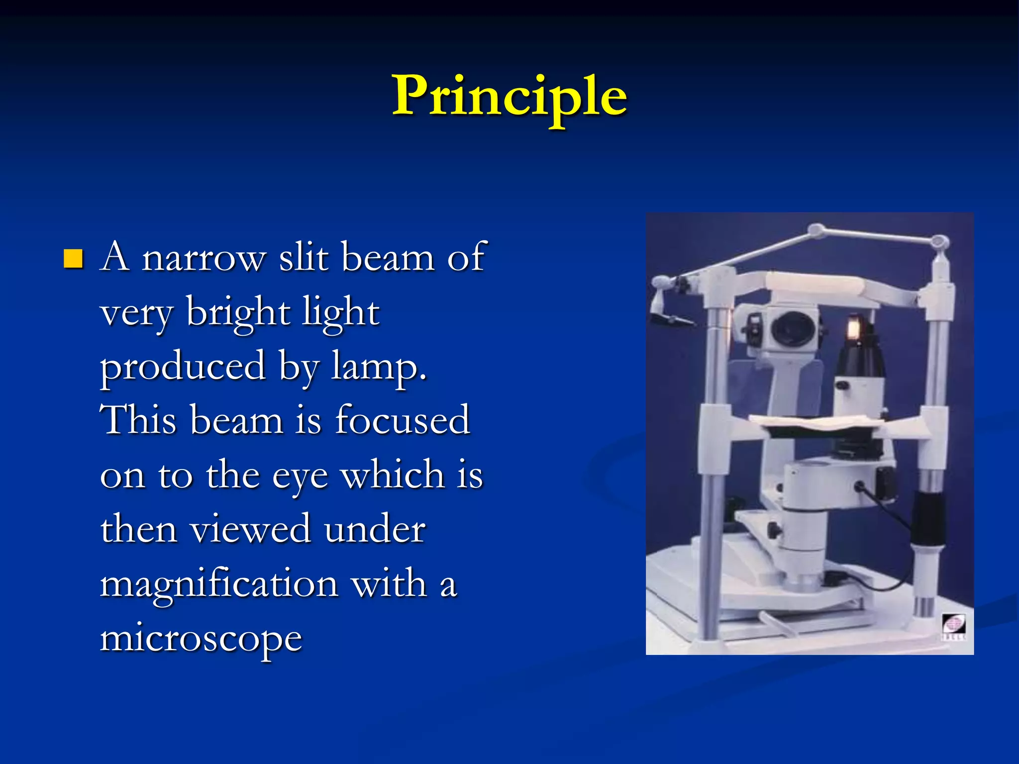
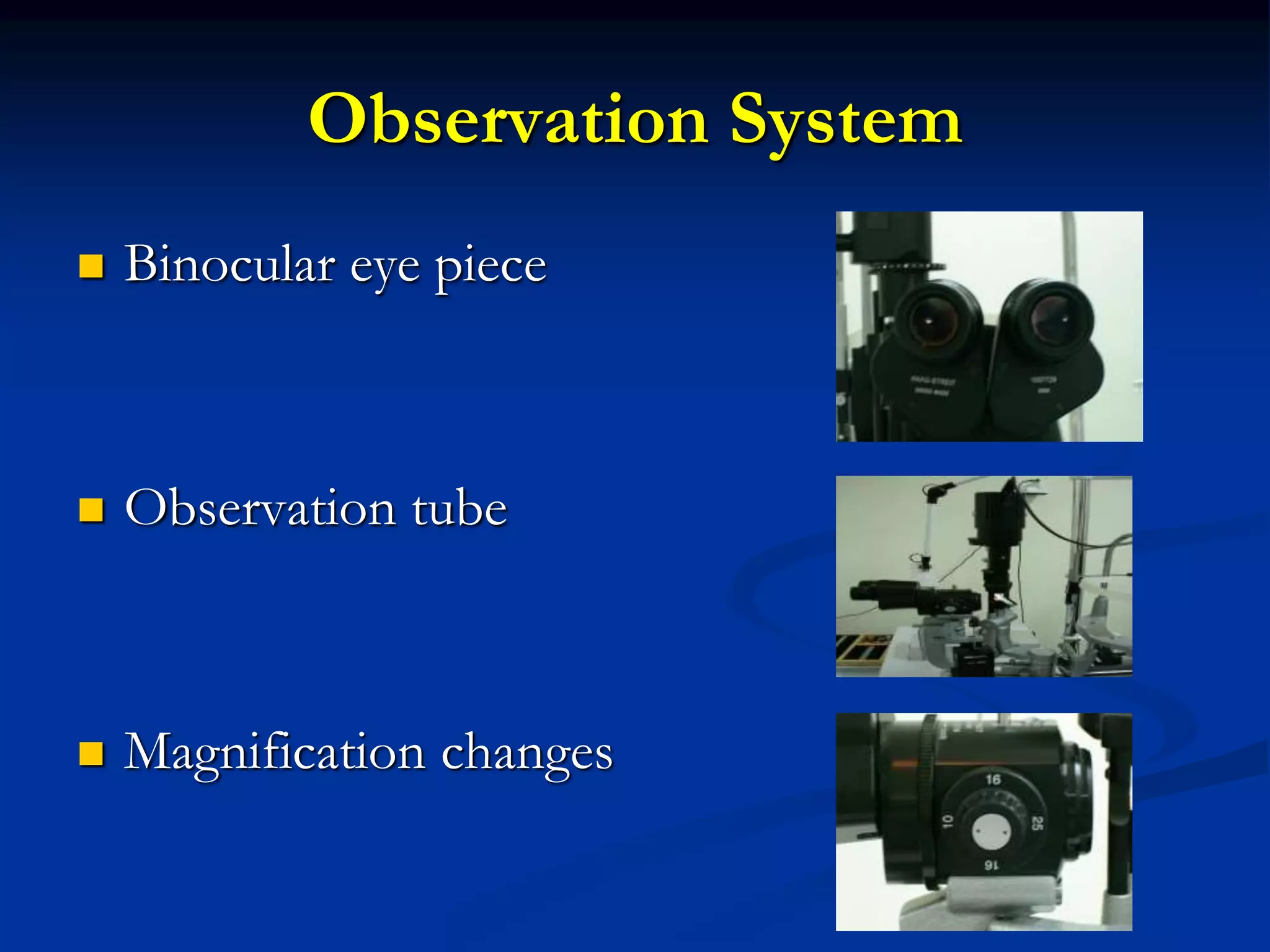
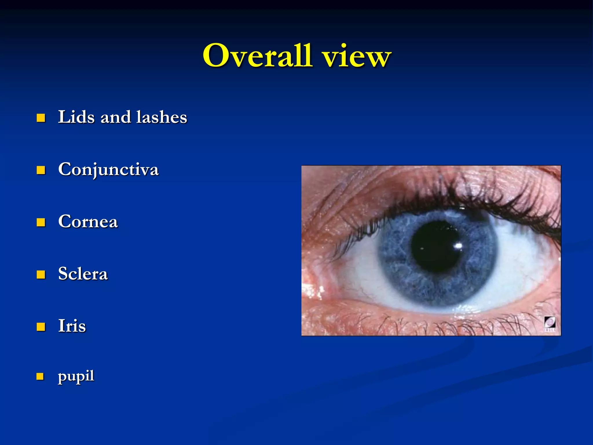
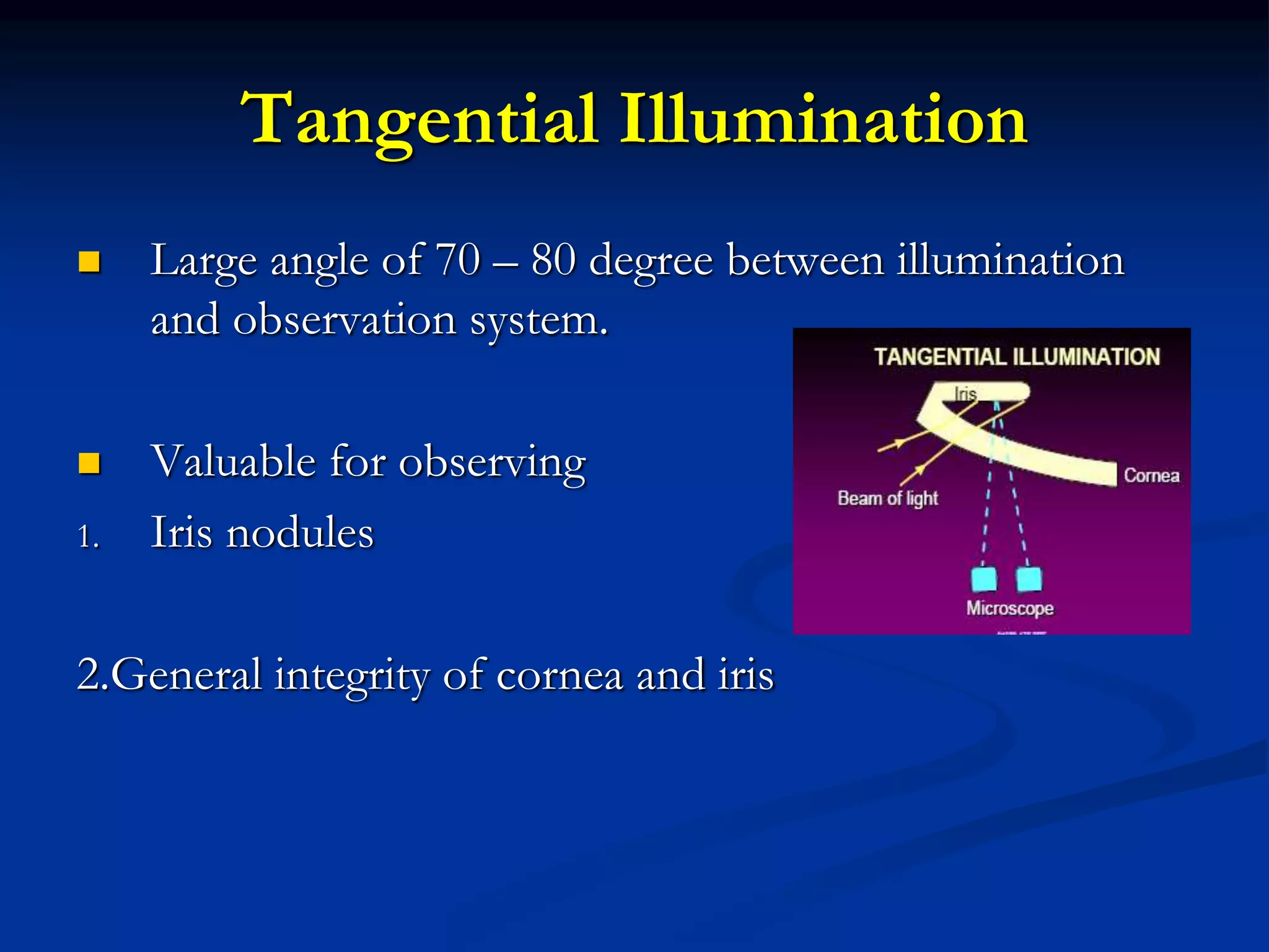
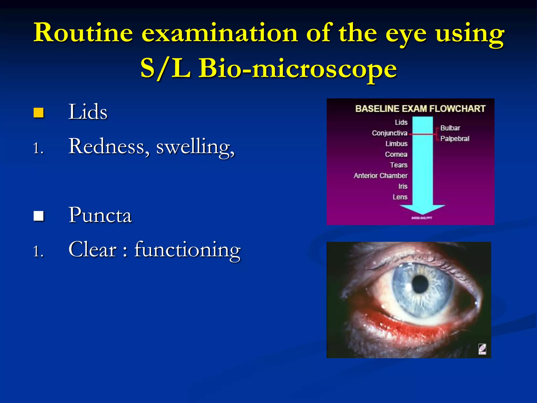
Slit lamp examination
Bhaktiprada Sahoo
Optometrist & Ocularist
History
Allivar Gullstrand
 Noble prize -1911
 Slit lamp
Principle
 A narrow slit beam of
very bright light
produced by lamp.
This beam is focused
on to the eye which is
then viewed under
magnification with a
microscope
Parts
 Mechanical support
 Observation system
 Illumination system
Mechanical Support
 Forehead rest
 Chin rest
 Fixation target
 Power supply unit
 Locking control system
Observation System
 Binocular eye piece
 Observation tube
 Magnification changes
Illumination System
 Lamp housing unit
 Slit width and height
control
 Neutral density filter
 Cobalt blue light filter
 Red-free filter
 Field size control
 Diffuser
 prism
Magnification Range
 Low magnification -7x-10x
 Medium magnification – 20x-25x
 High magnification – 30x-40x
Low Magnification
 Lids
 Bulbar conjunctiva/ sclera
 Cornea / limbus
 Tear
 Anterior chamber
Medium Magnification
 Epithelium
 Stroma
 Endothelium
 Contact lens fit / lens condition
High Magnification
 Epitheium
1. Vacuoles
2. Microcysts
3. dystrophies
 Stroma
1. Straie
2. Folds
 Endothelium
1. Polymegathism
2. Guttata
3. Blebs
4. Dystrophies
Illumination Techniques
 Diffuse
 Direct
 Indirect
 Retro illumination
 Specular reflection
 Sclerotic scatter
 Tangential
Diffuse Illumination
 45° angle between light
and microscope
 Fully open slit
 Diffusing filter
Overall view
 Lids and lashes
 Conjunctiva
 Cornea
 Sclera
 Iris
 pupil
Direct Illumination
 Observation and illumination
system focused at same point
 Vary angle of illumination
 Low to high magnification
 Vary width and height of
light source
Optic section (Narrow, focused light)
 Indicates depth
 Localise
1. Nerve fibers
2. Blood vessels
3. Infiltrate
4. Cataracts
5. Anterior chamber angle
Parallelepiped (Wider, focused light)
 Wider field of view
 Illuminated block of
cornea
 More extensive
examination
Conical beam
 Inflammatory cells /flare in AC
 Darked room
Indirect Illumination
 Observation and
illumination systems are
not focused at same
point
 Vary angle of
illumination
 Vary beam width
Valuables for observing
 Epithelial erosions
 Epithelial Dystrophies
Retro Illumination
 Object is illuminated by
reflected light
 Moderately wide beam
 Medium to high
magnification
 Reflected light from iris or
fundus
Valuables for observing
 Vascularisation
 Limabal Injection
 Epithelial oedema
 Microcysts
 Vacuoles
Specular reflection
 Angle of incidence equal to angle of reflection
 Valuable for observing
 Endothelial cell
 Tear film lipid layer
Sclerotic Scatter
 Total internal reflection
of wide angle light source
 Valuable for observing
 Localized epithelial
Oedema
 Corneal Scar
 FB in Cornea
Tangential Illumination
 Large angle of 70 – 80 degree between illumination
and observation system.
 Valuable for observing
1. Iris nodules
2.General integrity of cornea and iris
Routine examination of the eye using
S/L Bio-microscope
 Lids
1. Redness, swelling,
 Puncta
1. Clear : functioning
Continuation
 Conjunctiva
1. Redness
2. Inflammation
Continuation
 Limbus
1. Redness
2. Neovascularisation
3. staining
 Cornea
1. Transparency
1. Tissues damage
 Tear
1. Quality
2. Lipid layer
3. debris
 Anterior chamber
1. Cells and flare
1. Persistent pupillary
membranes
Iris examination
 Inflammation
 Pigmentation
 Iridectomy
 coloboma
Crystalline lens examination
 Opacities
 Pigment on capsule
 Subluxation
 Iris attachments
Thank you

Slit lamp examination.ppt