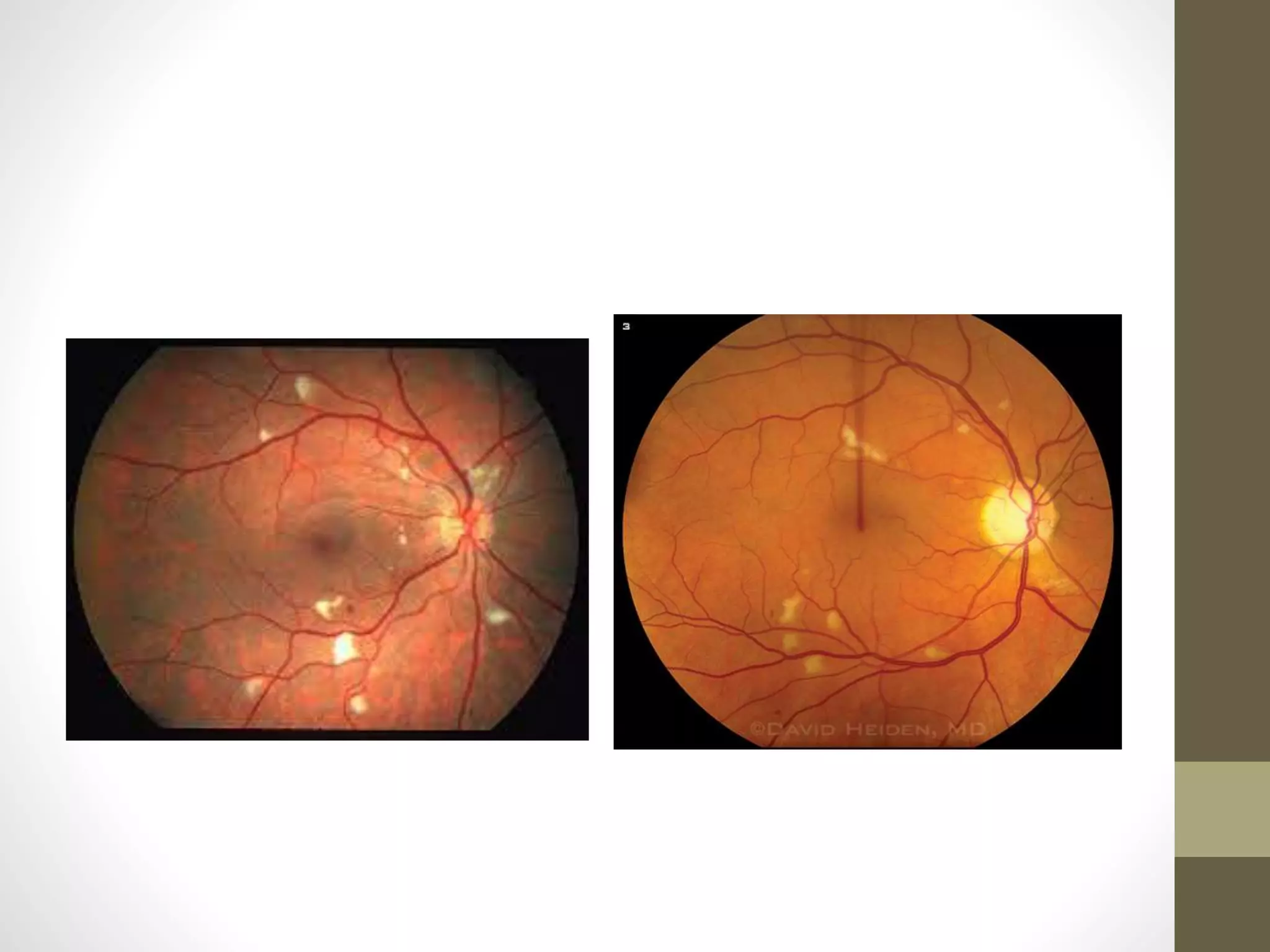
SlideShare a Scribd company logo
Uveitis.
Part 3
By
Dr. Amr Mounir.MD
Lecturer of Ophthalmology
Sohag University
PARASITIC UVEITIS
PARASITIC UVEITIS
1-Toxoplasmosis
2-Toxocariasis
3- Onchocerciasis
4- Cysticercosis
Toxoplasmosis
Toxoplasmosis
Toxoplasmosis is caused by Toxoplasma gondii, an obligate
intracellular protozoan.
The cat is the definitive host, with intermediate hosts including
mice, livestock, birds and humans.
Oocysts are excreted in cat faeces and then ingested by
intermediate hosts including via contaminated water supplies.
Occasionally; infection may be transmitted via organ
transplantation or blood transfusion
Systemic features:
1) Congenital toxoplasmosis
Neurological and visceral involvement may be very severe,
Retinochoroiditis may occur in over 75%
2) Postnatal childhood acquisition
50% of cases of childhood toxoplasmosis
3) Acquired toxoplasmosis in immunocompetent adults
subclinical in 80–90%, early retinitis may occur in up to 20%
4) Toxoplasmosis in immunocompromised patients
meningoencephalitis, pneumonitis, retinochoroiditis
Ocular features:
Toxoplasmosis constitutes 20–60% of all posterior uveitis
Reactivation at previously inactive cyst-containing scars is the
rule in the immunocompetent
Recurrent episodes of inflammation are common and occur
when the cysts rupture and release hundreds of tachyzoites into
normal retinal cells
Ocular involvement from congenital infection may only be
detected later in life with the incidental discovery of typical
retinochoroidal scars, though occasionally macular or optic
nerve damage may impair vision in childhood.
C/P:
Symptoms:
Unilateral acute or subacute onset of floaters, blurring and photophobia.
Signs:
- ‘Spill-over’ anterior uveitis is common
-- A single inflammatory focus of fluffy white retinitis or retinochoroiditis
associated with a pigmented scar (‘satellite lesion’) is typical.
-- De novo foci not associated with an old scar, and multiple
-lesions
- Vitritis may be severe and impair fundus visualization. ‘Headlight in the fog’
- Vasculitis
- Optic disc oedema
- Extensive and fulminant retinal involvement
- Retinochoroiditis may be absent
- Neuroretinitis
- Punctate outer retinal toxoplasmosis
- Visual loss: Causes of permanently reduced vision
(around 25% of eyes) include macular inflammatory lesions
and oedema, optic nerve involvement , vascular occlusion ,
serous, rhegmatogenous and tractional retinal detachment
and late secondary choroidal neovascularization
Healing: in immunocompetent hosts usually occurs
spontaneously within 6–8 weeks, although vitreous opacities
take longer to clear.
The inflammatory focus is replaced by a sharply demarcated
atrophic scar that develops a pigmented border
Recurrence: The average number of recurrent attacks per
patient
Investigation:
Serology:
Toxoplasma IgG antibodies are detectable in the
serum within 1–2 weeks of initial infection.
Positivity to IgM antibodies usually means that infection has
been acquired within the last year.
PCR testing of intraocular fluid is variably sensitive (16–67%) but
highly specific.
Ocular fluid antibody assessment. Calculating the ratio
(Goldmann–Witmer coefficient) of specific IgG in aqueous
humour to that in serum seems to be a reasonably sensitive
(48–90%) investigation.
Treatment:
-Evidence for the efficacy of current regimens is
limited.
-Eradication of the parasite has not been
demonstrated but parasite activity and
multiplication may be reduced.
- Spontaneous resolution generally occurs,
treatment is not administered in every case.
Indications for treatment:
Sight-threatening lesion involving the macula, papillomacular bundle,
optic nerve head or a major blood vessel, for severe vitritis and in the
immunocompromised.
Prednisolone (1 mg/kg) is given initially and tapered according to
clinical response,
Should always be used in conjunction with a specific anti-Toxoplasma
agent, most frequently pyrimethamine combined with sulfadiazine
(‘classic’ or ‘triple’ therapy, sometimes supplemented with
clindamycin).
Intravitreal therapy with clindamycin (1 mg) and dexamethasone (400
μg) may be as effective as triple therapy in reactivated infection; two
to three injections (two-weekly intervals) may be required. It may be
preferred in recurrent infection in pregnancy
Other lines of therapy:
Azithromycin 250–500 mg daily shows evidence of reducing
the rate of recurrence of retinochoroiditis and its use in
combination with pyrimethamine, folinic acid and
prednisolone is a promising newer regimen. Clarithromycin
may be a good alternative to azithromycin.
Co-trimoxazole (trimethoprim 160 mg/sulfamethoxazole
800 mg) twice daily in combination with prednisolone is a
lower-cost and better-tolerated option that might not be
quite as effective as classic therapy.
Clindamycin 300 mg four times daily may be added to triple
therapy (see above) or used instead of pyrimethamine.
Pseudomembranous colitis is a potential adverse effect.
Atovaquone 750 mg two to four times daily.
Topical steroid and mydriatic may be given for anterior
uveitis.
Antimicrobial maintenance therapy is used in immunocompromised patients.
Toxocariasis
Toxocariasis is caused by infestation with a common intestinal
ascarid (roundworm) of dogs, Toxocara canis; puppies are more
commonly infected than adult dogs, and are also more likely to
spread the organism.
Human infestation is by ingestion of soil or food contaminated
with ova shed in canine faeces; young children are at particular
risk from the soil of parks and playgrounds.
Once ingested, ova develop into larvae, which penetrate the
intestinal wall and travel to various organs such as the liver,
lungs, skin, brain and eyes, with resultant inflammation
C/P:
- Asymptomatic infestation is common.
- Visceral toxocariasis (VT), also known as visceral larva
migrans (VLM) is systemic infection of variable severity that
usually occurs in a child aged 2–7 years. Fever, abdominal
pain, pneumonitis, lymphadenopathy, hepatomegaly and
myocarditis are some of the possible features.
- Covert toxocariasis: mild systemic form
Ocular toxocariasis:
(ocular larva migrans – OLM) generally occurs independently of
VLM, and is associated with a lower parasitic load. It is typically
unilateral, and in around two-thirds causes some degree of
permanent visual impairment.
a) Chronic endophthalmitis typically presents with leukocoria,
strabismus, floaters or unilateral visual loss. Features may
include anterior uveitis, vitritis, chorioretinitis, papillitis and a
fundus granuloma.
b) Posterior pole or peripheral granuloma without
Inflammation
c) Chorioretinal scar
d) Diffuse unilateral subacute neuroretinitis (DUSN).
Investigations:
It is particularly important to distinguish a Toxocara granuloma
from retinoblastoma.
Full blood count. Eosinophilia may be present, particularly in
VLM, and can become chronic
Hypergammaglobulinaemia especially IgE.
• Serology. Antibodies to Toxocara canis are detectable in only
about 50% of ocular cases. Positivity is common in the
general population (14% overall in the USA).
• Ultrasonography may be useful if the media are hazy.
• Aqueous or vitreous sampling for eosinophilia, antibody
detection and PCR.
• Biopsy of a granuloma of the skin or elsewhere for larvae is
sometimes possible.
Ocular treatment
- Prevention by good hygiene practices and deworming of pets.
- Steroids. Topical, regional and systemic as indicated.
- Anthelmintic agents such as mebendazole and
thiabendazole can be considered in OT, noting that worm
death may promote inflammation.
- Vitrectomy for sight-threatening tractional sequelae
Onchocerciasis
Onchocerciasis, which affects the eyes and skin, is the second
most common cause of infectious blindness in the world. It is
endemic in areas of Africa and other regions.
The parasitic helminth Onchocerca volvulus is causative; the
vector is the Simulium blackfly, which breeds in fast-flowing
water, hence the colloquial term “river blindness”
Larvae are transmitted when the fly bites to obtain blood; they
migrate to subcutaneous sites to form onchocercomas, where
microfilariae are produced by adult worms.
Degenerating microfilariae excite an intense inflammatory
reaction accounting for most of the clinical manifestations of the
disease.
C/P:
Systemic features are principally dermatological and include
pruritus, a maculopapular rash (onchodermatitis ) involving the
buttocks and extremities, and areas of hypo- and
hyperpigmentation on the shins (‘leopard skin)
Eye manifestations:
Live microfilariae may be seen in the cornea, vitreous and
suspended in the anterior chamber after the patient has postured
face-down for a few minutes followed by immediate slit lamp
examination.
Anterior uveitis is an early feature. Pear-shaped pupillary
dilatation may be seen, and is due to posterior synechiae.
Keratitis.
Punctate keratitis (snowflake opacities) affects a
third of patients and consists of infiltrates surrounding dead
microfilariae; initial lesions are most commonly located
at 3 and 9 o’clock in the anterior third of the stroma
Slowly progressing sclerosing keratitis may
eventually involve the entire cornea.
Chorioretinitis is usually bilateral and predominantly
involves the temporal fundus, sparing the macula until late.
Treatment
Ivermectin (supplied in many countries by Merck at no
charge) kills microfilariae (but not adult worms) and is
given at least annually for many years. Ivermectin occasionally
precipitates inflammation, so prophylactic prednisolone may be
considered in patients with visible anterior chamber.
Moxidectin is a newer drug that may be superior to
ivermectin.
Doxycycline 100–200 mg per day for six weeks targets
Suramin is effective against adult worms. It is given
intravenously.
Steroids. Anterior uveitis is responsive
Cysticercosis
Cysticercosis refers to infection by Cysticercus cellulosae, the
larval form of the pork tapeworm Taenia solium.
Ingesting cysts of T. solium in undercooked pork leads to
intestinal tapeworm development (taeniasis); the infested
human then sheds eggs that lead to larval infection
(cysticercosis) when ingested by the same or another individual.
Inflammation develops in response to antigens released by dead
organisms.
Clinical features
Systemic disease may involve the lungs, muscle and CNS
(neurocysticercosis).
MRI and CT imaging are effective at
demonstrating cysts; plain X-rays may show calcified cysts.
Serology and stool analysis are useful for diagnosis.
Ocular features include cysts of the conjunctiva and
occasionally the orbit and eyelids.
The anterior chamber may show a free-floating cyst .
Larvae entering the subretinal space can cause exudative retinal
detachment
They can also pass into the vitreous where released toxins can
incite an intense vision-threatening inflammatory reaction.
Treatment
Systemic steroids to control inflammation are combined with
Surgical removal of the larvae from the anterior chamber,
vitreous and subretinal space.
Anthelmintic agents such as albendazole may be appropriate in
systemic disease, but should be used with
caution under specialist guidance, and often with steroid
co-administration.
Diffuse unilateral subacute
neuroretinitis (DUSN)
DUSN is a clinical syndrome due to the presence of a single
motile subretinal nematode such as Toxocara canis, Baylisascaris
procyonis and Ancylostoma caninum
Misdiagnosis (e.g. multifocal choroiditis) is common as the
worm is often small and may be overlooked.
Clinical features
Presentation is with insidious monocular visual decrease;
diagnosis is essentially clinical.
Electroretinography (ERG) is subnormal, even in early disease.
Acute disease.
Crops of grey–white outer retinal lesions
vitritis, papillitis and retinal vasculitis.
End-stage disease. Optic atrophy, retinal vascular
attenuation and diffuse RPE degeneration
Treatment
Photocoagulation (200 μm, 0.2–0.5 s, 150–300 mW) is the
treatment of choice when a worm can be visualized (<50%); if
necessary a slit beam or very light laser burns are first used to
shepherd (sometimes lure) the photosensitive nematode away
from the fovea.
Systemic albendazole (400 mg for 30 days) or vitrectomy
may be appropriate in some cases.
Steroid cover may be prudent with all treatment modalities.
VIRAL UVEITIS
Uveitis in human immunodeficiency
virus infection
Human immunodeficiency virus infection/acquired
immunodeficiency syndrome (HIV/AIDS) is transmitted by
unprotected sexual intercourse (heterosexual contact overall
globally but sex between men in wealthier countries), via
contaminated blood or needles, and vertically from mother to
child transplacentally, during birth or breastfeeding. HIV
depletes CD4+ T cells, which are vital to the initiation of the
immune response to pathogens
Systemic features:
Ocular features:
Eyelid: Blepharitis, Kaposi sarcoma, multiple molluscum
lesions and herpes zoster ophthalmicus.
Orbit: Cellulitis (e.g. aspergillosis, contiguous sinus
infection), B-cell lymphoma.
Conjunctiva: Kaposi sarcoma, squamous cell carcinoma and
microvasculopathy (up to 80%).
Cornea: Keratoconjunctivitis sicca and an increased
incidence of keratitis, e.g. herpes simplex and zoster, and
fungal.
Anterior uveitis: associated with ocular infections, or
(commonly) with drug toxicity, e.g. rifabutin, cidofovir.
HIV-related retinal microangiopathy: Retinal microangiopathy is
the most frequent retinopathy in patients with AIDS, developing
in up to 70% of patients.
HIV infection of the retinal vascular endothelium, and
abnormalities of flow. It manifests with cotton-wool spots
and/or retinal haemorrhages,
In contrast to CMV retinitis, lesions are usually asymptomatic
and almost invariably disappear spontaneously after several
weeks
Other viral retinitis: cytomegalovirus retinitis (most
common), progressive retinal necrosis, acute retinal necrosis.
Protozoal: toxoplasmic retinochoroiditis, often atypical.
Fungal: Pneumocystis choroiditis, Histoplasma
chorioretinitis, cryptococcal choroiditis, candidiasis.
Bacterial: syphilis, tuberculosis.
Neoplastic: B-cell intraocular lymphoma.
Neuro-ophthalmological. Usually secondary to meningitis
or encephalopathy due to an opportunistic infection (e.g.
toxoplasmosis, cryptococcosis, neurosyphilis) or neoplastic
process (e.g. CNS lymphoma).
Cytomegalovirus retinitis
Infection with cytomegalovirus (CMV), a herpes virus, is very
common in the general population, causing no or minimal
constitutional symptoms in most healthy individuals.
CMV retinitis is seen in patients immunocompromised from a
variety of causes; it is a common opportunistic ocular infection
in patients with AIDS, in whom it may represent reactivation of
latent infection.
Without treatment, severe visual loss is essentially inevitable
Ocular features
Presentation is with reduced vision from macular involvement
or with floaters from vitritis.
- Anterior uveitis can occur but is usually mild with little
or no injection; it is considered separately later in this
chapter.
- Cataract is a common later-stage finding.
- Vitritis is typically mild, except in immune recovery
- Retinitis. The characteristic appearance is of one or two
areas of dense white retinal infiltration associated
prominently with flame-shaped retinal hemorrhages
(‘pizza pie’ or ‘Margherita pizza’), beginning peripherally
(centrally in 10%) and extending along the course of the
vascular arcades
Optic neuritis may result from direct spread or
from primary involvement.
Retinal necrosis is evident in areas where active
inflammation has settled, leaving irregular pigmentation,
atrophy and holes frequently leading to retinal detachment
a major cause of visual morbidity (up to 50%).
Frosted branch angiitis (FBA) describes marked vascular
sheathing that occurs in about 6% this appearance is seen in
other conditions, and the term is also used for a distinct
idiopathic disorder (primary FBA)
Immune recovery uveitis (IRU).
This is a cause of limited visual outcome in CMV retinitis,
thought to be due to a rejuvenated immune response against
residual viral antigen following immune reconstitution with
HAART.
Manifestations can be severe, progressing to phthisis in some
cases.
Treatment
Highly active antiretroviral therapy (HAART) is also sometimes called
combination antiretroviral therapy, or antiretroviral therapy is the
mainstay of management, restoring the patient’s innate ability to
suppress CMV activity.
• Valganciclovir.
• Ganciclovir, foscarnet and cidofovir given intravenously
were formerly key therapeutic agents, but substantial side
effects have largely led to their relegation to reserve status.
• Ganciclovir slow-release intravitreal implant.
• Vitrectomy with endolaser demarcation and silicone oil
tamponade is successful in around 75% of CMV-related
retinal detachments.
• Steroids may be required for IRU, though intravitreal and
systemic administration should be used with caution.
Rubella:
Latent rubella virus may cause chronic anterior uveitis relatively
unresponsive to steroids, and has been implicated in the causation of
Fuchs uveitis syndrome. Reported ocular features of congenital rubella
include cataract, anterior uveitis, (salt and pepper) pigmentary
retinopathy glaucoma and microphthalmos.
Measles:
Infection acquired in childhood typically features conjunctivitis
and epithelial keratitis; occasionally retinitis with macular and
disc oedema can occur. Subacute sclerosing panencephalitis
(SSPE) is a late complication of measles infection, manifesting
with chronic progressive neurodegenerative and usually fatal
disease of childhood caused by the measles virus. Posterior
uveitis is common, and may be the presenting feature.
FUNGAL UVEITIS
Presumed ocular histoplasmosis
syndrome (POHS)
Histoplasma capsulatum infection occurs following inhalation of
the yeast form of this dimorphic fungus, and can lead to the
systemic
mycosis histoplasmosis – pulmonary involvement is the
most common feature. It is common in AIDS. POHS is relatively
common in areas of endemic histoplasmosis (e.g. the Mississippi
river valley in the USA), implicating H. capsulatum in its
aetiology
Ocular features
Sixty per cent have bilateral signs.
Presentation:
POHS is usually asymptomatic unless macular
choroidal neovascularization supervenes; signs may be
discovered at a routine eye examination.
Classic triad:
(i) multiple white atrophic chorioretinal ‘histo’ spots about 200
μm in diameter
(ii) peripapillary atrophy
(iii) vitritis is absent. Linear midperipheral scars also occur (5%).
Choroidal neovascularization (CNV)
is a late manifestation
occurring in less than 5% of affected eyes. It is usually
associated with a pre-existing macular histo spot. Associated
subretinal fluid and haemorrhage lead to a fall in vision.
Acute chorioretinitis
is almost always asymptomatic and
rarely identified, but discrete oval–round whitish lesions
<400 μm in diameter that may develop into classic punchedout
histo spots have been described.
Investigation
HLA testing: POHS is associated with HLA-B7 and DRw2.
Serological testing:
is helpful if positive, but is usually
negative in the absence of systemic mycosis.
FA and OCT
when CNV is suspected.
Treatment
Spontaneous regression of CNV may occasionally occur, but
without treatment 60% of eyes with CNV have a final visual
acuity of less than 6/60.
Intravitreal anti-vascular endothelial growth factor (VEGF)
injection for CNV.
Amsler grid testing of the fellow eye at least weekly,
particularly if a macular histospot is present (25% risk of
CNV).
Pneumocystis choroiditis
The fungus Pneumocystis jirovecii, a pulmonary commensal, is a
major cause of mortality in uncontrolled AIDS.
Systemic antimicrobial prophylaxis has replaced pulmonary-only
preventative treatment with inhaled pentamidine, and along
with immune reconstitution has dramatically reduced the
incidence of Pneumocystis choroiditis.
Multiple slowly progressing deep round yellow–
orange lesions commonly bilateral, are characteristic.
There is minimal vitritis, and visual loss is often negligible.
Cryptococcal choroiditis
Cryptococcus neoformans, a dimorphic yeast, enters the body
through inhalation, and can spread to the eye in the
bloodstream or from the CNS via the optic nerve.
As with pneumocystosis, cryptococcosis was formerly
responsible for much morbidity
Endogenous Candida
endophthalmitis
Candida (usually the commensal C. albicans) can be introduced
into the eye from the external environment by trauma or
surgery or can spread from fungal keratitis, but endogenous
infection is an important alternative route.
Risk factors for metastatic spread include intravenous drug
abuse, a septic focus associated with an indwelling catheter,
chronic lung disease such as cystic fibrosis, general debilitation
and diabetes
Clinical features
Systemic candidiasis may already have been
diagnosed; up to a third of patients with untreated
candidaemia will develop ocular involvement.
Peripheral fundus lesions may cause little or no visual
disturbance while central lesions or severe vitritis will manifest
earlier.
Bilateral involvement is common.
Anterior uveitis is uncommon or mild in early disease but
may become prominent later.
Vitritis: May be marked with fluffy ‘cotton
ball’ or ‘string of pearls’ colonies, sometimes
progressing to abscess formation.
Chorioretinitis: one or more small creamy white lesions
with overlying vitritis .
Retinal necrosis: may lead to retinal detachment, with severe
proliferative vitreoretinopathy.
Investigation
Vitreous biopsy (preferably using a vitreous cutter rather
than a needle) to identify the organism (PCR and culture)
and identify sensitivities.
Systemic investigation, e.g. blood and urine cultures
Treatment :
Antifungal treatment:
Intravenous amphotericin-B in combination with oral
flucytosine, but resistance is a concern.
Voriconazole orally or intravenously
has a broad spectrum of antifungal action with low reported
resistance and high ocular penetration; adjunctive
(intravitreal treatment may be given (100 μg in 0.1 ml
Pars plana vitrectomy should be considered at an early stage,
especially for severe or unresponsive disease
Aspergillus endophthalmitis
Aspergillus species are common environmental fungi, but cause
disease in humans less commonly than Candida.
Spores undergo airborne spread, and risk factors for infection
include intravenous drug abuse, chronic lung disease, organ
transplantation, and blood disorders; neutropenia may be of
particular importance.
Iridocyclitis and vitritis are common.
Yellowish retinal and subretinal infiltrates tend towards macular
involvement at an earlier stage than Candida infection.
Occlusive retinal vasculitis is common
Coccidioidomycosis
Coccidioides immitis acquired by inhalation usually causes a mild
pulmonary infection, but wider systemic involvement can occur
and reinfection can lead to chronic lung disease.
Ocular features include severe granulomatous anterior uveitis
and multifocal choroiditis.
BACTERIAL UVEITIS
Tuberculosis
Tuberculosis (TB) is a chronic granulomatous infection usually
caused in humans by Mycobacterium tuberculosis.
TB is primarily a pulmonary disease but may spread by the
bloodstream to other sites; ocular involvement commonly
occurs without clinically overt systemic disease.
Immune deficiency is a risk factor
Ocular features
Anterior uveitis
Vitritis is very common
Choroidal granuloma : A very large abscess-like
tubercle is termed a tuberculoma.
Choroiditis independent of tubercles, typically multifocal
and in a centrifugally spreading serpiginous pattern
(serpiginoid), has increasingly been recognized.
Choroiditis that tracks retinal vessels may have reasonably
specificity for TB.
Retinal vasculitis
Other manifestations include reddish-brown eyelid nodules
(lupus vulgaris), conjunctivitis, phlyctenulosis, interstitial
keratitis, scleritis, exudative retinal detachment and optic
neuropathy including neuroretinitis.
Choroidal granuloma
Serpiginous pattern
Occlusive vasculitis
Investigation
Systemic assessment
Ocular:
Aqueous or vitreous sampling
Acquired syphilis
Syphilis is caused by the spirochaete bacterium Treponema
pallidum.
In adults the disease is usually sexually acquired when
organisms enter through a skin or mucous membrane abrasion.
Ocular features
Anterior uveitis occurs in about 4% of patients with secondary
syphilis
Chorioretinitis is often multifocal
Acute syphilitic posterior placoid chorioretinopathy (ASPPC) is
characterized by large pale-yellowish subretinal lesions in the
posterior pole
Retinitis has a ‘ground glass’ appearance; associated vasculitis
may be occlusive
Optic neuritis and neuroretinitis
Other features include conjunctivitis, episcleritis and scleritis,
intermediate uveitis, glaucoma, cataract and miscellaneous
neuro-ophthalmic features related to CNS involvement including
Argyll Robertson pupils
Investigation
Serology is the mainstay and is discussed under ‘Anterior
uveitis’ earlier in this chapter.
Systemic assessment by an appropriate specialist, including
lumbar puncture to rule out neurosyphilis. HIV status
should be established.
Aqueous and/or vitreous sampling for PCR
Endogenous bacterial
endophthalmitis
A wide range of organisms can be responsible – Gram-positives
predominate in North America and Europe and Gram-negatives
in eastern Asian.
Risk factors include debilitating disease of many types as well as
other factors such as intravenous drug abuse; spread can occur
from any potential focus such as an indwelling catheter or septic
joint.
Ocular features:
similar to those of postoperative endophthalmitis
Lyme disease
Lyme disease (borreliosis), like syphilis, is caused by a
spirochaete. The responsible organism, Borrelia burgdorferi, is
transmitted through tick bites; deer are important vectors.
The disease is endemic in regions of North America, Europe and
Asia, but can be difficult to diagnose. Several days after a bite an
annular skin lesion, erythema chronicum migrans.
Ocular features
Uveitis is relatively uncommon but can be anterior
(granulomatous or non-granulomatous.
Other manifestations include early transient conjunctivitis,
bilateral stromal keratitis, episcleritis , scleritis, orbital myositis,
optic neuritis, papilloedema and ocular motor and facial nerve
palsy.
Investigations:
Serology should be performed at least a month after infection
Cat-scratch disease
Cat-scratch disease (bartonellosis) is caused by Bartonella
henselae, a Gram-negative rod.
Infection is usually mediated transmitted by the scratch (or bite)
of an apparently healthy cat.
Ocular features:
The eyes are affected in 5–10%: neuroretinitis is the most
common manifestation and consists of disc oedema with
macular exudate in a star conformation; intermediate uveitis,
focal retinochoroiditis, vasculitis, and conjunctivitis with a 2–4
mm conjunctival granuloma
Leprosy
Leprosy (Hansen disease) is a chronic granulomatous infection
caused by Mycobacterium leprae and M. lepromatosis.
Ocular features:
Ocular signs are principally due to direct bacterial invasion.
Anterior uveitis: chronic and low-grade; classically
‘plasmoid’ (prominent fibrin).
Iris pearls (pathognomonic): usually under 0.5 mm in
diameter .
Keratitis: thickened, beaded corneal nerves, punctate
subepithelial lesions, pannus and vascularization
MISCELLANEOUS IDIOPATHIC
CHORIORETINOPATHIES
Multiple evanescent white dot syndrome (MEWDS)
Acute idiopathic blind spot enlargement syndrome (AIBSE)
Acute posterior multifocal placoid pigment epitheliopathy (APMPPE)
Serpiginous choroidopathy
Relentless placoid chorioretinitis (RPC)
Persistent placoid maculopathy (PPM)
Acute macular neuroretinopathy (AMN)
Acute zonal occult outer retinopathy (AZOOR)
Punctate inner choroidopathy (PIC)
Multifocal choroiditis and panuveitis
Progressive subretinal fibrosis and uveitis syndrome (SFU)
Birdshot retinochoroidopathy
Acute retinal pigment epitheliitis
(Unilateral) acute idiopathic maculopathy (AIM)
Acute multifocal retinitis
Frosted branch angiitis (FBA)
Idiopathic retinal vasculitis, aneurysms and neuroretinitis syndrome (IRVAN)
Thank You

More Related Content

What's hot

Post operative endophthalmitis
Post operative endophthalmitisPost operative endophthalmitis
Post operative endophthalmitisSamuel Ponraj
 
Uveitis
UveitisUveitis
Optic nerve and its disorders
Optic nerve and its disorders Optic nerve and its disorders
Optic nerve and its disorders
Vidyashree H T Krishna
 
neovascular glaucoma
neovascular glaucomaneovascular glaucoma
neovascular glaucoma
SSSIHMS-PG
 
Strabismus - Dr. Halal
Strabismus - Dr. HalalStrabismus - Dr. Halal
Strabismus - Dr. Halal
Dr-Ahmed Halal
 
Hyphema
HyphemaHyphema
Hyphema
Ali Faris
 
Optic neuropathy
Optic neuropathyOptic neuropathy
Optic neuropathy
Niwar Ameen
 
Bacterial keratitis
Bacterial keratitisBacterial keratitis
Bacterial keratitis
Othman Al-Abbadi
 
Bacterial corneal ulcer
Bacterial corneal ulcerBacterial corneal ulcer
Bacterial corneal ulcer
drkvasantha
 
Primary angle closure glaucoma
Primary angle closure glaucomaPrimary angle closure glaucoma
Primary angle closure glaucoma
Mutahir Shah
 
Bacterial keratitis
Bacterial keratitisBacterial keratitis
Bacterial keratitis
Dinesh Madduri
 
Ocular parasitic infection
Ocular parasitic infectionOcular parasitic infection
Ocular parasitic infection
9925752690
 
Diabetic macular edema
Diabetic macular edemaDiabetic macular edema
Diabetic macular edema
SAMEEKSHA AGRAWAL
 
Ectopia lentis edit
Ectopia lentis editEctopia lentis edit
Ectopia lentis edit
Laxmi Eye Institute
 
MCQs for revision ophthalmology
 MCQs for revision ophthalmology MCQs for revision ophthalmology
MCQs for revision ophthalmologyDrAfiqahMF
 
Ophthalmology
OphthalmologyOphthalmology
Ophthalmology
laraib jameel
 
Blunt Ocular Trauma
Blunt Ocular TraumaBlunt Ocular Trauma
Blunt Ocular Trauma
Abdul Munim Khan Suri
 
Ocular micriobiology
Ocular micriobiologyOcular micriobiology
Ocular micriobiology
Kiflom hagos
 
Episcleritis and scleritis
Episcleritis and scleritisEpiscleritis and scleritis
Episcleritis and scleritis
Panit Cherdchu
 

What's hot (20)

Post operative endophthalmitis
Post operative endophthalmitisPost operative endophthalmitis
Post operative endophthalmitis
 
Uveitis
UveitisUveitis
Uveitis
 
Optic nerve and its disorders
Optic nerve and its disorders Optic nerve and its disorders
Optic nerve and its disorders
 
neovascular glaucoma
neovascular glaucomaneovascular glaucoma
neovascular glaucoma
 
Strabismus - Dr. Halal
Strabismus - Dr. HalalStrabismus - Dr. Halal
Strabismus - Dr. Halal
 
Hyphema
HyphemaHyphema
Hyphema
 
Optic neuropathy
Optic neuropathyOptic neuropathy
Optic neuropathy
 
Bacterial keratitis
Bacterial keratitisBacterial keratitis
Bacterial keratitis
 
Bacterial corneal ulcer
Bacterial corneal ulcerBacterial corneal ulcer
Bacterial corneal ulcer
 
Primary angle closure glaucoma
Primary angle closure glaucomaPrimary angle closure glaucoma
Primary angle closure glaucoma
 
Corneal Allograft Rejection
Corneal Allograft RejectionCorneal Allograft Rejection
Corneal Allograft Rejection
 
Bacterial keratitis
Bacterial keratitisBacterial keratitis
Bacterial keratitis
 
Ocular parasitic infection
Ocular parasitic infectionOcular parasitic infection
Ocular parasitic infection
 
Diabetic macular edema
Diabetic macular edemaDiabetic macular edema
Diabetic macular edema
 
Ectopia lentis edit
Ectopia lentis editEctopia lentis edit
Ectopia lentis edit
 
MCQs for revision ophthalmology
 MCQs for revision ophthalmology MCQs for revision ophthalmology
MCQs for revision ophthalmology
 
Ophthalmology
OphthalmologyOphthalmology
Ophthalmology
 
Blunt Ocular Trauma
Blunt Ocular TraumaBlunt Ocular Trauma
Blunt Ocular Trauma
 
Ocular micriobiology
Ocular micriobiologyOcular micriobiology
Ocular micriobiology
 
Episcleritis and scleritis
Episcleritis and scleritisEpiscleritis and scleritis
Episcleritis and scleritis
 

Similar to Uveitis part 3

Choroiditis
ChoroiditisChoroiditis
Choroiditis
Dr Samarth Mishra
 
endophthalmitis Denis kamara.pptx
endophthalmitis Denis kamara.pptxendophthalmitis Denis kamara.pptx
endophthalmitis Denis kamara.pptx
MURHEC
 
Toxoplasmosis
ToxoplasmosisToxoplasmosis
Toxoplasmosis
Vinitkumar MJ
 
Herpetic Keratitis.docx
Herpetic Keratitis.docxHerpetic Keratitis.docx
Herpetic Keratitis.docx
Iddi Ndyabawe
 
Chlamydia trachomatis.pptx microbiology
Chlamydia  trachomatis.pptx microbiologyChlamydia  trachomatis.pptx microbiology
Chlamydia trachomatis.pptx microbiology
Sujit Shah
 
Endophthalmitis
EndophthalmitisEndophthalmitis
Endophthalmitis
SharubanaaArun
 
Mucormycosis
MucormycosisMucormycosis
Mucormycosis
SREENIVAS KAMATH
 
fungal / mycotic corneal ulcer power point presentation for O.A 2nd year stud...
fungal / mycotic corneal ulcer power point presentation for O.A 2nd year stud...fungal / mycotic corneal ulcer power point presentation for O.A 2nd year stud...
fungal / mycotic corneal ulcer power point presentation for O.A 2nd year stud...
Vinitkumar MJ
 
Monkeypox.pdf
Monkeypox.pdfMonkeypox.pdf
Monkeypox.pdf
Naya Hassan
 
Nitin endophthalmitis prevention and management
Nitin   endophthalmitis prevention and managementNitin   endophthalmitis prevention and management
Nitin endophthalmitis prevention and management
Nitin Renge
 
Parisitic infection
Parisitic infectionParisitic infection
Parisitic infection
Shruti Laddha
 
Infections and salivary gland disease in pediatric age: how to manage - Slide...
Infections and salivary gland disease in pediatric age: how to manage - Slide...Infections and salivary gland disease in pediatric age: how to manage - Slide...
Infections and salivary gland disease in pediatric age: how to manage - Slide...
WAidid
 
Ocular toxoplasmosis
Ocular toxoplasmosisOcular toxoplasmosis
Ocular toxoplasmosis
pragyarai53
 
Ocular Toxoplasmosis
Ocular Toxoplasmosis Ocular Toxoplasmosis
Ocular Toxoplasmosis
Leo Francis Pacquing
 
Trachoma
TrachomaTrachoma
Trachoma
DrRahulMahala
 
Med Helmenths Kalazar
Med Helmenths KalazarMed Helmenths Kalazar
Med Helmenths Kalazar
Shaikhani.
 
MALARIA AND THE EYE
MALARIA AND THE EYEMALARIA AND THE EYE
MALARIA AND THE EYE
AVURUCHUKWUNALUJAMES1
 
Leishmaniasis for 3rd
Leishmaniasis for 3rdLeishmaniasis for 3rd
Leishmaniasis for 3rd
Shaikhani.
 
Trachoma
TrachomaTrachoma
Trachoma
SSSIHMS-PG
 
100 mc qsintropicalmedicine
100 mc qsintropicalmedicine100 mc qsintropicalmedicine
100 mc qsintropicalmedicine
Elyas Mohammed
 

Similar to Uveitis part 3 (20)

Choroiditis
ChoroiditisChoroiditis
Choroiditis
 
endophthalmitis Denis kamara.pptx
endophthalmitis Denis kamara.pptxendophthalmitis Denis kamara.pptx
endophthalmitis Denis kamara.pptx
 
Toxoplasmosis
ToxoplasmosisToxoplasmosis
Toxoplasmosis
 
Herpetic Keratitis.docx
Herpetic Keratitis.docxHerpetic Keratitis.docx
Herpetic Keratitis.docx
 
Chlamydia trachomatis.pptx microbiology
Chlamydia  trachomatis.pptx microbiologyChlamydia  trachomatis.pptx microbiology
Chlamydia trachomatis.pptx microbiology
 
Endophthalmitis
EndophthalmitisEndophthalmitis
Endophthalmitis
 
Mucormycosis
MucormycosisMucormycosis
Mucormycosis
 
fungal / mycotic corneal ulcer power point presentation for O.A 2nd year stud...
fungal / mycotic corneal ulcer power point presentation for O.A 2nd year stud...fungal / mycotic corneal ulcer power point presentation for O.A 2nd year stud...
fungal / mycotic corneal ulcer power point presentation for O.A 2nd year stud...
 
Monkeypox.pdf
Monkeypox.pdfMonkeypox.pdf
Monkeypox.pdf
 
Nitin endophthalmitis prevention and management
Nitin   endophthalmitis prevention and managementNitin   endophthalmitis prevention and management
Nitin endophthalmitis prevention and management
 
Parisitic infection
Parisitic infectionParisitic infection
Parisitic infection
 
Infections and salivary gland disease in pediatric age: how to manage - Slide...
Infections and salivary gland disease in pediatric age: how to manage - Slide...Infections and salivary gland disease in pediatric age: how to manage - Slide...
Infections and salivary gland disease in pediatric age: how to manage - Slide...
 
Ocular toxoplasmosis
Ocular toxoplasmosisOcular toxoplasmosis
Ocular toxoplasmosis
 
Ocular Toxoplasmosis
Ocular Toxoplasmosis Ocular Toxoplasmosis
Ocular Toxoplasmosis
 
Trachoma
TrachomaTrachoma
Trachoma
 
Med Helmenths Kalazar
Med Helmenths KalazarMed Helmenths Kalazar
Med Helmenths Kalazar
 
MALARIA AND THE EYE
MALARIA AND THE EYEMALARIA AND THE EYE
MALARIA AND THE EYE
 
Leishmaniasis for 3rd
Leishmaniasis for 3rdLeishmaniasis for 3rd
Leishmaniasis for 3rd
 
Trachoma
TrachomaTrachoma
Trachoma
 
100 mc qsintropicalmedicine
100 mc qsintropicalmedicine100 mc qsintropicalmedicine
100 mc qsintropicalmedicine
 

More from Amr Mounir

Corneal Causes of Decreased Vision.pptx
Corneal Causes of Decreased Vision.pptxCorneal Causes of Decreased Vision.pptx
Corneal Causes of Decreased Vision.pptx
Amr Mounir
 
Secondary Corneal Ectasia.pptx
Secondary Corneal Ectasia.pptxSecondary Corneal Ectasia.pptx
Secondary Corneal Ectasia.pptx
Amr Mounir
 
Refractive surgery for beginners which surgery for which patien.pptx
Refractive surgery for beginners  which surgery for which patien.pptxRefractive surgery for beginners  which surgery for which patien.pptx
Refractive surgery for beginners which surgery for which patien.pptx
Amr Mounir
 
References & Legends.pptx
References & Legends.pptxReferences & Legends.pptx
References & Legends.pptx
Amr Mounir
 
H-index.pptx
H-index.pptxH-index.pptx
H-index.pptx
Amr Mounir
 
Management of Cataract.pptx
Management of Cataract.pptxManagement of Cataract.pptx
Management of Cataract.pptx
Amr Mounir
 
Noncontact Meibography.pptx
Noncontact Meibography.pptxNoncontact Meibography.pptx
Noncontact Meibography.pptx
Amr Mounir
 
Discussion.pptx
Discussion.pptxDiscussion.pptx
Discussion.pptx
Amr Mounir
 
Retinal detachment
Retinal detachmentRetinal detachment
Retinal detachment
Amr Mounir
 
Eyelid malposition
Eyelid malpositionEyelid malposition
Eyelid malposition
Amr Mounir
 
Blepharitis
BlepharitisBlepharitis
Blepharitis
Amr Mounir
 
Eye and covid 19
Eye and covid 19Eye and covid 19
Eye and covid 19
Amr Mounir
 
Ptosis
PtosisPtosis
Ptosis
Amr Mounir
 
Chalazion
ChalazionChalazion
Chalazion
Amr Mounir
 
Non infectious corneal ulcers
Non infectious corneal ulcersNon infectious corneal ulcers
Non infectious corneal ulcers
Amr Mounir
 
Keratoconus mangment
Keratoconus mangmentKeratoconus mangment
Keratoconus mangment
Amr Mounir
 
Infectious corneal ulcers
Infectious corneal ulcersInfectious corneal ulcers
Infectious corneal ulcers
Amr Mounir
 
Glaucoma medication
Glaucoma medicationGlaucoma medication
Glaucoma medication
Amr Mounir
 
Congenital glaucoma
Congenital glaucomaCongenital glaucoma
Congenital glaucoma
Amr Mounir
 
Femtolasik...Indications and limitations
Femtolasik...Indications and limitationsFemtolasik...Indications and limitations
Femtolasik...Indications and limitations
Amr Mounir
 

More from Amr Mounir (20)

Corneal Causes of Decreased Vision.pptx
Corneal Causes of Decreased Vision.pptxCorneal Causes of Decreased Vision.pptx
Corneal Causes of Decreased Vision.pptx
 
Secondary Corneal Ectasia.pptx
Secondary Corneal Ectasia.pptxSecondary Corneal Ectasia.pptx
Secondary Corneal Ectasia.pptx
 
Refractive surgery for beginners which surgery for which patien.pptx
Refractive surgery for beginners  which surgery for which patien.pptxRefractive surgery for beginners  which surgery for which patien.pptx
Refractive surgery for beginners which surgery for which patien.pptx
 
References & Legends.pptx
References & Legends.pptxReferences & Legends.pptx
References & Legends.pptx
 
H-index.pptx
H-index.pptxH-index.pptx
H-index.pptx
 
Management of Cataract.pptx
Management of Cataract.pptxManagement of Cataract.pptx
Management of Cataract.pptx
 
Noncontact Meibography.pptx
Noncontact Meibography.pptxNoncontact Meibography.pptx
Noncontact Meibography.pptx
 
Discussion.pptx
Discussion.pptxDiscussion.pptx
Discussion.pptx
 
Retinal detachment
Retinal detachmentRetinal detachment
Retinal detachment
 
Eyelid malposition
Eyelid malpositionEyelid malposition
Eyelid malposition
 
Blepharitis
BlepharitisBlepharitis
Blepharitis
 
Eye and covid 19
Eye and covid 19Eye and covid 19
Eye and covid 19
 
Ptosis
PtosisPtosis
Ptosis
 
Chalazion
ChalazionChalazion
Chalazion
 
Non infectious corneal ulcers
Non infectious corneal ulcersNon infectious corneal ulcers
Non infectious corneal ulcers
 
Keratoconus mangment
Keratoconus mangmentKeratoconus mangment
Keratoconus mangment
 
Infectious corneal ulcers
Infectious corneal ulcersInfectious corneal ulcers
Infectious corneal ulcers
 
Glaucoma medication
Glaucoma medicationGlaucoma medication
Glaucoma medication
 
Congenital glaucoma
Congenital glaucomaCongenital glaucoma
Congenital glaucoma
 
Femtolasik...Indications and limitations
Femtolasik...Indications and limitationsFemtolasik...Indications and limitations
Femtolasik...Indications and limitations
 

Recently uploaded

Ocular injury ppt Upendra pal optometrist upums saifai etawah
Ocular injury  ppt  Upendra pal  optometrist upums saifai etawahOcular injury  ppt  Upendra pal  optometrist upums saifai etawah
Ocular injury ppt Upendra pal optometrist upums saifai etawah
pal078100
 
Evaluation of antidepressant activity of clitoris ternatea in animals
Evaluation of antidepressant activity of clitoris ternatea in animalsEvaluation of antidepressant activity of clitoris ternatea in animals
Evaluation of antidepressant activity of clitoris ternatea in animals
Shweta
 
Phone Us ❤85270-49040❤ #ℂall #gIRLS In Surat By Surat @ℂall @Girls Hotel With...
Phone Us ❤85270-49040❤ #ℂall #gIRLS In Surat By Surat @ℂall @Girls Hotel With...Phone Us ❤85270-49040❤ #ℂall #gIRLS In Surat By Surat @ℂall @Girls Hotel With...
Phone Us ❤85270-49040❤ #ℂall #gIRLS In Surat By Surat @ℂall @Girls Hotel With...
Savita Shen $i11
 
Triangles of Neck and Clinical Correlation by Dr. RIG.pptx
Triangles of Neck and Clinical Correlation by Dr. RIG.pptxTriangles of Neck and Clinical Correlation by Dr. RIG.pptx
Triangles of Neck and Clinical Correlation by Dr. RIG.pptx
Dr. Rabia Inam Gandapore
 
Report Back from SGO 2024: What’s the Latest in Cervical Cancer?
Report Back from SGO 2024: What’s the Latest in Cervical Cancer?Report Back from SGO 2024: What’s the Latest in Cervical Cancer?
Report Back from SGO 2024: What’s the Latest in Cervical Cancer?
bkling
 
The POPPY STUDY (Preconception to post-partum cardiovascular function in prim...
The POPPY STUDY (Preconception to post-partum cardiovascular function in prim...The POPPY STUDY (Preconception to post-partum cardiovascular function in prim...
The POPPY STUDY (Preconception to post-partum cardiovascular function in prim...
Catherine Liao
 
Prix Galien International 2024 Forum Program
Prix Galien International 2024 Forum ProgramPrix Galien International 2024 Forum Program
Prix Galien International 2024 Forum Program
Levi Shapiro
 
BENIGN PROSTATIC HYPERPLASIA.BPH. BPHpdf
BENIGN PROSTATIC HYPERPLASIA.BPH. BPHpdfBENIGN PROSTATIC HYPERPLASIA.BPH. BPHpdf
BENIGN PROSTATIC HYPERPLASIA.BPH. BPHpdf
DR SETH JOTHAM
 
Hemodialysis: Chapter 3, Dialysis Water Unit - Dr.Gawad
Hemodialysis: Chapter 3, Dialysis Water Unit - Dr.GawadHemodialysis: Chapter 3, Dialysis Water Unit - Dr.Gawad
Hemodialysis: Chapter 3, Dialysis Water Unit - Dr.Gawad
NephroTube - Dr.Gawad
 
Lung Cancer: Artificial Intelligence, Synergetics, Complex System Analysis, S...
Lung Cancer: Artificial Intelligence, Synergetics, Complex System Analysis, S...Lung Cancer: Artificial Intelligence, Synergetics, Complex System Analysis, S...
Lung Cancer: Artificial Intelligence, Synergetics, Complex System Analysis, S...
Oleg Kshivets
 
MANAGEMENT OF ATRIOVENTRICULAR CONDUCTION BLOCK.pdf
MANAGEMENT OF ATRIOVENTRICULAR CONDUCTION BLOCK.pdfMANAGEMENT OF ATRIOVENTRICULAR CONDUCTION BLOCK.pdf
MANAGEMENT OF ATRIOVENTRICULAR CONDUCTION BLOCK.pdf
Jim Jacob Roy
 
Ozempic: Preoperative Management of Patients on GLP-1 Receptor Agonists
Ozempic: Preoperative Management of Patients on GLP-1 Receptor Agonists  Ozempic: Preoperative Management of Patients on GLP-1 Receptor Agonists
Ozempic: Preoperative Management of Patients on GLP-1 Receptor Agonists
Saeid Safari
 
Antiulcer drugs Advance Pharmacology .pptx
Antiulcer drugs Advance Pharmacology .pptxAntiulcer drugs Advance Pharmacology .pptx
Antiulcer drugs Advance Pharmacology .pptx
Rohit chaurpagar
 
TEST BANK for Operations Management, 14th Edition by William J. Stevenson, Ve...
TEST BANK for Operations Management, 14th Edition by William J. Stevenson, Ve...TEST BANK for Operations Management, 14th Edition by William J. Stevenson, Ve...
TEST BANK for Operations Management, 14th Edition by William J. Stevenson, Ve...
kevinkariuki227
 
heat stroke and heat exhaustion in children
heat stroke and heat exhaustion in childrenheat stroke and heat exhaustion in children
heat stroke and heat exhaustion in children
SumeraAhmad5
 
The Normal Electrocardiogram - Part I of II
The Normal Electrocardiogram - Part I of IIThe Normal Electrocardiogram - Part I of II
The Normal Electrocardiogram - Part I of II
MedicoseAcademics
 
KDIGO 2024 guidelines for diabetologists
KDIGO 2024 guidelines for diabetologistsKDIGO 2024 guidelines for diabetologists
KDIGO 2024 guidelines for diabetologists
د.محمود نجيب
 
Couples presenting to the infertility clinic- Do they really have infertility...
Couples presenting to the infertility clinic- Do they really have infertility...Couples presenting to the infertility clinic- Do they really have infertility...
Couples presenting to the infertility clinic- Do they really have infertility...
Sujoy Dasgupta
 
Superficial & Deep Fascia of the NECK.pptx
Superficial & Deep Fascia of the NECK.pptxSuperficial & Deep Fascia of the NECK.pptx
Superficial & Deep Fascia of the NECK.pptx
Dr. Rabia Inam Gandapore
 
ARTHROLOGY PPT NCISM SYLLABUS AYURVEDA STUDENTS
ARTHROLOGY PPT NCISM SYLLABUS AYURVEDA STUDENTSARTHROLOGY PPT NCISM SYLLABUS AYURVEDA STUDENTS
ARTHROLOGY PPT NCISM SYLLABUS AYURVEDA STUDENTS
Dr. Vinay Pareek
 

Recently uploaded (20)

Ocular injury ppt Upendra pal optometrist upums saifai etawah
Ocular injury  ppt  Upendra pal  optometrist upums saifai etawahOcular injury  ppt  Upendra pal  optometrist upums saifai etawah
Ocular injury ppt Upendra pal optometrist upums saifai etawah
 
Evaluation of antidepressant activity of clitoris ternatea in animals
Evaluation of antidepressant activity of clitoris ternatea in animalsEvaluation of antidepressant activity of clitoris ternatea in animals
Evaluation of antidepressant activity of clitoris ternatea in animals
 
Phone Us ❤85270-49040❤ #ℂall #gIRLS In Surat By Surat @ℂall @Girls Hotel With...
Phone Us ❤85270-49040❤ #ℂall #gIRLS In Surat By Surat @ℂall @Girls Hotel With...Phone Us ❤85270-49040❤ #ℂall #gIRLS In Surat By Surat @ℂall @Girls Hotel With...
Phone Us ❤85270-49040❤ #ℂall #gIRLS In Surat By Surat @ℂall @Girls Hotel With...
 
Triangles of Neck and Clinical Correlation by Dr. RIG.pptx
Triangles of Neck and Clinical Correlation by Dr. RIG.pptxTriangles of Neck and Clinical Correlation by Dr. RIG.pptx
Triangles of Neck and Clinical Correlation by Dr. RIG.pptx
 
Report Back from SGO 2024: What’s the Latest in Cervical Cancer?
Report Back from SGO 2024: What’s the Latest in Cervical Cancer?Report Back from SGO 2024: What’s the Latest in Cervical Cancer?
Report Back from SGO 2024: What’s the Latest in Cervical Cancer?
 
The POPPY STUDY (Preconception to post-partum cardiovascular function in prim...
The POPPY STUDY (Preconception to post-partum cardiovascular function in prim...The POPPY STUDY (Preconception to post-partum cardiovascular function in prim...
The POPPY STUDY (Preconception to post-partum cardiovascular function in prim...
 
Prix Galien International 2024 Forum Program
Prix Galien International 2024 Forum ProgramPrix Galien International 2024 Forum Program
Prix Galien International 2024 Forum Program
 
BENIGN PROSTATIC HYPERPLASIA.BPH. BPHpdf
BENIGN PROSTATIC HYPERPLASIA.BPH. BPHpdfBENIGN PROSTATIC HYPERPLASIA.BPH. BPHpdf
BENIGN PROSTATIC HYPERPLASIA.BPH. BPHpdf
 
Hemodialysis: Chapter 3, Dialysis Water Unit - Dr.Gawad
Hemodialysis: Chapter 3, Dialysis Water Unit - Dr.GawadHemodialysis: Chapter 3, Dialysis Water Unit - Dr.Gawad
Hemodialysis: Chapter 3, Dialysis Water Unit - Dr.Gawad
 
Lung Cancer: Artificial Intelligence, Synergetics, Complex System Analysis, S...
Lung Cancer: Artificial Intelligence, Synergetics, Complex System Analysis, S...Lung Cancer: Artificial Intelligence, Synergetics, Complex System Analysis, S...
Lung Cancer: Artificial Intelligence, Synergetics, Complex System Analysis, S...
 
MANAGEMENT OF ATRIOVENTRICULAR CONDUCTION BLOCK.pdf
MANAGEMENT OF ATRIOVENTRICULAR CONDUCTION BLOCK.pdfMANAGEMENT OF ATRIOVENTRICULAR CONDUCTION BLOCK.pdf
MANAGEMENT OF ATRIOVENTRICULAR CONDUCTION BLOCK.pdf
 
Ozempic: Preoperative Management of Patients on GLP-1 Receptor Agonists
Ozempic: Preoperative Management of Patients on GLP-1 Receptor Agonists  Ozempic: Preoperative Management of Patients on GLP-1 Receptor Agonists
Ozempic: Preoperative Management of Patients on GLP-1 Receptor Agonists
 
Antiulcer drugs Advance Pharmacology .pptx
Antiulcer drugs Advance Pharmacology .pptxAntiulcer drugs Advance Pharmacology .pptx
Antiulcer drugs Advance Pharmacology .pptx
 
TEST BANK for Operations Management, 14th Edition by William J. Stevenson, Ve...
TEST BANK for Operations Management, 14th Edition by William J. Stevenson, Ve...TEST BANK for Operations Management, 14th Edition by William J. Stevenson, Ve...
TEST BANK for Operations Management, 14th Edition by William J. Stevenson, Ve...
 
heat stroke and heat exhaustion in children
heat stroke and heat exhaustion in childrenheat stroke and heat exhaustion in children
heat stroke and heat exhaustion in children
 
The Normal Electrocardiogram - Part I of II
The Normal Electrocardiogram - Part I of IIThe Normal Electrocardiogram - Part I of II
The Normal Electrocardiogram - Part I of II
 
KDIGO 2024 guidelines for diabetologists
KDIGO 2024 guidelines for diabetologistsKDIGO 2024 guidelines for diabetologists
KDIGO 2024 guidelines for diabetologists
 
Couples presenting to the infertility clinic- Do they really have infertility...
Couples presenting to the infertility clinic- Do they really have infertility...Couples presenting to the infertility clinic- Do they really have infertility...
Couples presenting to the infertility clinic- Do they really have infertility...
 
Superficial & Deep Fascia of the NECK.pptx
Superficial & Deep Fascia of the NECK.pptxSuperficial & Deep Fascia of the NECK.pptx
Superficial & Deep Fascia of the NECK.pptx
 
ARTHROLOGY PPT NCISM SYLLABUS AYURVEDA STUDENTS
ARTHROLOGY PPT NCISM SYLLABUS AYURVEDA STUDENTSARTHROLOGY PPT NCISM SYLLABUS AYURVEDA STUDENTS
ARTHROLOGY PPT NCISM SYLLABUS AYURVEDA STUDENTS
 

Uveitis part 3

  • 1. Uveitis. Part 3 By Dr. Amr Mounir.MD Lecturer of Ophthalmology Sohag University
  • 5. Toxoplasmosis Toxoplasmosis is caused by Toxoplasma gondii, an obligate intracellular protozoan. The cat is the definitive host, with intermediate hosts including mice, livestock, birds and humans. Oocysts are excreted in cat faeces and then ingested by intermediate hosts including via contaminated water supplies. Occasionally; infection may be transmitted via organ transplantation or blood transfusion
  • 6.
  • 7. Systemic features: 1) Congenital toxoplasmosis Neurological and visceral involvement may be very severe, Retinochoroiditis may occur in over 75% 2) Postnatal childhood acquisition 50% of cases of childhood toxoplasmosis 3) Acquired toxoplasmosis in immunocompetent adults subclinical in 80–90%, early retinitis may occur in up to 20% 4) Toxoplasmosis in immunocompromised patients meningoencephalitis, pneumonitis, retinochoroiditis
  • 8.
  • 9. Ocular features: Toxoplasmosis constitutes 20–60% of all posterior uveitis Reactivation at previously inactive cyst-containing scars is the rule in the immunocompetent Recurrent episodes of inflammation are common and occur when the cysts rupture and release hundreds of tachyzoites into normal retinal cells Ocular involvement from congenital infection may only be detected later in life with the incidental discovery of typical retinochoroidal scars, though occasionally macular or optic nerve damage may impair vision in childhood.
  • 10. C/P: Symptoms: Unilateral acute or subacute onset of floaters, blurring and photophobia. Signs: - ‘Spill-over’ anterior uveitis is common -- A single inflammatory focus of fluffy white retinitis or retinochoroiditis associated with a pigmented scar (‘satellite lesion’) is typical. -- De novo foci not associated with an old scar, and multiple -lesions - Vitritis may be severe and impair fundus visualization. ‘Headlight in the fog’ - Vasculitis
  • 11. - Optic disc oedema - Extensive and fulminant retinal involvement - Retinochoroiditis may be absent - Neuroretinitis - Punctate outer retinal toxoplasmosis - Visual loss: Causes of permanently reduced vision (around 25% of eyes) include macular inflammatory lesions and oedema, optic nerve involvement , vascular occlusion , serous, rhegmatogenous and tractional retinal detachment and late secondary choroidal neovascularization
  • 12. Healing: in immunocompetent hosts usually occurs spontaneously within 6–8 weeks, although vitreous opacities take longer to clear. The inflammatory focus is replaced by a sharply demarcated atrophic scar that develops a pigmented border Recurrence: The average number of recurrent attacks per patient
  • 13.
  • 14. Investigation: Serology: Toxoplasma IgG antibodies are detectable in the serum within 1–2 weeks of initial infection. Positivity to IgM antibodies usually means that infection has been acquired within the last year. PCR testing of intraocular fluid is variably sensitive (16–67%) but highly specific. Ocular fluid antibody assessment. Calculating the ratio (Goldmann–Witmer coefficient) of specific IgG in aqueous humour to that in serum seems to be a reasonably sensitive (48–90%) investigation.
  • 15. Treatment: -Evidence for the efficacy of current regimens is limited. -Eradication of the parasite has not been demonstrated but parasite activity and multiplication may be reduced. - Spontaneous resolution generally occurs, treatment is not administered in every case.
  • 16. Indications for treatment: Sight-threatening lesion involving the macula, papillomacular bundle, optic nerve head or a major blood vessel, for severe vitritis and in the immunocompromised. Prednisolone (1 mg/kg) is given initially and tapered according to clinical response, Should always be used in conjunction with a specific anti-Toxoplasma agent, most frequently pyrimethamine combined with sulfadiazine (‘classic’ or ‘triple’ therapy, sometimes supplemented with clindamycin). Intravitreal therapy with clindamycin (1 mg) and dexamethasone (400 μg) may be as effective as triple therapy in reactivated infection; two to three injections (two-weekly intervals) may be required. It may be preferred in recurrent infection in pregnancy
  • 17. Other lines of therapy: Azithromycin 250–500 mg daily shows evidence of reducing the rate of recurrence of retinochoroiditis and its use in combination with pyrimethamine, folinic acid and prednisolone is a promising newer regimen. Clarithromycin may be a good alternative to azithromycin. Co-trimoxazole (trimethoprim 160 mg/sulfamethoxazole 800 mg) twice daily in combination with prednisolone is a lower-cost and better-tolerated option that might not be quite as effective as classic therapy. Clindamycin 300 mg four times daily may be added to triple therapy (see above) or used instead of pyrimethamine. Pseudomembranous colitis is a potential adverse effect. Atovaquone 750 mg two to four times daily. Topical steroid and mydriatic may be given for anterior uveitis. Antimicrobial maintenance therapy is used in immunocompromised patients.
  • 19. Toxocariasis is caused by infestation with a common intestinal ascarid (roundworm) of dogs, Toxocara canis; puppies are more commonly infected than adult dogs, and are also more likely to spread the organism. Human infestation is by ingestion of soil or food contaminated with ova shed in canine faeces; young children are at particular risk from the soil of parks and playgrounds. Once ingested, ova develop into larvae, which penetrate the intestinal wall and travel to various organs such as the liver, lungs, skin, brain and eyes, with resultant inflammation
  • 20.
  • 21. C/P: - Asymptomatic infestation is common. - Visceral toxocariasis (VT), also known as visceral larva migrans (VLM) is systemic infection of variable severity that usually occurs in a child aged 2–7 years. Fever, abdominal pain, pneumonitis, lymphadenopathy, hepatomegaly and myocarditis are some of the possible features. - Covert toxocariasis: mild systemic form
  • 22. Ocular toxocariasis: (ocular larva migrans – OLM) generally occurs independently of VLM, and is associated with a lower parasitic load. It is typically unilateral, and in around two-thirds causes some degree of permanent visual impairment. a) Chronic endophthalmitis typically presents with leukocoria, strabismus, floaters or unilateral visual loss. Features may include anterior uveitis, vitritis, chorioretinitis, papillitis and a fundus granuloma. b) Posterior pole or peripheral granuloma without Inflammation c) Chorioretinal scar d) Diffuse unilateral subacute neuroretinitis (DUSN).
  • 23.
  • 24.
  • 25. Investigations: It is particularly important to distinguish a Toxocara granuloma from retinoblastoma. Full blood count. Eosinophilia may be present, particularly in VLM, and can become chronic Hypergammaglobulinaemia especially IgE. • Serology. Antibodies to Toxocara canis are detectable in only about 50% of ocular cases. Positivity is common in the general population (14% overall in the USA). • Ultrasonography may be useful if the media are hazy. • Aqueous or vitreous sampling for eosinophilia, antibody detection and PCR. • Biopsy of a granuloma of the skin or elsewhere for larvae is sometimes possible.
  • 26.
  • 27. Ocular treatment - Prevention by good hygiene practices and deworming of pets. - Steroids. Topical, regional and systemic as indicated. - Anthelmintic agents such as mebendazole and thiabendazole can be considered in OT, noting that worm death may promote inflammation. - Vitrectomy for sight-threatening tractional sequelae
  • 29. Onchocerciasis, which affects the eyes and skin, is the second most common cause of infectious blindness in the world. It is endemic in areas of Africa and other regions. The parasitic helminth Onchocerca volvulus is causative; the vector is the Simulium blackfly, which breeds in fast-flowing water, hence the colloquial term “river blindness” Larvae are transmitted when the fly bites to obtain blood; they migrate to subcutaneous sites to form onchocercomas, where microfilariae are produced by adult worms. Degenerating microfilariae excite an intense inflammatory reaction accounting for most of the clinical manifestations of the disease.
  • 30.
  • 31. C/P: Systemic features are principally dermatological and include pruritus, a maculopapular rash (onchodermatitis ) involving the buttocks and extremities, and areas of hypo- and hyperpigmentation on the shins (‘leopard skin)
  • 32. Eye manifestations: Live microfilariae may be seen in the cornea, vitreous and suspended in the anterior chamber after the patient has postured face-down for a few minutes followed by immediate slit lamp examination. Anterior uveitis is an early feature. Pear-shaped pupillary dilatation may be seen, and is due to posterior synechiae. Keratitis. Punctate keratitis (snowflake opacities) affects a third of patients and consists of infiltrates surrounding dead microfilariae; initial lesions are most commonly located at 3 and 9 o’clock in the anterior third of the stroma Slowly progressing sclerosing keratitis may eventually involve the entire cornea. Chorioretinitis is usually bilateral and predominantly involves the temporal fundus, sparing the macula until late.
  • 33.
  • 34.
  • 35. Treatment Ivermectin (supplied in many countries by Merck at no charge) kills microfilariae (but not adult worms) and is given at least annually for many years. Ivermectin occasionally precipitates inflammation, so prophylactic prednisolone may be considered in patients with visible anterior chamber. Moxidectin is a newer drug that may be superior to ivermectin. Doxycycline 100–200 mg per day for six weeks targets Suramin is effective against adult worms. It is given intravenously. Steroids. Anterior uveitis is responsive
  • 36. Cysticercosis Cysticercosis refers to infection by Cysticercus cellulosae, the larval form of the pork tapeworm Taenia solium. Ingesting cysts of T. solium in undercooked pork leads to intestinal tapeworm development (taeniasis); the infested human then sheds eggs that lead to larval infection (cysticercosis) when ingested by the same or another individual. Inflammation develops in response to antigens released by dead organisms.
  • 37.
  • 38. Clinical features Systemic disease may involve the lungs, muscle and CNS (neurocysticercosis). MRI and CT imaging are effective at demonstrating cysts; plain X-rays may show calcified cysts. Serology and stool analysis are useful for diagnosis. Ocular features include cysts of the conjunctiva and occasionally the orbit and eyelids. The anterior chamber may show a free-floating cyst . Larvae entering the subretinal space can cause exudative retinal detachment They can also pass into the vitreous where released toxins can incite an intense vision-threatening inflammatory reaction.
  • 39.
  • 40. Treatment Systemic steroids to control inflammation are combined with Surgical removal of the larvae from the anterior chamber, vitreous and subretinal space. Anthelmintic agents such as albendazole may be appropriate in systemic disease, but should be used with caution under specialist guidance, and often with steroid co-administration.
  • 41. Diffuse unilateral subacute neuroretinitis (DUSN) DUSN is a clinical syndrome due to the presence of a single motile subretinal nematode such as Toxocara canis, Baylisascaris procyonis and Ancylostoma caninum Misdiagnosis (e.g. multifocal choroiditis) is common as the worm is often small and may be overlooked.
  • 42. Clinical features Presentation is with insidious monocular visual decrease; diagnosis is essentially clinical. Electroretinography (ERG) is subnormal, even in early disease. Acute disease. Crops of grey–white outer retinal lesions vitritis, papillitis and retinal vasculitis. End-stage disease. Optic atrophy, retinal vascular attenuation and diffuse RPE degeneration
  • 43.
  • 44. Treatment Photocoagulation (200 μm, 0.2–0.5 s, 150–300 mW) is the treatment of choice when a worm can be visualized (<50%); if necessary a slit beam or very light laser burns are first used to shepherd (sometimes lure) the photosensitive nematode away from the fovea. Systemic albendazole (400 mg for 30 days) or vitrectomy may be appropriate in some cases. Steroid cover may be prudent with all treatment modalities.
  • 46. Uveitis in human immunodeficiency virus infection Human immunodeficiency virus infection/acquired immunodeficiency syndrome (HIV/AIDS) is transmitted by unprotected sexual intercourse (heterosexual contact overall globally but sex between men in wealthier countries), via contaminated blood or needles, and vertically from mother to child transplacentally, during birth or breastfeeding. HIV depletes CD4+ T cells, which are vital to the initiation of the immune response to pathogens
  • 47. Systemic features: Ocular features: Eyelid: Blepharitis, Kaposi sarcoma, multiple molluscum lesions and herpes zoster ophthalmicus. Orbit: Cellulitis (e.g. aspergillosis, contiguous sinus infection), B-cell lymphoma. Conjunctiva: Kaposi sarcoma, squamous cell carcinoma and microvasculopathy (up to 80%). Cornea: Keratoconjunctivitis sicca and an increased incidence of keratitis, e.g. herpes simplex and zoster, and fungal. Anterior uveitis: associated with ocular infections, or (commonly) with drug toxicity, e.g. rifabutin, cidofovir.
  • 48. HIV-related retinal microangiopathy: Retinal microangiopathy is the most frequent retinopathy in patients with AIDS, developing in up to 70% of patients. HIV infection of the retinal vascular endothelium, and abnormalities of flow. It manifests with cotton-wool spots and/or retinal haemorrhages, In contrast to CMV retinitis, lesions are usually asymptomatic and almost invariably disappear spontaneously after several weeks
  • 49.
  • 50. Other viral retinitis: cytomegalovirus retinitis (most common), progressive retinal necrosis, acute retinal necrosis. Protozoal: toxoplasmic retinochoroiditis, often atypical. Fungal: Pneumocystis choroiditis, Histoplasma chorioretinitis, cryptococcal choroiditis, candidiasis. Bacterial: syphilis, tuberculosis. Neoplastic: B-cell intraocular lymphoma. Neuro-ophthalmological. Usually secondary to meningitis or encephalopathy due to an opportunistic infection (e.g. toxoplasmosis, cryptococcosis, neurosyphilis) or neoplastic process (e.g. CNS lymphoma).
  • 51. Cytomegalovirus retinitis Infection with cytomegalovirus (CMV), a herpes virus, is very common in the general population, causing no or minimal constitutional symptoms in most healthy individuals. CMV retinitis is seen in patients immunocompromised from a variety of causes; it is a common opportunistic ocular infection in patients with AIDS, in whom it may represent reactivation of latent infection. Without treatment, severe visual loss is essentially inevitable
  • 52. Ocular features Presentation is with reduced vision from macular involvement or with floaters from vitritis. - Anterior uveitis can occur but is usually mild with little or no injection; it is considered separately later in this chapter. - Cataract is a common later-stage finding. - Vitritis is typically mild, except in immune recovery - Retinitis. The characteristic appearance is of one or two areas of dense white retinal infiltration associated prominently with flame-shaped retinal hemorrhages (‘pizza pie’ or ‘Margherita pizza’), beginning peripherally (centrally in 10%) and extending along the course of the vascular arcades
  • 53.
  • 54. Optic neuritis may result from direct spread or from primary involvement. Retinal necrosis is evident in areas where active inflammation has settled, leaving irregular pigmentation, atrophy and holes frequently leading to retinal detachment a major cause of visual morbidity (up to 50%). Frosted branch angiitis (FBA) describes marked vascular sheathing that occurs in about 6% this appearance is seen in other conditions, and the term is also used for a distinct idiopathic disorder (primary FBA)
  • 55.
  • 56. Immune recovery uveitis (IRU). This is a cause of limited visual outcome in CMV retinitis, thought to be due to a rejuvenated immune response against residual viral antigen following immune reconstitution with HAART. Manifestations can be severe, progressing to phthisis in some cases.
  • 57. Treatment Highly active antiretroviral therapy (HAART) is also sometimes called combination antiretroviral therapy, or antiretroviral therapy is the mainstay of management, restoring the patient’s innate ability to suppress CMV activity. • Valganciclovir. • Ganciclovir, foscarnet and cidofovir given intravenously were formerly key therapeutic agents, but substantial side effects have largely led to their relegation to reserve status. • Ganciclovir slow-release intravitreal implant. • Vitrectomy with endolaser demarcation and silicone oil tamponade is successful in around 75% of CMV-related retinal detachments. • Steroids may be required for IRU, though intravitreal and systemic administration should be used with caution.
  • 58. Rubella: Latent rubella virus may cause chronic anterior uveitis relatively unresponsive to steroids, and has been implicated in the causation of Fuchs uveitis syndrome. Reported ocular features of congenital rubella include cataract, anterior uveitis, (salt and pepper) pigmentary retinopathy glaucoma and microphthalmos. Measles: Infection acquired in childhood typically features conjunctivitis and epithelial keratitis; occasionally retinitis with macular and disc oedema can occur. Subacute sclerosing panencephalitis (SSPE) is a late complication of measles infection, manifesting with chronic progressive neurodegenerative and usually fatal disease of childhood caused by the measles virus. Posterior uveitis is common, and may be the presenting feature.
  • 60. Presumed ocular histoplasmosis syndrome (POHS) Histoplasma capsulatum infection occurs following inhalation of the yeast form of this dimorphic fungus, and can lead to the systemic mycosis histoplasmosis – pulmonary involvement is the most common feature. It is common in AIDS. POHS is relatively common in areas of endemic histoplasmosis (e.g. the Mississippi river valley in the USA), implicating H. capsulatum in its aetiology
  • 61.
  • 62. Ocular features Sixty per cent have bilateral signs. Presentation: POHS is usually asymptomatic unless macular choroidal neovascularization supervenes; signs may be discovered at a routine eye examination. Classic triad: (i) multiple white atrophic chorioretinal ‘histo’ spots about 200 μm in diameter (ii) peripapillary atrophy (iii) vitritis is absent. Linear midperipheral scars also occur (5%).
  • 63.
  • 64. Choroidal neovascularization (CNV) is a late manifestation occurring in less than 5% of affected eyes. It is usually associated with a pre-existing macular histo spot. Associated subretinal fluid and haemorrhage lead to a fall in vision. Acute chorioretinitis is almost always asymptomatic and rarely identified, but discrete oval–round whitish lesions <400 μm in diameter that may develop into classic punchedout histo spots have been described.
  • 65.
  • 66. Investigation HLA testing: POHS is associated with HLA-B7 and DRw2. Serological testing: is helpful if positive, but is usually negative in the absence of systemic mycosis. FA and OCT when CNV is suspected.
  • 67. Treatment Spontaneous regression of CNV may occasionally occur, but without treatment 60% of eyes with CNV have a final visual acuity of less than 6/60. Intravitreal anti-vascular endothelial growth factor (VEGF) injection for CNV. Amsler grid testing of the fellow eye at least weekly, particularly if a macular histospot is present (25% risk of CNV).
  • 68. Pneumocystis choroiditis The fungus Pneumocystis jirovecii, a pulmonary commensal, is a major cause of mortality in uncontrolled AIDS. Systemic antimicrobial prophylaxis has replaced pulmonary-only preventative treatment with inhaled pentamidine, and along with immune reconstitution has dramatically reduced the incidence of Pneumocystis choroiditis. Multiple slowly progressing deep round yellow– orange lesions commonly bilateral, are characteristic. There is minimal vitritis, and visual loss is often negligible.
  • 69.
  • 70. Cryptococcal choroiditis Cryptococcus neoformans, a dimorphic yeast, enters the body through inhalation, and can spread to the eye in the bloodstream or from the CNS via the optic nerve. As with pneumocystosis, cryptococcosis was formerly responsible for much morbidity
  • 71.
  • 72. Endogenous Candida endophthalmitis Candida (usually the commensal C. albicans) can be introduced into the eye from the external environment by trauma or surgery or can spread from fungal keratitis, but endogenous infection is an important alternative route. Risk factors for metastatic spread include intravenous drug abuse, a septic focus associated with an indwelling catheter, chronic lung disease such as cystic fibrosis, general debilitation and diabetes
  • 73. Clinical features Systemic candidiasis may already have been diagnosed; up to a third of patients with untreated candidaemia will develop ocular involvement. Peripheral fundus lesions may cause little or no visual disturbance while central lesions or severe vitritis will manifest earlier. Bilateral involvement is common. Anterior uveitis is uncommon or mild in early disease but may become prominent later.
  • 74.
  • 75. Vitritis: May be marked with fluffy ‘cotton ball’ or ‘string of pearls’ colonies, sometimes progressing to abscess formation. Chorioretinitis: one or more small creamy white lesions with overlying vitritis . Retinal necrosis: may lead to retinal detachment, with severe proliferative vitreoretinopathy.
  • 76.
  • 77. Investigation Vitreous biopsy (preferably using a vitreous cutter rather than a needle) to identify the organism (PCR and culture) and identify sensitivities. Systemic investigation, e.g. blood and urine cultures
  • 78. Treatment : Antifungal treatment: Intravenous amphotericin-B in combination with oral flucytosine, but resistance is a concern. Voriconazole orally or intravenously has a broad spectrum of antifungal action with low reported resistance and high ocular penetration; adjunctive (intravitreal treatment may be given (100 μg in 0.1 ml Pars plana vitrectomy should be considered at an early stage, especially for severe or unresponsive disease
  • 79. Aspergillus endophthalmitis Aspergillus species are common environmental fungi, but cause disease in humans less commonly than Candida. Spores undergo airborne spread, and risk factors for infection include intravenous drug abuse, chronic lung disease, organ transplantation, and blood disorders; neutropenia may be of particular importance. Iridocyclitis and vitritis are common. Yellowish retinal and subretinal infiltrates tend towards macular involvement at an earlier stage than Candida infection. Occlusive retinal vasculitis is common
  • 80.
  • 81. Coccidioidomycosis Coccidioides immitis acquired by inhalation usually causes a mild pulmonary infection, but wider systemic involvement can occur and reinfection can lead to chronic lung disease. Ocular features include severe granulomatous anterior uveitis and multifocal choroiditis.
  • 83. Tuberculosis Tuberculosis (TB) is a chronic granulomatous infection usually caused in humans by Mycobacterium tuberculosis. TB is primarily a pulmonary disease but may spread by the bloodstream to other sites; ocular involvement commonly occurs without clinically overt systemic disease. Immune deficiency is a risk factor
  • 84. Ocular features Anterior uveitis Vitritis is very common Choroidal granuloma : A very large abscess-like tubercle is termed a tuberculoma. Choroiditis independent of tubercles, typically multifocal and in a centrifugally spreading serpiginous pattern (serpiginoid), has increasingly been recognized. Choroiditis that tracks retinal vessels may have reasonably specificity for TB. Retinal vasculitis Other manifestations include reddish-brown eyelid nodules (lupus vulgaris), conjunctivitis, phlyctenulosis, interstitial keratitis, scleritis, exudative retinal detachment and optic neuropathy including neuroretinitis.
  • 89. Acquired syphilis Syphilis is caused by the spirochaete bacterium Treponema pallidum. In adults the disease is usually sexually acquired when organisms enter through a skin or mucous membrane abrasion.
  • 90. Ocular features Anterior uveitis occurs in about 4% of patients with secondary syphilis Chorioretinitis is often multifocal Acute syphilitic posterior placoid chorioretinopathy (ASPPC) is characterized by large pale-yellowish subretinal lesions in the posterior pole Retinitis has a ‘ground glass’ appearance; associated vasculitis may be occlusive Optic neuritis and neuroretinitis Other features include conjunctivitis, episcleritis and scleritis, intermediate uveitis, glaucoma, cataract and miscellaneous neuro-ophthalmic features related to CNS involvement including Argyll Robertson pupils
  • 91.
  • 92. Investigation Serology is the mainstay and is discussed under ‘Anterior uveitis’ earlier in this chapter. Systemic assessment by an appropriate specialist, including lumbar puncture to rule out neurosyphilis. HIV status should be established. Aqueous and/or vitreous sampling for PCR
  • 93. Endogenous bacterial endophthalmitis A wide range of organisms can be responsible – Gram-positives predominate in North America and Europe and Gram-negatives in eastern Asian. Risk factors include debilitating disease of many types as well as other factors such as intravenous drug abuse; spread can occur from any potential focus such as an indwelling catheter or septic joint. Ocular features: similar to those of postoperative endophthalmitis
  • 94. Lyme disease Lyme disease (borreliosis), like syphilis, is caused by a spirochaete. The responsible organism, Borrelia burgdorferi, is transmitted through tick bites; deer are important vectors. The disease is endemic in regions of North America, Europe and Asia, but can be difficult to diagnose. Several days after a bite an annular skin lesion, erythema chronicum migrans.
  • 95. Ocular features Uveitis is relatively uncommon but can be anterior (granulomatous or non-granulomatous. Other manifestations include early transient conjunctivitis, bilateral stromal keratitis, episcleritis , scleritis, orbital myositis, optic neuritis, papilloedema and ocular motor and facial nerve palsy. Investigations: Serology should be performed at least a month after infection
  • 96. Cat-scratch disease Cat-scratch disease (bartonellosis) is caused by Bartonella henselae, a Gram-negative rod. Infection is usually mediated transmitted by the scratch (or bite) of an apparently healthy cat. Ocular features: The eyes are affected in 5–10%: neuroretinitis is the most common manifestation and consists of disc oedema with macular exudate in a star conformation; intermediate uveitis, focal retinochoroiditis, vasculitis, and conjunctivitis with a 2–4 mm conjunctival granuloma
  • 97. Leprosy Leprosy (Hansen disease) is a chronic granulomatous infection caused by Mycobacterium leprae and M. lepromatosis. Ocular features: Ocular signs are principally due to direct bacterial invasion. Anterior uveitis: chronic and low-grade; classically ‘plasmoid’ (prominent fibrin). Iris pearls (pathognomonic): usually under 0.5 mm in diameter . Keratitis: thickened, beaded corneal nerves, punctate subepithelial lesions, pannus and vascularization
  • 98.
  • 99. MISCELLANEOUS IDIOPATHIC CHORIORETINOPATHIES Multiple evanescent white dot syndrome (MEWDS) Acute idiopathic blind spot enlargement syndrome (AIBSE) Acute posterior multifocal placoid pigment epitheliopathy (APMPPE) Serpiginous choroidopathy Relentless placoid chorioretinitis (RPC) Persistent placoid maculopathy (PPM) Acute macular neuroretinopathy (AMN) Acute zonal occult outer retinopathy (AZOOR) Punctate inner choroidopathy (PIC) Multifocal choroiditis and panuveitis Progressive subretinal fibrosis and uveitis syndrome (SFU) Birdshot retinochoroidopathy Acute retinal pigment epitheliitis (Unilateral) acute idiopathic maculopathy (AIM) Acute multifocal retinitis Frosted branch angiitis (FBA) Idiopathic retinal vasculitis, aneurysms and neuroretinitis syndrome (IRVAN)