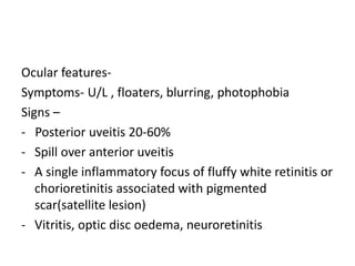
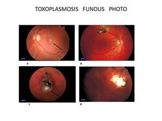
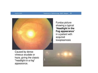
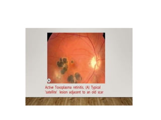
TOXOPLASMOSIS
DR. ANITA KUMARI
ANTERIOR SEGMENT FELLOW
SCEH, LAHAN
INTRODUCTION
• Toxoplasmosis is caused by Toxoplasma gondii
• Derived from Greek word “Toxon” means bow
• An obligate intracellular protozoan
• Cat - definitive host
• Intermediate hosts including mice, livestock, birds
and humans
PATHOGENESIS
TOXOPLASMOSIS LIFE CYCLE
CLINICAL FEATURES
• Systemic features-
1. Congenital toxoplasmosis
- Mother –symptom free/mild constitutional
manifestations
- Severity of fetal involvement related to the duration
of gestation at the time of maternal infection
- Fetal abnormalities range from chorioretinitis,
hydrocephalus, convulsions and intracerebral
calcifications to only mild effects, such as slightly
diminished vision
• Strabismus, nystagmus and microphthalmia can also
be seen, usually in infants infected early in
development
• Infants infected late in gestation may have a fever,
rash, hepatomegaly, splenomegaly, pneumonia,
chorioretinitis or a generalized infection.
• Many infected infants are asymptomatic at birth;
however, some will develop learning and visual
disabilities
CONGENITAL TOXOPLASMOSIS
2. Postnatal childhood acquisition
- 50% of of cases of childhood toxoplasmosis
3. Acquired toxoplasmosis in immunocompetent patients
- Subclinical in 80-90%
- Cervical lymphadenopathy, fever, malaise, pharyngitis are
common
- Early retinitis- upto 20%
4. Toxoplasmosis in immunocompromised patients
- Acquired or reactivation of pre-existing disease
- Meningoencephalitis, pneumonitis, retinochoroiditis
Ocular features-
Symptoms- U/L , floaters, blurring, photophobia
Signs –
- Posterior uveitis 20-60%
- Spill over anterior uveitis
- A single inflammatory focus of fluffy white retinitis or
chorioretinitis associated with pigmented
scar(satellite lesion)
- Vitritis, optic disc oedema, neuroretinitis
TOXOPLASMOSIS FUNDUS PHOTO
MANAGEMENT
Investigation
• Diagnosis is usually based on clinical examination
findings.
1. Serology - Toxoplasma IgG antibodies are detectable in
the serum within 1–2 weeks of initial infection
 Positivity to IgM antibodies usually means that infection
has been acquired within the last year, and so helps to
distinguish between acute (newly acquired) and chronic
infection
 Occasionally IgM is positive due to persistence or
reactivation following earlier infection
2. PCR testing of intraocular fluid sensitive (16– 67%) but highly
specific
 can be diagnostic in clinically uncertain cases
 Aqueous and vitreous probably give similar yields
3. Ocular fluid antibody assessment - Calculating the ratio
(Goldmann–Witmer coefficient) of specific IgG in aqueous
humour to that in serum seems to be a reasonably sensitive (48–
90%) investigation
4. Imaging
 Macular OCT will demonstrate any macular oedema if
vitritis is not preventative
 B-scan ultrasonic imaging can be used to exclude retinal
detachment in the presence of severe vitritis
 FAF may facilitate monitoring of inflammatory activity
Treatment
• Prednisolone (1 mg/kg) is given initially and tapered according to
clinical response
• Pyrimethamine - loading dose of 75–100 mg for 1–2 days
followed by 25–50 mg daily for 4 weeks in combination with oral
folinic (not folic) acid 5 mg thrice a week to retard
thrombocytopenia, leukopenia and folate deficiency.
• Sulfadiazine 1 g 4t/daily for 3–4 weeks is usually given in
combination with pyrimethamine.
• Intravitreal therapy with clindamycin (1 mg) and dexamethasone
(400 µg) may be as effective as triple therapy in reactivated
infection; two to three injections (two-weekly intervals) may be
required
• Azithromycin 250–500 mg daily- reducing the rate of recurrence of
retinochoroiditis
• Clarithromycin may be a good alternative to azithromycin
• Co-trimoxazole (trimethoprim 160 mg/sulfamethoxazole 800 mg)
twice daily in combination with prednisolone
• Clindamycin 300 mg four times daily may be added to triple
therapy or used instead of pyrimethamine
• Atovaquone theoretically attacks encysted bradyzoites, but does
not seem to prevent recurrence in vivo; dosing is 750 mg two to
four times daily
• Topical steroid and mydriatic may be given for anterior uveitis
• Antimicrobial maintenance therapy is used in
immunocompromised patients
COMPLICATION
• Serous rhegmatogenous /exudative RD
• Optic neuritis
• Choroidal neovascularization
• Periarteritis
• Serous macular detachment
TOXOPLASMOSIS VITREOUS HAZE WITH RD
Home work
• What are the parasites which affect eyes?
• What is triple therapy in toxoplasmosis?
• Define oocyst, bradyzoites, tachyzoites?
• Life cycle of toxoplasma gondii ?
• How to prevent toxoplasmosis infection ?
Thank you

Toxoplasmosis

  • 1.
    TOXOPLASMOSIS DR. ANITA KUMARI ANTERIORSEGMENT FELLOW SCEH, LAHAN
  • 2.
    INTRODUCTION • Toxoplasmosis iscaused by Toxoplasma gondii • Derived from Greek word “Toxon” means bow • An obligate intracellular protozoan • Cat - definitive host • Intermediate hosts including mice, livestock, birds and humans
  • 3.
  • 4.
    CLINICAL FEATURES • Systemicfeatures- 1. Congenital toxoplasmosis - Mother –symptom free/mild constitutional manifestations - Severity of fetal involvement related to the duration of gestation at the time of maternal infection - Fetal abnormalities range from chorioretinitis, hydrocephalus, convulsions and intracerebral calcifications to only mild effects, such as slightly diminished vision
  • 5.
    • Strabismus, nystagmusand microphthalmia can also be seen, usually in infants infected early in development • Infants infected late in gestation may have a fever, rash, hepatomegaly, splenomegaly, pneumonia, chorioretinitis or a generalized infection. • Many infected infants are asymptomatic at birth; however, some will develop learning and visual disabilities
  • 7.
  • 8.
    2. Postnatal childhoodacquisition - 50% of of cases of childhood toxoplasmosis 3. Acquired toxoplasmosis in immunocompetent patients - Subclinical in 80-90% - Cervical lymphadenopathy, fever, malaise, pharyngitis are common - Early retinitis- upto 20% 4. Toxoplasmosis in immunocompromised patients - Acquired or reactivation of pre-existing disease - Meningoencephalitis, pneumonitis, retinochoroiditis
  • 9.
    Ocular features- Symptoms- U/L, floaters, blurring, photophobia Signs – - Posterior uveitis 20-60% - Spill over anterior uveitis - A single inflammatory focus of fluffy white retinitis or chorioretinitis associated with pigmented scar(satellite lesion) - Vitritis, optic disc oedema, neuroretinitis
  • 10.
  • 13.
    MANAGEMENT Investigation • Diagnosis isusually based on clinical examination findings. 1. Serology - Toxoplasma IgG antibodies are detectable in the serum within 1–2 weeks of initial infection  Positivity to IgM antibodies usually means that infection has been acquired within the last year, and so helps to distinguish between acute (newly acquired) and chronic infection  Occasionally IgM is positive due to persistence or reactivation following earlier infection
  • 14.
    2. PCR testingof intraocular fluid sensitive (16– 67%) but highly specific  can be diagnostic in clinically uncertain cases  Aqueous and vitreous probably give similar yields
  • 15.
    3. Ocular fluidantibody assessment - Calculating the ratio (Goldmann–Witmer coefficient) of specific IgG in aqueous humour to that in serum seems to be a reasonably sensitive (48– 90%) investigation 4. Imaging  Macular OCT will demonstrate any macular oedema if vitritis is not preventative  B-scan ultrasonic imaging can be used to exclude retinal detachment in the presence of severe vitritis  FAF may facilitate monitoring of inflammatory activity
  • 16.
    Treatment • Prednisolone (1mg/kg) is given initially and tapered according to clinical response • Pyrimethamine - loading dose of 75–100 mg for 1–2 days followed by 25–50 mg daily for 4 weeks in combination with oral folinic (not folic) acid 5 mg thrice a week to retard thrombocytopenia, leukopenia and folate deficiency. • Sulfadiazine 1 g 4t/daily for 3–4 weeks is usually given in combination with pyrimethamine. • Intravitreal therapy with clindamycin (1 mg) and dexamethasone (400 µg) may be as effective as triple therapy in reactivated infection; two to three injections (two-weekly intervals) may be required
  • 17.
    • Azithromycin 250–500mg daily- reducing the rate of recurrence of retinochoroiditis • Clarithromycin may be a good alternative to azithromycin • Co-trimoxazole (trimethoprim 160 mg/sulfamethoxazole 800 mg) twice daily in combination with prednisolone • Clindamycin 300 mg four times daily may be added to triple therapy or used instead of pyrimethamine • Atovaquone theoretically attacks encysted bradyzoites, but does not seem to prevent recurrence in vivo; dosing is 750 mg two to four times daily • Topical steroid and mydriatic may be given for anterior uveitis • Antimicrobial maintenance therapy is used in immunocompromised patients
  • 18.
    COMPLICATION • Serous rhegmatogenous/exudative RD • Optic neuritis • Choroidal neovascularization • Periarteritis • Serous macular detachment
  • 19.
  • 21.
    Home work • Whatare the parasites which affect eyes? • What is triple therapy in toxoplasmosis? • Define oocyst, bradyzoites, tachyzoites? • Life cycle of toxoplasma gondii ? • How to prevent toxoplasmosis infection ?
  • 22.