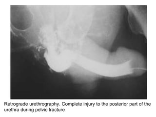
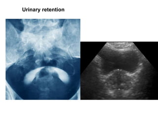
Emergency Urology
Traumatology of the urinary tract and
genitals
Introduction
• Urogenital trauma - 2% of all organ trauma cases.
• The most common of these are kidney trauma.
Kidney trauma
• Open: Plot-perforated or gunshot wound
• Closed: Car accidents (50-70%),
iatrogenic, sports, industrial, life traumatic
• Isolated
• Combined with other organ trauma
Mechanism of closed kidney injury. Left: A direct blow to the
abdominal area gives damaging force to the renal hilum. Right:
Falling from a height on the sitting causes renal hilum damage.
Classification of closed kidney injury
Grade I
• Renal contusion
• Subcapsular isolated
hematoma
• The wholeness of the
parenchyma is not
violated
• Micro or
macrohematuria
Grade II
• Isolated perirenal
hematoma
• Damage less than 1
cm deep in the
parenchyma
• No urinary
extravasation
Grade III
• Damage greater than
1 cm of the
parenchyma
• The collective system
is not damaged
• No urinary
extravasation
Grade IV
• Parenchymal damage
involves the cortical,
medullar layers, and the
collective system
• Damaged nutritious
blood vessels with
development of
thrombosis, hematoma
and urinoma.
Grade V
• Complete, multiple
abnormalities of renal
wholeness
• Shattered or
devascularized kidney
with active bleeding;
main renal vascular
laceration or avulsion
• The basic symptoms
– Pain
– Development of volumetric formation in the
lumbar region (hematoma, uro-hematoma)
– Micro or Macrohematuria
– Hemodynamic changes: blood pressure
variability, tachycardia (grade III-IV)
– Shock (class IV)
– Open trauma: Wounds in the lumbar region,
hematuria, secreting the urine from the wound
with blood.
• Diagnostics:
– Ultrasonography
• Injuries to the parenchyma, hematoma, urinoma.
– Radiography
• Skeletal system injuries
– Excretory urography
• Anatomical and functional status of the kidney
• Contralateral kidney condition
– Angiography
• Grade of parenchymal contrast, vascular wholeness
abnormalities, contrast extravasation
– Computed tomography
• ”The gold standard”
• Evaluating damaged kidney shape, location, blood
supply, type of injury.
– Dynamic scintigraphy
• In case of maintaining renal function: observation in
dynamics.
Computed tomography: Right kidney injury. Excessive
extravasation of the urine and large-size
retroperitoneal hematoma
Treatment of renal traumas
• Hospitalization
• Absolute indications of surgical intervention :
– Massive bleeding
– Multiple injuries of the renal parenchyma
– Damage of the magistral blood vessels
– All open, penetrated damage
Treatment according to the grade of injury :
• I-II grades
– Conservative (bed rest, antibiotic therapy, analgesic and
hemostatic therapy)
• III-IV grades
– Conservative if the patient is hemodynamically stable
– Surgical if the patient is hemodynamically unstable
• V grade
– Surgical
Surgery can be an organ-saving or a nephrectomy.
Multiple injuries of the renal parenchyma, shattered or
devascularized kidney with active bleeding, main renal
vascular laceration or avulsion are an evidence for
nephrectomy
Ureteral injury
• Open or closed
• The external factors of the ureteral injury is often
combined with damage to other organs
• Isolated trauma is mainly iatrogenic
– Pelvic surgery
• Including hysterectomy 54%
– Healing or diagnostic endoscopic manipulations
(catheterization, stent, extraction of the stone)
• Iatrogenic trauma of the ureter during gynecological operations
is due to the close relationship of the ureter with the uterine
blood vessels.
• Classification of ureter injuries :
– I grade: Only hematoma
– II grade: Less than 50% damage to the ureteral
wall circumference
– III grade: More than 50% damage to the ureteral
wall circumference
– IV grade: Complete disruption of ureteral
wholeness with its ends less than 2 cm
inaccuracy
– V grade: Complete disruption of ureteral
wholeness with its ends more than 2 cm
inaccuracy
• Symptoms
– Hematuria
– Pain
– Temperature rise
– Upper urinary tract obstruction
– In bilateral trauma: posttraumatic anuria
• Diagnostics
– Cystoscopy: blood flow from the ureter,
inability to pass a urethral catheter
– Excretory urography: Extravasation of contrast
into the damaged area
– Retrograde ureteropyelography : The catheter
faces a blockage, contrast is extravasated.
Excretory urography.
Middle third of right
ureteral injury by
extravasation of
contrast
• Complications in delayed diagnosis
– Stricture formation and hydronephrosis
– Uretero-vaginal or uretero-uterine fistula
• Treatment
– I-II grades: Retrograde catheterization or
stenting
– III grade: Surgery (Restoration of the wall of
the ureter with nodal sutures)
– IV-V grades: Uretero-ureteral anastomosis
– Urinoma and hematoma drainage
– In large size defects : Ureteroneocystotstomy
(Boari’s or Psoas-hitch), Plastic with ileum
Urinary bladder trauma
• Open
• Closed
• Iatrogenic
• Often combined with other traumas of the
internal organs
• Closed urinary bladder injuries
– Intraperitoneal
• Develops with the damage caused by blows to the
full bladder
• Penetrated trauma of the peritoneum-covered peak
• Pouring urine into the abdominal cavity
– Extraperitoneal
• Develops during fracture of the pelvic bones
• Mostly damaged inferolateral wall
• Infiltration of the pelvic cavity with urine
• Symptoms
– Pain in the lower abdomen and suprapubic area
with irradiation in the perineum, rectum and
urethra
– Difficult urination / acute urinary retention
– Hematuria
– Formation of urinoma
– A clinical picture of peritonitis during
intraperitoneal injury is also provided
• Diagnosis
– Excretory Urography
– Ascending urethrocystography
Extraperitoneal (left) and intraperitoneal (right) urinary bladder
injury on the cystogram.
• Treatment
– Anti-shock measures
– Drainage and restoration of bladder wholeness
– During intraperitoneal injury: Abdominal
drainage
– During extraperitoneal Injury: Drainage of the
small pelvic cavity and paravesical space
Urethral injury
• Mostly found in men
• Mechanism :
– Impact of external force (blowing to the
perineal area or falling on hard surface)
– Pelvic fractures
– Iatrogenic: Urethral instrumental examination
– Open traumatic injury
• Symptoms
–Urethrorrhagia
–Urinary retention
–Perineal, scrotal hematoma
• Diagnosis
–Retrograde urethrography
Retrograde urethrography. Complete injury to the posterior part of the
urethra during pelvic fracture
Retrograde urethrography. Bulbar urethral stricture
• Treatment
– Urinary bladder drainage
– During incomplete damage: Conservative
treatment (antibiotic therapy, local
hypothermia)
– During complete damage : Surgical treatment
– In advanced cases: Post-traumatic stricture
treatment (optical urethrotomy or open
reconstruction of the urethra)
External genital trauma
• Penile injury
– Open or closed
– Penile fracture: Rupture of one or both of the tunica
albuginea, the fibrous coverings that envelop the
penis's corpora cavernosa. It is caused by rapid blunt
force to an erect penis, usually during vaginal
intercourse.
– Clinical manifestation : Pain, swelling, changed colour,
formation of hematoma.
– Treatment
• Penile dislocation: Conservative
• Penile fracture: Surgical
• Open fracture: Wound debridement,
necrectomy
• Urinary bladder drainage with suprapubic
cystostomy, if the urethra is damaged.
Penile fracture
Emergency surgeon interference
Traumatic amputation of the penis
Paraphimosis
• Scrotal, testicular and epididymal injury
– Open or closed
– Clinical manifestations: Pain, increased
scrotum size, swelling, blue colored skin.
– Diagnosis: Ultrasonography.
– Differential diagnosis with testicular torsion.
Scrotal hematoma
Testicular trauma. Hematocele with ultrasonography
– Treatment
• Closed trauma: Conservative (bed rest, scrotal
fixation, antibiotic therapy, analgesic therapy)
• When developing a large-sized hematoma in a
scrotum: opening scrotum, revision, drainage.
• Breaking continuity of testis or epididymis:
Resection and wholeness restoration or
orchiectomy.
• Open trauma: Surgical intervention according to the
degree of injury (organ-saving operation or
orchiectomy).
Testicular torsion
Critical time of intervention : 6 hours!
3 hours
Testicular torsion Doppler-ultrasonography
Normal Torsion
Correction of the torsion + orchidopexy and contralateral
orchidopexy!
Testicular torsion
Testicular Torsion
> 6 hrs of Torsion
Orchiectomy
Manual correction of the torsion (detorsion)
70 %
Urinary retention
Urinary retention
Urinary retention
Urinary bladder catheterization
• Transurethral
• Suprapubic cystostomy

urology trauma (1).pptx

  • 1.
    Emergency Urology Traumatology ofthe urinary tract and genitals
  • 2.
    Introduction • Urogenital trauma- 2% of all organ trauma cases. • The most common of these are kidney trauma.
  • 3.
    Kidney trauma • Open:Plot-perforated or gunshot wound • Closed: Car accidents (50-70%), iatrogenic, sports, industrial, life traumatic • Isolated • Combined with other organ trauma
  • 4.
    Mechanism of closedkidney injury. Left: A direct blow to the abdominal area gives damaging force to the renal hilum. Right: Falling from a height on the sitting causes renal hilum damage.
  • 5.
  • 6.
    Grade I • Renalcontusion • Subcapsular isolated hematoma • The wholeness of the parenchyma is not violated • Micro or macrohematuria
  • 7.
    Grade II • Isolatedperirenal hematoma • Damage less than 1 cm deep in the parenchyma • No urinary extravasation
  • 8.
    Grade III • Damagegreater than 1 cm of the parenchyma • The collective system is not damaged • No urinary extravasation
  • 9.
    Grade IV • Parenchymaldamage involves the cortical, medullar layers, and the collective system • Damaged nutritious blood vessels with development of thrombosis, hematoma and urinoma.
  • 10.
    Grade V • Complete,multiple abnormalities of renal wholeness • Shattered or devascularized kidney with active bleeding; main renal vascular laceration or avulsion
  • 11.
    • The basicsymptoms – Pain – Development of volumetric formation in the lumbar region (hematoma, uro-hematoma) – Micro or Macrohematuria – Hemodynamic changes: blood pressure variability, tachycardia (grade III-IV) – Shock (class IV) – Open trauma: Wounds in the lumbar region, hematuria, secreting the urine from the wound with blood.
  • 12.
    • Diagnostics: – Ultrasonography •Injuries to the parenchyma, hematoma, urinoma. – Radiography • Skeletal system injuries – Excretory urography • Anatomical and functional status of the kidney • Contralateral kidney condition – Angiography • Grade of parenchymal contrast, vascular wholeness abnormalities, contrast extravasation
  • 13.
    – Computed tomography •”The gold standard” • Evaluating damaged kidney shape, location, blood supply, type of injury. – Dynamic scintigraphy • In case of maintaining renal function: observation in dynamics.
  • 14.
    Computed tomography: Rightkidney injury. Excessive extravasation of the urine and large-size retroperitoneal hematoma
  • 15.
    Treatment of renaltraumas • Hospitalization • Absolute indications of surgical intervention : – Massive bleeding – Multiple injuries of the renal parenchyma – Damage of the magistral blood vessels – All open, penetrated damage
  • 16.
    Treatment according tothe grade of injury : • I-II grades – Conservative (bed rest, antibiotic therapy, analgesic and hemostatic therapy) • III-IV grades – Conservative if the patient is hemodynamically stable – Surgical if the patient is hemodynamically unstable • V grade – Surgical Surgery can be an organ-saving or a nephrectomy. Multiple injuries of the renal parenchyma, shattered or devascularized kidney with active bleeding, main renal vascular laceration or avulsion are an evidence for nephrectomy
  • 17.
    Ureteral injury • Openor closed • The external factors of the ureteral injury is often combined with damage to other organs • Isolated trauma is mainly iatrogenic – Pelvic surgery • Including hysterectomy 54% – Healing or diagnostic endoscopic manipulations (catheterization, stent, extraction of the stone)
  • 18.
    • Iatrogenic traumaof the ureter during gynecological operations is due to the close relationship of the ureter with the uterine blood vessels.
  • 19.
    • Classification ofureter injuries : – I grade: Only hematoma – II grade: Less than 50% damage to the ureteral wall circumference – III grade: More than 50% damage to the ureteral wall circumference – IV grade: Complete disruption of ureteral wholeness with its ends less than 2 cm inaccuracy – V grade: Complete disruption of ureteral wholeness with its ends more than 2 cm inaccuracy
  • 20.
    • Symptoms – Hematuria –Pain – Temperature rise – Upper urinary tract obstruction – In bilateral trauma: posttraumatic anuria
  • 21.
    • Diagnostics – Cystoscopy:blood flow from the ureter, inability to pass a urethral catheter – Excretory urography: Extravasation of contrast into the damaged area – Retrograde ureteropyelography : The catheter faces a blockage, contrast is extravasated.
  • 22.
    Excretory urography. Middle thirdof right ureteral injury by extravasation of contrast
  • 23.
    • Complications indelayed diagnosis – Stricture formation and hydronephrosis – Uretero-vaginal or uretero-uterine fistula
  • 24.
    • Treatment – I-IIgrades: Retrograde catheterization or stenting – III grade: Surgery (Restoration of the wall of the ureter with nodal sutures) – IV-V grades: Uretero-ureteral anastomosis – Urinoma and hematoma drainage – In large size defects : Ureteroneocystotstomy (Boari’s or Psoas-hitch), Plastic with ileum
  • 25.
    Urinary bladder trauma •Open • Closed • Iatrogenic • Often combined with other traumas of the internal organs
  • 27.
    • Closed urinarybladder injuries – Intraperitoneal • Develops with the damage caused by blows to the full bladder • Penetrated trauma of the peritoneum-covered peak • Pouring urine into the abdominal cavity – Extraperitoneal • Develops during fracture of the pelvic bones • Mostly damaged inferolateral wall • Infiltration of the pelvic cavity with urine
  • 28.
    • Symptoms – Painin the lower abdomen and suprapubic area with irradiation in the perineum, rectum and urethra – Difficult urination / acute urinary retention – Hematuria – Formation of urinoma – A clinical picture of peritonitis during intraperitoneal injury is also provided
  • 29.
    • Diagnosis – ExcretoryUrography – Ascending urethrocystography Extraperitoneal (left) and intraperitoneal (right) urinary bladder injury on the cystogram.
  • 30.
    • Treatment – Anti-shockmeasures – Drainage and restoration of bladder wholeness – During intraperitoneal injury: Abdominal drainage – During extraperitoneal Injury: Drainage of the small pelvic cavity and paravesical space
  • 31.
    Urethral injury • Mostlyfound in men • Mechanism : – Impact of external force (blowing to the perineal area or falling on hard surface) – Pelvic fractures – Iatrogenic: Urethral instrumental examination – Open traumatic injury
  • 32.
    • Symptoms –Urethrorrhagia –Urinary retention –Perineal,scrotal hematoma • Diagnosis –Retrograde urethrography
  • 33.
    Retrograde urethrography. Completeinjury to the posterior part of the urethra during pelvic fracture
  • 34.
  • 35.
    • Treatment – Urinarybladder drainage – During incomplete damage: Conservative treatment (antibiotic therapy, local hypothermia) – During complete damage : Surgical treatment – In advanced cases: Post-traumatic stricture treatment (optical urethrotomy or open reconstruction of the urethra)
  • 36.
    External genital trauma •Penile injury – Open or closed – Penile fracture: Rupture of one or both of the tunica albuginea, the fibrous coverings that envelop the penis's corpora cavernosa. It is caused by rapid blunt force to an erect penis, usually during vaginal intercourse. – Clinical manifestation : Pain, swelling, changed colour, formation of hematoma.
  • 37.
    – Treatment • Peniledislocation: Conservative • Penile fracture: Surgical • Open fracture: Wound debridement, necrectomy • Urinary bladder drainage with suprapubic cystostomy, if the urethra is damaged.
  • 38.
  • 39.
  • 40.
  • 41.
    • Scrotal, testicularand epididymal injury – Open or closed – Clinical manifestations: Pain, increased scrotum size, swelling, blue colored skin. – Diagnosis: Ultrasonography. – Differential diagnosis with testicular torsion.
  • 42.
  • 43.
    Testicular trauma. Hematocelewith ultrasonography
  • 44.
    – Treatment • Closedtrauma: Conservative (bed rest, scrotal fixation, antibiotic therapy, analgesic therapy) • When developing a large-sized hematoma in a scrotum: opening scrotum, revision, drainage. • Breaking continuity of testis or epididymis: Resection and wholeness restoration or orchiectomy. • Open trauma: Surgical intervention according to the degree of injury (organ-saving operation or orchiectomy).
  • 45.
    Testicular torsion Critical timeof intervention : 6 hours! 3 hours
  • 46.
  • 47.
    Correction of thetorsion + orchidopexy and contralateral orchidopexy! Testicular torsion
  • 48.
    Testicular Torsion > 6hrs of Torsion Orchiectomy Manual correction of the torsion (detorsion) 70 %
  • 49.
  • 50.
  • 51.
  • 52.
    Urinary bladder catheterization •Transurethral • Suprapubic cystostomy