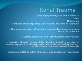
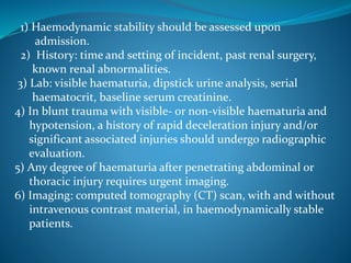
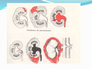
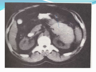
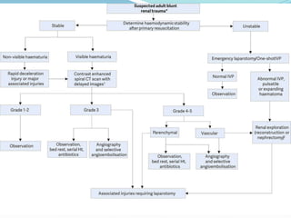
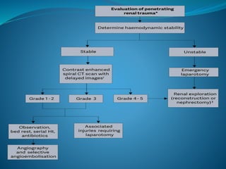
Table 1: Injury severity scale for the kidney*#
Grade
Description
1 Contusion or non-expanding subcapsular haematoma, no laceration
2 Non-expanding perirenal haematoma, cortical laceration < 1 cm deep
without extravasation
3 Cortical laceration > 1 cm without urinary extra-vasation
4 Laceration: through corticomedullary junction into collecting system or
vascular: segmental renal artery or vein injury with contained haematoma,
or partial vessel laceration, or vessel thrombosis
5Laceration: shattered kidney or vascular: renal pedicle injury or avulsion
1) Haemodynamic stability should be assessed upon
admission.
2) History: time and setting of incident, past renal surgery,
known renal abnormalities.
3) Lab: visible haematuria, dipstick urine analysis, serial
haematocrit, baseline serum creatinine.
4) In blunt trauma with visible- or non-visible haematuria and
hypotension, a history of rapid deceleration injury and/or
significant associated injuries should undergo radiographic
evaluation.
5) Any degree of haematuria after penetrating abdominal or
thoracic injury requires urgent imaging.
6) Imaging: computed tomography (CT) scan, with and without
intravenous contrast material, in haemodynamically stable
patients.
Ureteral Trauma
 Ureteral injuries are quite rare - most are iatrogenic. They
are often missed intra-operatively, usually involve the lower
ureter, and may result in severe sequelae.
 Risk factors include advanced malignancy, prior surgery or
irradiation - i.e. conditions which alter the normal
anatomy.
 Preoperative prophylactic stents do not prevent ureteral
injury, but may assist in its detection.
 External ureteral trauma usually accompanies severe
abdominal and pelvic injuries.
 Gunshot wounds account for the majority of penetrating
ureteral trauma, while motor vehicle accidents account for
most blunt injuries.
Diagnostic evaluation
 A high index of suspicion of ureteral injury should be
maintained as the majority of cases are diagnosed late,
predisposing the patient to pain, infection, and renal
function impairment.
 Haematuria is an unreliable indicator.
 Extravasation of contrast material in computed
tomography (CT) is the hallmark sign of ureteral
trauma.
 In unclear cases, a retrograde or antegrade urography
is required for confirmation
•Partial injury can be managed with ureteral stenting or
urinary diversion by a nephrostomy.
• In complete injuries, ureteral reconstruction following
temporary urinary diversion is required.
• The type of repair procedure depends on the site of the
injury
• Proximal- and mid-ureteral injuries can often be
managed by primary uretero-ureterostomy, while a distal
injury can be treated with ureteral reimplantation
Ureteral reconstruction options by
site of injury
 Site of injury Reconstruction options
 Upper ureter
 Uretero-ureterostomy
 Transuretero-ureterostomy
 Uretero-calycostomy
 Mid ureter
 Uretero-ureterostomy
 Transuretero-ureterostomy
 Ureteral re-implantation and a Boari flap
 Lower ureter
 Ureteral re-implantation
 Ureteral re-implantation with a psoas hitch
 Complete
 Ileal interposition graft
 Autotransplantation
 Bladder injuries can be due to external (blunt or
penetrating) or iatrogenic trauma.
 Iatrogenic trauma is caused by external laceration or
internal perforation (mainly during transurethral
resection of the bladder)
 Blunt bladder injuries are strongly associated with
pelvic fractures.
 Bladder injuries are classified as extraperitoneal,
intraperitoneal or combined.
External trauma
 Clinical signs and symptoms
 Cardinal sign: visible haematuria.
 Others: abdominal tenderness, inability to void,
bruises over the suprapubic region, and abdominal
distension (in case of urinary ascites).
 Penetrating bladder injury: entrance- and exit wounds
in lower abdomen or perineum.
 Bloody urethrorrhagia: suspect concomitant urethral
injury.
External perforation: extravasation of urine, visible
laceration, clear fluid in the surgical field,
appearance of the bladder catheter, and blood
and/or gas (in case of laparoscopy) in the urine bag.
Internal perforation: fatty tissue or bowel between
detrusor muscle fibres, inability to distend the
bladder, low return of irrigation fluid and/or
abdominal distension.
Post-operative symptoms of unrecognised bladder
perforation: haematuria, lower abdominal pain,
abdominal distension, ileus, peritonitis, sepsis, urine
imaging
 Cystography (conventional or CT-cystography)
 Fill the bladder with at least 350 mL of dilute contrast
material.
 Computed tomography cystography is preferred in case of
other possible abdominal injuries or causes of abdominal
pain.
 Standard evaluation for external trauma and in case of
suspicion of an iatrogenic bladder injury in the post-
operative setting.
 Imperative in case of visible haematuria combined with
pelvic fracture
Cystoscopy
 To detect intra-operative bladder injuries.
 Recommended after minimally invasive synthetic sub-
urethral sling operations by retropubic route.
 Optional after any other type of sling procedure or
transvaginal mesh procedure.
Surgical repair (two-layer vesicorraphy)
Conservative management
(urinary catheter)
 Conservative management is an option for small,
uncomplicated, iatrogenic intraperitoneal bladder
perforations.
 In the absence of bladder neck involvement and/or
associated injuries that require surgical intervention,
extraperitoneal bladder ruptures caused by blunt trauma
are managed conservatively.
 Post-operative recognised extraperitoneal perforation.
 Blunt extraperitoneal perforation.
 Iatrogenic internal extraperitoneal perforation.
 Small internal intraperitoneal perforation in absence of
ileus and peritonitis. Placement of an intraperitoneal drain
is optional.
Injuries to the anterior urethra (AU) are caused by trauma
during sexual intercourse (associated with penile fracture),
penetrating trauma, placement of penile constriction bands,
and from iatrogenic trauma e.g. endoscopic instruments,
catheterisation.
Injuries to the posterior urethra (PU) occur with pelvic
fractures, mostly as a result of motor vehicle accidents. The
male PU is injured in 4-19% of pelvic fractures, and the female
urethra in 0-6% of all pelvic fractures.
The combination of straddle fractures with diastasis of the
sacroiliac joint has the highest risk of urethral injury.
Injuries can vary from simple stretching to partial rupture to
complete disruptions.
Urethral injuries in women are rare.
Diagnostic evaluation
 Blood at the external urethral meatus is the most common clinical sign, and indicates the
need for further diagnostic work up.
 Although non-specific, haematuria on a first voided specimen may indicate urethral
injury. The amount of urethral bleeding correlates poorly with the severity of injury.
 Pain on urination or inability to void may indicate disruption.
 Blood at the vaginal introitus is present in more than 80% of female patients with pelvic
fractures and co-existing urethral injuries.
 Rectal examination may reveal a “high riding” prostate. However, this is an unreliable
finding. Blood on the examination finger is suggestive of a rectal injury associated with
pelvic fracture.
 Urethral bleeding or urinary extravasation can cause penile and scrotal swelling and
haematoma.
 Retrograde urethrography is the gold standard for evaluating urethral injury and urethral
catheterisation should be avoided until the urethra is imaged.
 In an unstable patient, however, an attempt can be made to pass a urethral catheter
(gently, by someone with urological experience). If this is not possible, a suprapubic
catheter is inserted and a retrograde urethrogram is performed later.
 In females, urethroscopy may be an important adjunct for the identification and staging
of urethral injuries.
Management
 While intervention should be guided by the clinical
circumstances, the following treatment is suggested:
 Retrograde urethrography is the gold standard for
evaluating urethral injuries;
 Delayed formal urethroplasty is the procedure of
choice for the treatment of posterior urethral
distraction defects;
 Partial posterior urethral ruptures should be treated
by urethral or suprapubic catheterisation;
 Blunt anterior urethral injuries should be treated by
suprapubic diversion.
Iatrogenic urethral trauma
 Most commonly caused by urethral instrumentation,
and results in stricture formation.
 Due to their variable location and severity, they often
require different management strategies.
 Short and flimsy strictures can be treated by
urethrotomy.
 If the stricture is longer or denser, urethroplasty
should be considered.
Genital Trauma
Penile fracture
 Usually results from trauma to the erect penis during sexual intercourse or
masturbation.
 Sudden cracking or popping sound, pain and immediate detumescence.
 Local swelling of the penile shaft is seen and this may extend to the lower
abdominal wall.
 The rupture of the tunica may be palpable.
 Thorough history and examination confirms diagnosis.
 Imaging ultrasound or magnetic resonance imaging may be useful.
 Management
 Subcutaneous haematoma, without associated rupture of the cavernosal tunica
albuginea does not require surgical intervention. Non-steroidal analgesics and
ice-packs are recommended.
 In penile fracture, early surgical intervention with closure of the tunica
albuginea is recommended.
 Intra-operative flexible cystoscopy is useful to diagnose urethral injury and to
further localise tunical damage.
 Conservative management of penile fracture is not recommended.
?
urology trauma english.pptx

urology trauma english.pptx

  • 1.
    Table 1: Injuryseverity scale for the kidney*# Grade Description 1 Contusion or non-expanding subcapsular haematoma, no laceration 2 Non-expanding perirenal haematoma, cortical laceration < 1 cm deep without extravasation 3 Cortical laceration > 1 cm without urinary extra-vasation 4 Laceration: through corticomedullary junction into collecting system or vascular: segmental renal artery or vein injury with contained haematoma, or partial vessel laceration, or vessel thrombosis 5Laceration: shattered kidney or vascular: renal pedicle injury or avulsion
  • 2.
    1) Haemodynamic stabilityshould be assessed upon admission. 2) History: time and setting of incident, past renal surgery, known renal abnormalities. 3) Lab: visible haematuria, dipstick urine analysis, serial haematocrit, baseline serum creatinine. 4) In blunt trauma with visible- or non-visible haematuria and hypotension, a history of rapid deceleration injury and/or significant associated injuries should undergo radiographic evaluation. 5) Any degree of haematuria after penetrating abdominal or thoracic injury requires urgent imaging. 6) Imaging: computed tomography (CT) scan, with and without intravenous contrast material, in haemodynamically stable patients.
  • 7.
    Ureteral Trauma  Ureteralinjuries are quite rare - most are iatrogenic. They are often missed intra-operatively, usually involve the lower ureter, and may result in severe sequelae.  Risk factors include advanced malignancy, prior surgery or irradiation - i.e. conditions which alter the normal anatomy.  Preoperative prophylactic stents do not prevent ureteral injury, but may assist in its detection.  External ureteral trauma usually accompanies severe abdominal and pelvic injuries.  Gunshot wounds account for the majority of penetrating ureteral trauma, while motor vehicle accidents account for most blunt injuries.
  • 8.
    Diagnostic evaluation  Ahigh index of suspicion of ureteral injury should be maintained as the majority of cases are diagnosed late, predisposing the patient to pain, infection, and renal function impairment.  Haematuria is an unreliable indicator.  Extravasation of contrast material in computed tomography (CT) is the hallmark sign of ureteral trauma.  In unclear cases, a retrograde or antegrade urography is required for confirmation
  • 9.
    •Partial injury canbe managed with ureteral stenting or urinary diversion by a nephrostomy. • In complete injuries, ureteral reconstruction following temporary urinary diversion is required. • The type of repair procedure depends on the site of the injury • Proximal- and mid-ureteral injuries can often be managed by primary uretero-ureterostomy, while a distal injury can be treated with ureteral reimplantation
  • 10.
    Ureteral reconstruction optionsby site of injury  Site of injury Reconstruction options  Upper ureter  Uretero-ureterostomy  Transuretero-ureterostomy  Uretero-calycostomy  Mid ureter  Uretero-ureterostomy  Transuretero-ureterostomy  Ureteral re-implantation and a Boari flap  Lower ureter  Ureteral re-implantation  Ureteral re-implantation with a psoas hitch  Complete  Ileal interposition graft  Autotransplantation
  • 12.
     Bladder injuriescan be due to external (blunt or penetrating) or iatrogenic trauma.  Iatrogenic trauma is caused by external laceration or internal perforation (mainly during transurethral resection of the bladder)  Blunt bladder injuries are strongly associated with pelvic fractures.  Bladder injuries are classified as extraperitoneal, intraperitoneal or combined.
  • 13.
    External trauma  Clinicalsigns and symptoms  Cardinal sign: visible haematuria.  Others: abdominal tenderness, inability to void, bruises over the suprapubic region, and abdominal distension (in case of urinary ascites).  Penetrating bladder injury: entrance- and exit wounds in lower abdomen or perineum.  Bloody urethrorrhagia: suspect concomitant urethral injury.
  • 16.
    External perforation: extravasationof urine, visible laceration, clear fluid in the surgical field, appearance of the bladder catheter, and blood and/or gas (in case of laparoscopy) in the urine bag. Internal perforation: fatty tissue or bowel between detrusor muscle fibres, inability to distend the bladder, low return of irrigation fluid and/or abdominal distension. Post-operative symptoms of unrecognised bladder perforation: haematuria, lower abdominal pain, abdominal distension, ileus, peritonitis, sepsis, urine
  • 17.
    imaging  Cystography (conventionalor CT-cystography)  Fill the bladder with at least 350 mL of dilute contrast material.  Computed tomography cystography is preferred in case of other possible abdominal injuries or causes of abdominal pain.  Standard evaluation for external trauma and in case of suspicion of an iatrogenic bladder injury in the post- operative setting.  Imperative in case of visible haematuria combined with pelvic fracture
  • 18.
    Cystoscopy  To detectintra-operative bladder injuries.  Recommended after minimally invasive synthetic sub- urethral sling operations by retropubic route.  Optional after any other type of sling procedure or transvaginal mesh procedure.
  • 19.
  • 20.
    Conservative management (urinary catheter) Conservative management is an option for small, uncomplicated, iatrogenic intraperitoneal bladder perforations.  In the absence of bladder neck involvement and/or associated injuries that require surgical intervention, extraperitoneal bladder ruptures caused by blunt trauma are managed conservatively.  Post-operative recognised extraperitoneal perforation.  Blunt extraperitoneal perforation.  Iatrogenic internal extraperitoneal perforation.  Small internal intraperitoneal perforation in absence of ileus and peritonitis. Placement of an intraperitoneal drain is optional.
  • 21.
    Injuries to theanterior urethra (AU) are caused by trauma during sexual intercourse (associated with penile fracture), penetrating trauma, placement of penile constriction bands, and from iatrogenic trauma e.g. endoscopic instruments, catheterisation. Injuries to the posterior urethra (PU) occur with pelvic fractures, mostly as a result of motor vehicle accidents. The male PU is injured in 4-19% of pelvic fractures, and the female urethra in 0-6% of all pelvic fractures. The combination of straddle fractures with diastasis of the sacroiliac joint has the highest risk of urethral injury. Injuries can vary from simple stretching to partial rupture to complete disruptions. Urethral injuries in women are rare.
  • 22.
    Diagnostic evaluation  Bloodat the external urethral meatus is the most common clinical sign, and indicates the need for further diagnostic work up.  Although non-specific, haematuria on a first voided specimen may indicate urethral injury. The amount of urethral bleeding correlates poorly with the severity of injury.  Pain on urination or inability to void may indicate disruption.  Blood at the vaginal introitus is present in more than 80% of female patients with pelvic fractures and co-existing urethral injuries.  Rectal examination may reveal a “high riding” prostate. However, this is an unreliable finding. Blood on the examination finger is suggestive of a rectal injury associated with pelvic fracture.  Urethral bleeding or urinary extravasation can cause penile and scrotal swelling and haematoma.  Retrograde urethrography is the gold standard for evaluating urethral injury and urethral catheterisation should be avoided until the urethra is imaged.  In an unstable patient, however, an attempt can be made to pass a urethral catheter (gently, by someone with urological experience). If this is not possible, a suprapubic catheter is inserted and a retrograde urethrogram is performed later.  In females, urethroscopy may be an important adjunct for the identification and staging of urethral injuries.
  • 23.
    Management  While interventionshould be guided by the clinical circumstances, the following treatment is suggested:  Retrograde urethrography is the gold standard for evaluating urethral injuries;  Delayed formal urethroplasty is the procedure of choice for the treatment of posterior urethral distraction defects;  Partial posterior urethral ruptures should be treated by urethral or suprapubic catheterisation;  Blunt anterior urethral injuries should be treated by suprapubic diversion.
  • 24.
    Iatrogenic urethral trauma Most commonly caused by urethral instrumentation, and results in stricture formation.  Due to their variable location and severity, they often require different management strategies.  Short and flimsy strictures can be treated by urethrotomy.  If the stricture is longer or denser, urethroplasty should be considered.
  • 26.
    Genital Trauma Penile fracture Usually results from trauma to the erect penis during sexual intercourse or masturbation.  Sudden cracking or popping sound, pain and immediate detumescence.  Local swelling of the penile shaft is seen and this may extend to the lower abdominal wall.  The rupture of the tunica may be palpable.  Thorough history and examination confirms diagnosis.  Imaging ultrasound or magnetic resonance imaging may be useful.  Management  Subcutaneous haematoma, without associated rupture of the cavernosal tunica albuginea does not require surgical intervention. Non-steroidal analgesics and ice-packs are recommended.  In penile fracture, early surgical intervention with closure of the tunica albuginea is recommended.  Intra-operative flexible cystoscopy is useful to diagnose urethral injury and to further localise tunical damage.  Conservative management of penile fracture is not recommended.
  • 27.